Varieties
The main types of tumors are as follows:
- Epithelial - appear from epithelial cells, skin, and organ mucosa. This type is characterized by the appearance of a benign neoplasm (adenoma, papilloma), malignant (adenocarcinoma, squamous cell carcinoma, etc.).
- Nonepithelial - are the most extensive. Tumors can be of various natures: hemangioma, lipoma, chondrosarcoma, leiomyoma and others.
- Mixed - this type damages all layers of the walls of the organ, has instant disintegration with replacement of ulcers in that place. This type implies carcinoid formation, malignant lymphoma, carcinosarcoma.
Growing benign neoplasms are intraluminal and intramural. The first include polyps, adenomas, papillomas, the second - leiomyomas, esophageal cysts. Other types are observed extremely rarely.
Diseases characteristic of oncology
The development of a tumor can lead to the following pathologies:
- Hemangioma is a very rare disease, it has a soft consistency, with rather vague boundaries. The pathology is a non-epithelial vascular neoplasm of a benign nature, located along the wall of the organ, as well as tissues close to it. Hemangioma can expand to a large diameter, compress blood vessels and organs, disrupting the nutrition of cells.
- Blastoma is characterized by rapid tissue growth. It can be benign or malignant. It differs in that the first does not cover other tissues and does not expand metastases. Current medicine effectively cures non-cancerous tumors, but the malignant form is very severe due to hidden initial signs.
- Leiomyoma is a common type of tumor. Submucosal formation appears from the muscular lining of the esophagus. It has a round shape and makes its way into the lumen of the organ. It also appears asymptomatically for a long time.
- Hyperplasia is a precancerous condition, where tissue growth is observed with the replacement of the natural cellular structure with a pathological one. The main assumption for the occurrence of hyperplasia is its chemical disorder, which occurs due to the presence of a protracted GERD disease in a person. There is a disorder of the gastrointestinal tract functions, reflux of acid from the stomach into the esophagus.
In addition, dysplasia and metaplasia of the esophagus are distinguished. Pathologies of the organ are often associated with glycogen acanthosis (an oval-shaped white plaque located along the wall of the esophagus).
Classification
In medicine, experts distinguish between many different types of hyperplasia:
- Focal developing hyperplasia of the gastric mucosa. The first stage of development of the anomaly, when certain polyps begin to appear. Focal diagnosed hyperplasia of the stomach covers only some areas (“foci”), which is why it got its name. The lesions look like growths of various shapes and sizes, painted in a different color, so they are clearly visible during examination. Formations arise at the site of previously received damage or erosion.
- Follicular identified gastric hyperplasia. This type of pathology is often diagnosed. Lymphatic cells begin to grow. The reasons for the development of the anomaly are different: the influence of carcinogens, hormonal imbalance, the presence of the bacterium Helicobacter pylori, stressful situations and much more. A distinctive feature of this type of disease is the formation of follicles in the stomach.
- Hyperplasia of the integumentary pitted epithelium of the stomach. A dangerous type of pathology that can contribute to the appearance of a malignant tumor in the intestine. The structure of the epithelium changes under the influence of unfavorable factors: cells grow and become larger.
- Hyperplasia involving the antrum of the stomach. The antrum is the last part of the organ before it exits into the intestine. In this place, with the development of hyperplasia, multiple small growths, pits and ridges begin to form.
- Foveal hyperplasia of the gastric mucosa. This type of pathology is characterized by an increase in the length of the folds of the mucous membrane and an increase in their curvature. Pathology occurs due to prolonged inflammation or self-administration of anti-inflammatory drugs.
There are also other types of pathology: glandular, polypoid, lymphoid.
Hyperplasia of the antrum of the stomach
What is epithelium?
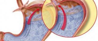
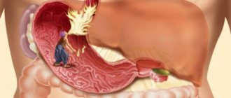
The gastric epithelium is a collection of columnar cells. They are called single-layer columnar epithelium. Against the background of prolonged irritation of the mucous membrane of the esophagus by gastric juice, for example, with gastroesophageal reflux, when the contents of the stomach are thrown into the esophagus, the risk of developing the gastric variant of esophageal metaplasia increases.
The essence of the disease is the replacement of the stratified squamous epithelium of the esophagus with a single-layer cylindrical epithelium, characteristic of the stomach. The part of the esophagus in which the mucous membrane of the organ passes into the stomach is most susceptible to tissue replacement. It is in the area of connection of the two organs that the most intense irritation with hydrochloric acid occurs against the background of gastroesophageal reflux.
Types of gastric hyperplasia
The classification of gastric hyperplasia is determined by the nature of the tissue confirmation and the type of cells that have undergone proliferation.
Focal gastric hyperplasia
Focal hyperplasia of the stomach is an early form of a polyp, manifests itself in the form of a benign tumor in one of the sectors of the stomach, in the so-called “focus”, hence the name. It can have a different size, usually resembles a small growth, with a modified structure, this can be seen especially well during a study with contrast, when paint gets on the foci of hyperplasia, they immediately change color and stand out against the background of normal tissues. The outgrowths may look like a tubercle or have a stalk; they can be single or multiple. They are also called wart hyperplasia.
Most often they are transformed from erosion of the mucous membrane. Detected by endoscopic examination.
Complications
A person refuses to eat, a paroxysmal cough occurs, and tracheal perforation and fistulas with subsequent expansion into blood vessels become complications. The state of health noticeably worsens when metastases spread to the liver, pancreas, lungs, and brain.
Replacing esophageal tissue with single-layer columnar epithelium can cause a number of serious complications, including:
- Transformation of foci of metaplasia into malignant neoplasms. The most common form of esophageal cancer is adenocarcinoma.
- Hemorrhagic syndrome localized in foci of metaplasia and damage to the esophagus.
- The appearance of peptic-type strictures. These are adhesions of connective tissue that narrow the lumen of the esophagus at their location and provoke the development of dysphagia, which consists in disrupting the process of swallowing food.
To prevent such complications, it is important to diagnose metaplasia in time and receive appropriate treatment aimed at reducing the size of the lesions and their number.
Prevention
To prevent the development of hyperplasia, you need to monitor your diet. It is imperative to follow a meal schedule and eat at the same time. The diet should be rich and balanced. Drink alcohol and carbonated drinks to a minimum.
It is also necessary to treat any other stomach diseases, strictly following the treatment regimen prescribed by the doctor. Avoid their chronicity, since chronic pathologies can also cause hyperplasia. Also, to prevent the disease, it is necessary to lead an active lifestyle and avoid stressful situations.
The main measure to prevent the appearance of hyperplasia is to control the diet and regimen. This is explained by the fact that very often the cause of the development of the pathological process is a passion for low-quality, fatty, heavy food. In addition, it is necessary to strictly follow the treatment regimen for other diseases, avoid getting into stressful situations and lead an active lifestyle.
Focal and diffuse metaplasia
In addition, there are such forms of esophageal metaplasia as diffuse and focal. The latter is characterized by the replacement of certain tubular glands, occurring as a result of an inflammatory process or cell renewal of the gastrointestinal tract. Diffuse metaplasia is determined by damage to the mucous membrane of the stomach and esophagus without cell death and disruption of tissue structure.
As already mentioned, it would be logical to consider Barrett's esophagus as a complication of reflux esophagitis. Essentially, the body is trying to protect the esophageal epithelium from erosion by gastric juice by replacing normal cells with cells typical of the duodenum, which are able to withstand high acidity.
However, a number of factors are well known that increase the risk of its development:
- Presence of gastroesophageal reflux.
- Transition to 60 years of age. Barrett's esophagus is rare in young people.
- Male gender. Men are more likely to develop the disease (twice as likely as women).
- Belonging to the Caucasian race.
- Consuming excessive amounts of alcohol.
- Smoking tobacco.
- Obesity. It increases the risk of reflux disease and therefore the formation of Barrett's esophagus.
Etiology
Gastric hyperplasia develops due to unfinished treatment of diseases of the gastrointestinal tract. As a result, active cell growth begins and polyps appear.
The main causes of hyperplasia:
- changes in hormonal balance, especially when the amount of estrogen is increased;
- genetic predisposition, in particular adenomatous polyposis (characteristic of polyps in the stomach) - if the pathology is diagnosed in a woman, the disease can be inherited by a daughter or granddaughter;
- long-term use of medications that can affect changes in the structure of the gastric mucosa;
- unfavorable environment - a pathological increase in the number of cells may begin.
The cause is the bacterium Helicobacter pylori and other infectious diseases.
Forecast
If a malignant tumor is not actively treated, the subsequent prognosis will be very poor. If there is no action, the patient will not live long, a maximum of 9 months.
When the pathology is revealed at stages 1-2 of development, surgical excision is performed, the patient’s condition returns to normal, and nothing threatens him.
Stage 3-4 of the disease has an uncertain prognosis due to the expansion of metastases. Here, current medicine can slow down the course of metastasis or stop it for a while. The prognosis for stage 3 cancer is quite unfavorable, the survival rate is no more than 15%.
Therefore, it is very important to recognize the disease in time, establish an accurate diagnosis, and determine a suitable treatment regimen in combination with current medications. All this will be the key to a successful recovery.
Risk factors
Cylindrical cell metaplasia of the esophagus occurs under the influence of the same provoking factors that lead to the appearance of gastroesophageal reflux disease. Experts identify the following risk factors:
- Decreased tone of the esophageal sphincter in the lower part. This occurs due to insufficient innervation, when the sphincter does not compress tightly and gastric juice is thrown back into the esophagus.
- Changes in the structure of the congenital type. Under the influence of genetic factors, the diameter of the lower part of the esophageal sphincter increases or decreases.
- Previously experienced inflammatory processes. Localized in the lower part of the esophagus, such processes provoke the appearance of scars in the organ and lead to incomplete compression of the sphincter.
- Varicose veins in severe form. In this case, it is the nodes formed during varicose veins that prevent the sphincter from completely closing.
- Disorder of the motor-evacuation function of the stomach and esophagus. Against the background of such a disorder, antiperistalsis is observed, when the walls of the organs begin to move, leading to the movement of food masses into the upper parts of the esophagus.
The listed factors can provoke the appearance of gastroesophageal reflux, which in turn can cause erosive type gastritis, which later develops into gastric and intestinal metaplasia of the esophagus.
Diagnostics
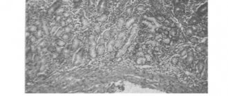
We figured out what hyperplasia is. It would be quite difficult to make a diagnosis based only on symptoms, so the patient is sent for gastroscopy. An endoscope, which has a light source and cameras, is inserted into the patient's stomach. The doctor can examine the walls of the stomach and note any changes. The doctor also performs a biopsy of the stomach walls. Histology helps to make an accurate diagnosis, exclude oncology, and also helps to identify the type of hyperplasia and the cause of its occurrence.
Treatment: general principles
The basic principles of treatment of esophageal metaplasia are the elimination and reduction of the size of foci of a single-layer columnar epithelial layer in the esophageal mucosa. Therapy is carried out comprehensively and includes both conservative treatment and surgical methods, and general recommendations on nutrition and lifestyle.
Experts believe that an important stage in the treatment of gastric metaplasia of the esophagus is the implementation of general recommendations that allow you to relieve the symptoms of the pathology. In addition, following the rules listed below increases the effectiveness of other treatment methods:
- Maintaining a balanced diet. Hot and cold dishes, as well as fried, fatty, smoked foods, marinades, pickles and spices are excluded.
- Supporting proper nutrition. You should eat at least five times a day in small portions. The final meal should be at least two hours before going to bed. You should try not to lie down after eating.
- Bringing body weight back to normal, subject to its deficiency or excess.
- Quitting bad habits, including alcohol and smoking. Such habits contribute to even greater irritation of the mucous membrane of the esophagus and increase the risk of increasing lesions of the disease.
- Physical activity should be moderate. Lifting heavy objects should be avoided as this increases the risk of developing pressure within the peritoneum.
The listed rules will increase the effectiveness of the treatment and prevent surgical intervention by eliminating small foci of gastric metaplasia of the esophageal mucosa.
Symptoms of esophageal polyps
Usually the disease is not accompanied by severe symptoms for a long period, since the size of neoplasias in the initial stages is small and they grow slowly. Over time, the patient begins to feel slight discomfort and pain when swallowing solid food.
The disease is characterized by nausea after eating and moderate pain
When tumors become larger, the following symptoms are added:
- breathing problems;
- belching;
- nausea and vomiting after eating;
- constant or periodic moderate pain in the chest.
If a tumor on a thin stalk is localized in the lower part of the esophagus, most often patients complain of sharp pain in the epigastrium. This symptom is a consequence of strangulation of the growth when the lower esophageal sphincter (cardia) closes.
Drug treatment
Conservative treatment of esophageal metaplasia involves taking medications whose action is aimed at reducing the amount of gastric juice reflux into the esophagus. For this purpose, specialists prescribe the following groups of medications to patients:
- Antacids. The effect of these drugs is to reduce the acidic qualities of gastric juice. Antacids include Maalox, Almagel, Phosphalugel, etc.
- Proton pump blockers. These drugs inhibit the production of hydrochloric acid. The most popular drug in this group for esophageal metaplasia is Omeprazole.
- H1-histamine receptor blockers. Also used to reduce the amount of acid produced. The most commonly prescribed drug is Famotidine.
- Prokinetics. These drugs have a stimulating effect on the peristalsis of the stomach and esophagus, which prevents the reflux of food mass into the upper part of the digestive organs. One of the brightest representatives of this group of medicines is Motilium.
The blockers mentioned above are prescribed only after checking the acidity of the gastric juice, as well as determining the intensity of its peristalsis. Only small foci of metaplasia of the esophageal epithelium can be treated with medication.