- It's a dull pain;
- lack of appetite;
- regular stomach upsets.
In later stages, more severe forms are observed:
- unpleasant belching;
- pain in the right hypochondrium;
- yellow coating on the tongue;
- as a result of the disease, jaundice develops;
- urine color is brown.
A person suffers from itching on the back or limbs. Due to swelling of the legs, varicose veins develop and there may be a risk of bleeding. At the terminal stage of the disease, metastases occur in the body, the person loses weight before our eyes and weakens. It is no longer possible to cure.
Diagnostics consists of notifying the patient’s place of residence and lifestyle. A clinical examination involves taking a general blood and urine test. At the first symptoms of a disorder in the body, it is necessary to undergo an ultrasound of the internal organs, X-rays, and MRI. Also, if alveococcosis is suspected, the attending physician prescribes a microscopic examination of the patient’s sputum.
The above steps will help you start treatment in a timely manner.
Etiology, pathogenesis and pathological anatomy
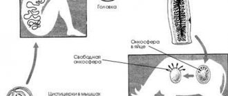
The causative agent of alveococcosis is the alveococcus Alveococcus multilocularis, which belongs to the genus Alveococcus, family Taeniidae, order Cyclophyllidea, class Cestoidea. There may be two subspecies - A. multilocularis multilocularis, common in Europe, and alveococcosis multilocularis sibiricensis, common in the Arctic, Siberia, and Japan.
Alveococcus is a small tapeworm, reaching a length of 1.3-2.2 mm. It consists of a head (scolex), equipped with 4 muscle suckers and 28-32 chitinous hooks arranged in two rows, a neck and 3-4 segments. Of these, the first two are asexual, the third is hermaphrodite, and the fourth (large) is mature and contains a sac-like uterus filled with eggs. The larval form is a conglomerate of small vesicles (parasitic node), connected by connective tissue and characterized by exogenous reproduction and infiltrating growth. The cavity of the bubbles contains a small amount of viscous liquid or thick dark mass. Some of the vesicles have scolex of the same structure as in adults. In cross-section, the parasitic node has a cellular structure with a decay cavity in the center. Alveococcus is a biohelminth that develops with a change of hosts. The definitive hosts are arctic fox, fox, dog, and less commonly wolf, coyote, and cat. Intermediate hosts are wild mouse-like rodents Cricetidae.
Adults parasitize in the small intestine of the definitive host, where after fertilization they produce eggs that are stored in the uterus of the helminth. Mature segments containing a uterus filled with eggs are rejected from the body of the parasite and are excreted along with excrement. Active crawling of segments from the anus is also possible. In this case, eggs are released from them, which remain on the fur of infected animals. Segments that fall onto the soil with feces can spread across its surface, leaving eggs behind. The latter do not need to develop in the external environment, since they contain a formed larva - oncosphere. Further development of the oncosphere occurs in the body of the intermediate host, which has swallowed a segment or eggs of the alveococcus. In his intestines, the oncosphere is freed from the egg membranes, with the help of 6 embryonic hooks it penetrates the capillaries of the intestinal wall and is carried by the blood stream into the liver, where a parasitic node is formed. Development in the body of rodents lasts about 2-3 months; The lifespan of different hosts varies; in humans, alveococcus remains viable for many years.
The definitive host becomes infected as a result of eating the liver of intermediate hosts containing formed alveococcal vesicles with scolex. The development of alveococcus in the body of dogs lasts 35-47 days, the lifespan is 3-31/2 months.
Alveococcus oncospheres are very resistant to external influences and can remain viable and invasive under snow for a long period of time, even at temperatures close to 40° below zero. They last especially long in the corpses of dead animals. In the conditions of Western Siberia (Baraba Lowland), eggs isolated from red foxes overwintered in the external environment and retained invasive properties. They also remained invasive in fox carcasses that were exposed to the external environment for 142 days (November–March) with air temperature fluctuations from -12 to -40°.
Alveococcosis of the liver
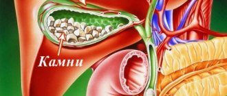
Echinococcus and alveococcus can cause severe damage to internal organs in humans. All these ailments are classified as cestodiases. Alveococcosis of the liver is a serious disease in which whitish cartilaginous nodes filled with dark burgundy contents form in the organ. This liquid contains the larvae of future alveococci. On a section of the liver, these nodes resemble cavities. Their size ranges from 5-300 mm.
There are no primary manifestations of the disease, and the disease itself has a chronic course. This is why patients turn to a specialist untimely; moreover, more often they go to a gastroenterologist, nephrologist, pulmonologist, therapist, and not to a parasitologist. Complications of alveococcosis are metastases to other organs and tissues. After multiple tumor-like formations form in one organ, the helminth easily grows through the walls of the organ and captures new tissue, moving further and further.
Attention! In the absence of timely and adequate treatment, the disease leads to the death of the patient.
Most often, tumor-like formations are localized inside the liver, but with strong growth they can also appear on the surface. If the parasite grows through the diaphragm, it will invade the lungs and kidneys. If the larvae enter the bloodstream, the possibility of damage to the brain and other organs cannot be ruled out. In people with immunodeficiency, the disease progresses rapidly.
Epidemiology
Alveococcosis is a natural focal disease, spread mainly between wild animals of the following types: arctic fox, fox - wild mouse-like rodents - arctic fox, fox. In a number of places, dogs are included in the epidemiological chain. A person does not actively participate in the transmission of invasion and is essentially a biological dead end for the parasite, since the parasitic nodes that form in his liver are, as a rule, inaccessible to the definitive host.
Human infection occurs directly from wild carnivores, through dogs, as well as through consumption of water and plants contaminated with eggs or segments of alveococcus. The first path is typical for areas with intensively developed hunting. During skinning, eggs stuck to the fur fall on the hands and are then carried into the mouth. When shaking and cutting the skins in a living room, the eggs end up on household items and food products, where they remain viable for a long time. This creates the possibility of infection. In the second route of infection, the helminth passes from the wild to humans through dogs, which become infected by eating rodents and then become a source of infection for people. In the third case, infection occurs as a result of eating wild berries and herbs, which may contain the excrement of infected carnivores, as well as drinking unboiled water from natural reservoirs that serve as watering places for wild animals.
Prevention or how to avoid infection
- It is necessary to observe the rules of personal hygiene, be sure to wash your hands while walking in the wild;
- Once every six months it is necessary to deworm pets, regardless of whether they are outside or not;
- do not eat berries from forest glades or any other fruits and vegetables from garden beds without washing;
- for the slightest problems with the liver, contact medical centers for an ultrasound examination;
- try to teach all of the above to your children.
If you follow these simple rules, you can protect yourself and your loved ones from the dangerous disease alveococcosis, which often leads to death.
Clinical picture
Alveococcosis in the first months and even years is almost asymptomatic. The first sign of the disease is an enlarged liver. Patients feel satisfactorily and often have no complaints. Later, there is a feeling of pressure in the right hypochondrium or epigastrium, and then heaviness and dull aching pain. By this time, it is possible to palpate a very dense (“iron” - N.M. Lyubimov) liver with an uneven surface. On palpation, pain is absent or insignificant. Exhaustion, jaundice, enlarged spleen and ascites do not occur during this period. After a few years, the liver enlarges even more, becomes lumpy and painful, patients develop weakness, decreased appetite and weight loss, subsequently developing jaundice and very rarely ascites. However, quite often, despite the long-standing process, patients do not have pronounced general disorders.
Complications include perihepatitis, proliferation of a parasitic “tumor” into neighboring organs and tissues, and decay in parasitic nodes. Metastases are predominantly in the lungs and brain. The growth of the parasitic “tumor” into the gallbladder, hepatoduodenal ligament, omentum, anterior abdominal wall, diaphragm, lung, pericardium, heart, spleen, pancreas, right adrenal gland and kidney, into the retroperitoneal tissue, inferior vena cava and spine was observed. Germination into the gates of the liver or compression of them is accompanied by jaundice.
During decay in the center of a parasitic tumor, sequestration is often observed, and sometimes profuse bleeding into the decay cavity. A breakthrough may occur into the free abdominal cavity, less often through the diaphragm into the pleural cavity, and when fused with lung tissue, into the bronchus, which leads to the formation of a bile-bronchial fistula.
The diagnosis is usually made in a late stage of the disease. Doctors who are familiar with this type of pathology and know the places of natural focality of alveococcosis are able to make a diagnosis at an early stage of the disease.
Alveococcosis of the liver must be differentiated from hydatid echinococcus, cirrhosis and liver neoplasms. It is difficult to distinguish alveococcosis from hydatid echinococcosis, since all the symptoms and laboratory tests characteristic of the latter (Casoni reactions and hemagglutination with latex, eosinophilia, etc.) can be present in both diseases. A test puncture is strictly prohibited at the slightest suspicion of hydatid echinococcosis. In the differential diagnosis of alveococcosis, it should be remembered that Lyubimov’s symptom of “iron density” is unreliable, since such density can occur with a calcified hydatid cyst.
Knowledge of the characteristics of the endemic focus helps to establish a diagnosis. It is easier to differentiate alveococcosis from cirrhosis, in which, as a rule, liver functions are impaired early, eosinophilia is absent, and the Casoni reaction and the hemagglutination reaction with latex are negative. When differentiating from malignant neoplasms, one should take into account the anamnesis, as well as the fact that with alveococcosis the liver is much denser, there is no cachexia, and specific reactions give a positive result.
For timely surgical treatment of alveococcosis, a correct diagnosis and clarification of the localization of parasitic nodes, their size, and so on are necessary. For this purpose, peritoneoscopy (see), hepatography (see), splenoportography (see), liver vasography, scanning with radioactive isotopes are used. Sometimes on plain radiographs you can see small foci of calcification - “lime splashes”.
Primary extrahepatic localizations of alveococcosis are extremely rare. The descriptions found in the literature are not reliable, since in most cases primary liver damage was not excluded during inspection of this organ or section. All described primary lesions of the lungs, brain, vertebrae, chest, and others were either metastases (lungs, brain) or germination of unrecognized alveococcosis of the liver. Alveococcosis of the retroperitoneal lymph nodes was reliably described by E. B. Orechkina (1961).
Symptoms, diagnosis of pulmonary alveococcosis
The helminth formed in the human body is localized in the lungs by lymphogenous route. More often, the lungs become the next site of damage after the liver as a result of the penetration of metastases. It is difficult to determine the pathogen in this organ. The stage of cyst formation lasts more than one year. Detection of alveococcosis in the early stages is the result of examining a patient for another reason.
Symptoms of alveococcosis in the lungs at the following stages of disease development:
- acute pain in the chest area;
- frequent cough, possibly with blood;
- discharge of pus.
Note that the lungs of children are more susceptible to the pathogen, so their disease develops faster in this organ.
Diagnosis is made using laboratory testing of sputum released by a patient with a cough. To determine the presence of the parasite in the body, fluoroscopy and computed tomography are performed. A general blood and urine test is also given.
Pulmonary alveococcosis is a fatal disease accompanied by unpleasant sensations and regular pain during breathing. It can be prevented in advance if detected in a timely manner. As a result of the opposite outcome, cancer develops.
Treatment
Radical surgery for alveococcosis can be performed in approximately 15% of patients. More than 80% of patients are admitted to surgical departments when radical removal of parasitic nodes is impossible due to the prevalence of the process.
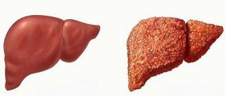
A large alveococcal node excised from the right lobe of the liver along with the gall bladder spread out on it (visible from above). The knot is cut
The node can be excised within healthy tissue - segmentectomy, liver lobectomy (see Hemihepatectomy), enucleated or partially resected and partially enucleated (resection - enucleation - A. N. Velikoretsky). If there are two or more nodes, and the patient’s general condition does not allow them to be removed simultaneously, the operation is performed in two or even three stages. Often the gallbladder is spread out on a parasitic tumor; in these cases, it is removed along with the alveococcal node (Fig.). If radical surgery is impossible (for example, when it grows into the inferior vena cava), and the parasitic tumor, having reached a large size, mechanically compresses neighboring organs, palliative resection is indicated, which is performed in order to delay the compression of the extrahepatic bile ducts by the growing tumor. The remaining unremoved areas of the alvesococcal node are subjected to local action of antiparasitic agents: injections at several points of 10-15 ml of trppaflavin solution (1:1000) or 3-4 ml of tepal (thymol ester of palmitic acid). In addition, treatment is carried out with steroid drugs and vitamins. The use of chemotherapy is being studied.
The technique of palliative resections developed by I. L. Bregadze does not present any particular difficulties, since the majority of parasitic nodes do not bleed. Bleeding from single large vessels is stopped by biological plugs from the omentum. After opening the abdominal cavity, a piece of the greater omentum is excised, from which biological tampons are immediately made. Pieces of omentum 0.5×0.5 cm are powdered with dry thrombin and fixed with two knots to the center of the ligature. At the moment of bleeding from a vessel gaping in the dense stroma, a sharply curved needle with a ligature with a biological swab threaded into its eye is inserted into its lumen. By tightening the ligature, it is possible to clog the lumen of the vessel and stop the bleeding. If there are decay cavities in the center of unremovable parasitic nodes, the cavities are drained, followed by systematic washing with antiparasitic drugs, and in case of infection - with antibiotics. In case of occlusive jaundice caused by germination of the porta hepatis, various bile drainage operations are performed.
Prevention
The fight against alveococcosis is difficult due to the fact that the main source of invasion is wild animals. In cases where human infection can occur through dogs, the destruction of strays and systematic deworming of commercial and domestic dogs is of great importance. In places where helminths are transmitted from wild animals, the main attention should be directed to sanitary educational work and instilling hygienic skills in the population. Personal prevention consists of taking precautions when interacting with dogs, thoroughly washing your hands, and not cutting skins in a residential area. In endemic areas, mass screening of the population using immunological reactions and clinical examination is of great importance. Their goal is the possible early detection of persons infected with alveococcosis, since advanced cases of the disease are not operable. First of all, it is necessary to examine people who are at increased risk of infection (hunters, shepherds and other people involved in cutting skins, people in constant contact with dogs, etc.), as well as family members of patients with alveococcosis due to the possibility of family infections. It is most advisable to use the latex agglutination reaction (see Agglutination) and the indirect hemagglutination reaction with echinococcal diagnosticum during examinations.
See also Echinococcosis.
Symptoms, diagnosis of alveococcosis of the kidneys
Kidney damage also occurs as a result of metastases from the liver to other organs. The natural functioning of the kidneys is disrupted as a result of the pathogen, and cytoplasmic proteins are separated. Alveococcosis is perceived as kidney necrosis, the symptoms will be the same:
- severe pain in the lower back;
- hyperthermia;
- pain when urinating;
- Pieces of renal papillae can be found in the urine.
With these symptoms, the patient self-medicates for kidney necrosis, not realizing that the real cause of the ailment lies elsewhere.
The terminal stage of renal alveococcosis occurs with terrible colic in the renal area. Even early detection of disease penetration may not bring a positive result.
Abdominal pain and weakness
When diagnosing the disease, the patient undergoes the following research methods:
- General laboratory tests of blood and urine.
- Microscopic examination of kidney biopsy.
- Isotope renography method, as a result of which the increased size of the kidney is established, which will help to draw attention to it.
After examining the kidneys, the doctor will prescribe both medicinal and surgical treatment. Extensive kidney metastases are a cancer definition of the third and fourth stages. It is difficult to cure them. Therefore, alveococcosis more often leads to death.
Bibliography
Alveolar echinococcosis and some issues of private surgery, ed. G. D. Zalessky, Novosibirsk, 1961; Alperovich B.I. Alveococcosis and its treatment. M., 1972, bibliogr.; Bregadze I. L. Experience in the treatment of alveolar echinococcus with thymol ester of palmitic acid, 8th Int. congr. on tropical medicine a. malaria, Abstr. a. Rev., p. 1133, Teheran, 1968; aka, Alveococcosis of the liver, in the book: Khir. hepatol., ed. B.V. Petrovsky, p. 269, M., 1972; Velikoretsky A. N. and Kasaikina T. N. About resection of liver tissue, Surgery, No. 5, p. 44, 1955; Deineka I. Ya. Human echinococcosis, M., 1968; Zorikhina V.I. Improvement of methods for immunodiagnosis of human echinococcosis and alveococcosis, Med. parasitol., t. 39, no. 2, p. 170, 1970; Leikina E. S. The most important helminthiases of humans, M., 1967; Lukashenko N.P. and Bregadze I.L. Echinococcosis and alveococcosis, Multivolume. manual on microcrystals, wedge, and epidemiology. infectious diseases, ed. N. N. Zhukova-Verezhnikova, vol. 9, p. 509, M., 1968; Stepankovskaya L.P. Study of the effectiveness of the indirect hemagglutination reaction with echinococcal diagnosticum, Med. parasitol., t. 41, no. 4, p. 400, 1972; Stucke K. Zur chirurgischen Therapie der Echinococcosis alveolaris, Ghirurg, Bd 34, S. 165, 1963, Bibliogr.
E. S. Leikina; I. L. Bregadze (cl. and treatment).