Research methods
If a patient has complaints about gastrointestinal disturbances, then he needs to undergo a full medical examination. There are several methods of medical diagnosis:
- Physical method. Based on a visual examination of the patient and collection of anamnesis.
- Laboratory research. Includes taking tests prescribed to confirm a preliminary diagnosis.
- Hardware methods. They provide the opportunity to examine the gastrointestinal tract and identify the presence of pathologies.
Only a doctor can select the optimal diagnostic option or prescribe a comprehensive examination. The choice will depend on the nature of the patient’s complaints, the collected medical history and preliminary diagnosis. We will talk about hardware research options.
Gastroscopy and sounding
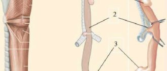
Gastroscopy refers to endoscopic examination methods. Provides comprehensive information about the inner surface of the duodenum and stomach. The manipulation is based on the oral insertion of a probe with a videoscope and a light bulb at the end into the cavity of the organ.
The study is unpleasant, but the most informative of all diagnostic measures existing today. During the examination, the doctor may remove single small polyps or take a tissue sample for a biopsy. The procedure is prescribed for chronic forms of gastritis and ulcerative pathology to confirm the diagnosis and select a treatment regimen.
A probe study is carried out in the same way as FGS. With the help of manipulation, the specialist receives information about gastric secretion, but does not see the mucous membrane.
Oral insertion of a probe is associated with extremely unpleasant sensations for the patient, which is why many are terrified of the procedure. How can you check your stomach without swallowing your intestines? Are there other research options?
How the research is carried out
To begin, the patient removes clothing above the waist. If a skin examination is planned, the person lies down on a couch. The doctor treats the area to be examined with gel and begins the diagnosis. The manipulation takes about 15 minutes.
If an ultrasound is performed through the oral cavity, the patient lies on his side, opens his mouth, the doctor sprays an anesthetic to reduce discomfort and inserts a probe to the level of the esophagus. The manipulation takes place very quickly, usually a few minutes are enough.
Alternative diagnostic methods
Any medical examination begins with collecting anamnesis during a personal conversation with the patient. The doctor then begins a visual examination of the patient. By palpating, the specialist finds out the localization of pain, the tension of the walls of the organ and the presence of dense structures.
The next stage of the examination will be to study the stomach using a hardware method. Modern medicine can offer several diagnostic options that can, to one degree or another, replace FGS:
- capsule gastroscopy;
- desmoid test according to Saly;
- radiography;
- ultrasonography;
- MRI (magnetic resonance imaging).
Advice. Before you go for a stomach check, you need to consult a specialist. For example, if a patient’s initial examination reveals possible gastritis, ultrasound will be useless in this case.
Below we will examine each diagnostic method in more detail.
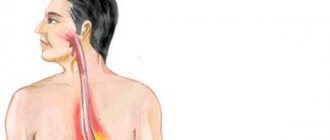
Dilatation of the esophagus
Dilation of the esophagus (Dilatatio oesophagi) is a persistent increase in the lumen of the esophagus, in which the lumen of the esophagus is increased evenly in all directions (Ectasia oesophagi) or in one limited place (Diverticulum oesophagi).
This disease is observed extremely rarely in animals and is more common in horses.
Etiology . Primary ectasia occurs in old animals due to general weakness or with sarcosporidiosis, secondary ectasia is recorded in animals with stenosis or prolonged blockage of the esophagus. The reason for the expansion of the esophagus in animals is the retraction of its wall by a lymph node fused to it, fastened by adhesions to the bronchus or skin (traction diverticulum). Less commonly, diverticula in animals develop as a result of hernial protrusion of the mucous membrane of the esophagus into the lumen between its separated or torn muscle fibers (pulsatile diverticulum). Prolonged spasms of the esophagus in animals can lead to prolonged dilatation of the esophagus.
Pathological changes . When opening a dead animal, we find the esophagus, which has an expansion in the form of ectasia or diverticulum; the wall of the expanded part of the esophagus is thinned. In some animals, it may fuse with adjacent organs.
Clinical picture . Animals greedily accept the food provided to them by the owner, but after some time, as the dilation fills and obstruction is created, they refuse the food and make repeated swallowing movements without a food coma with their heads lowered and convulsive contraction of the neck muscles. During a clinical examination of a sick animal on the left in the area of the jugular groove, the veterinarian notes a protrusion that has an oblong shape, which increases after eating food. When percussing this swelling, the veterinarian detects dullness, and through massage it is possible to induce belching in the animal with the release of food masses through the nasal cavity. The accumulation of food masses in the animal's diverticulum causes antiperistaltic contractions of its muscles, anxiety and pain. By probing the esophagus, a veterinarian can determine the obstruction of one or another segment of it. Periodically recurring obstruction of the esophagus causes convulsive contractions of its muscles, tense swallowing movements, stretching of the head, coughing and restlessness of the animal. At the same time, pressure on nearby organs (bronchi, trachea, carotid arteries, veins, vagal trunks) complicates the animal’s breathing and blood circulation. After removing dense feed masses from the diverticulum, the above functional disorders in the sick animal disappear.
Diagnosis . Frequently recurring disturbances in food intake and the passage of food through the esophagus, the formation of swelling in the jugular gutter and probing allow the veterinarian to make a correct diagnosis. In difficult cases, fluoroscopy with a contrast mass and endoscopy can help diagnose the disease.
Differential diagnosis . When conducting a differential diagnosis, the veterinarian must exclude diseases such as stomatitis, pharyngitis and other diseases of the esophagus.
Course and prognosis. The expansion of the esophagus in an animal develops slowly. The disturbance in the passage of food coma in the animal increases gradually. Developing stretching and necrosis of the muscle layer and mucous membrane can lead to rupture of the esophageal wall. When food is expelled through the nose and mouth, antiperistaltic contractions of the esophagus can cause aspiration pneumonia in the animal. A persistent disorder in the intake of food and water leads to exhaustion of the animal, reduces its productivity and can ultimately lead to the death of the animal.
Treatment . The sick animal is prescribed dietary feeding - flour and bran mash, crushed grain, green food, soft hay. When the cervical part is enlarged, bandages are recommended that compress the enlarged area of the esophagus, as well as periodic bougienage of the esophagus. Sometimes owners of high-value animals have to resort to surgical intervention by excision of the wall of the esophagus. It is advisable to cull less valuable productive animals from the main herd.
Prevention . Animal owners must comply with established rules for feeding and keeping animals.
Capsule gastroscopy
This research method is based on replacing the probe with a special capsule equipped with a video camera. The device allows for a thorough examination of the gastric mucosa and detects the disease in the early stages of development.
To make a diagnosis, the patient must swallow the capsule. For the inspection to be successful, you should prepare for it:
- The patient must adhere to a diet for 2 days before the procedure. It is recommended to exclude fatty, heavy foods, alcohol and dishes that cause flatulence from the diet. Food should be well chopped and steamed or boiled.
- The study is carried out in the morning, on an empty stomach. The capsule can be taken with ½ glass of plain liquid.
The process does not take much time and does not cause any discomfort to the person. During the examination, the patient can return to normal life, limiting physical activity. After 7–8 hours, the patient visits the doctor’s office again, where the doctor transfers the indicators recorded by the capsule into the computer and makes a diagnosis.
After a certain time, the device leaves the body naturally. The advantages of this procedure are obvious, but the method has not found wide application due to the rather high price of the device. In addition, such an examination does not allow you to perform a biopsy, remove polyps, or stop bleeding.
You can see how the stomach is examined using the capsule method in the video:
Desmoid test
Gastroenterologists often use a desmoid test to determine the degree of gastric juice activity. During the test, the patient swallows a bag filled with methylene blue powder and tied with catgut thread.
After the thread dissolves, the dye is gradually absorbed into the blood and no later than 18–20 hours later it is eliminated from the body. The study is based on assessing the intensity of urine staining. If the first portion of urine acquires a bright blue-green color, it means that the acidity of the stomach is increased.
Radiation research methods
Of the radiation methods, X-ray is the most widely used. Equipment for examination is available in almost every medical institution, so the examination is available to all segments of the population.
MRI and ultrasound are more modern research methods and pose less of a threat to the health of patients.
You can learn about the differences between these procedures from the video:
X-ray
Using radiography, a stomach ulcer is detected, its configuration is checked, and its size is assessed. R-graphy is carried out using a contrast agent - barium suspension. It is prescribed when the patient complains of rapid weight loss, the appearance of blood in the stool, frequent and debilitating diarrhea, and constant pain in the gastrointestinal tract.
The procedure is completely painless and not very complicated, but requires compliance with some rules:
- For 2–3 days before the examination, you should exclude alcohol, thick, fatty and solid foods from your diet.
- On the eve of the test, it is necessary to cleanse the intestines using an enema or special means with a laxative effect.
- Before the procedure, the patient is prohibited from eating or drinking colored drinks.
An X-ray of the stomach lasts 30–40 minutes. All this time, the doctor asks the patient to take certain positions and takes six pictures of the gastrointestinal tract in different projections.
Questioning and inspection
This is where any examination begins. Information about the patient’s complaints, the history of the disease and the patient’s life allows us to choose the optimal methods of further examination to make a diagnosis.
Patients with pathologies of the upper gastrointestinal tract often complain of swallowing problems and chest pain almost immediately after eating. Moreover, pain first appears when eating dense foods. In later stages, complaints arise even when drinking drinks.
The duration of the pathology, the characteristics of the treatment or its absence, and the result of the treatment used are important. The possible causes of pathologies in this part of the gastrointestinal tract are also clarified. The disease can be triggered by burns of the mucous membrane, injuries, dietary habits, and the patient’s profession. Information about concomitant pathologies in the body and allergic manifestations is important.
When examining a patient, pay attention to the general condition, skin color, bad breath, poor posture, the presence of tumor-like formations in the neck and other manifestations.
The doctor may auscultate the esophagus (listening with a stethoscope). Its information content is relative. It helps to suspect the presence of deviations from the norm. Physiologically, in the area of the xiphoid process of the sternum, the first noise is heard when food passes through the esophagus after swallowing, and the second when it reaches its ampullary part and goes into the stomach.
The doctor collects anamnesis from the patient
Examination, questioning and auscultation only allow you to choose the most optimal tactics for further hardware research methods. There is too little information available to make a definitive diagnosis.
There is a fairly wide selection of imaging methods for examining the esophagus: radiography, computed tomography, ultrasound, endoscopic and endoscopic ultrasound examination, esophagomanometry, esophagoscopy, Bernstein test, radionuclide and intraesophageal pH-metry. However, only a few of them are often used. This is an X-ray examination, esophagoscopy, ultrasound of the esophagus.
Easy diagnostics
Simple diagnostic methods are mandatory for use when a patient complains of an acute abdomen, nausea and other symptoms of gastric diseases.
Physical examination
Physical measures are carried out at a doctor’s appointment, the results depend on the qualifications of the medical specialist. The complex includes:
- studying the anamnesis, assessing symptoms from the patient’s words;
- visual examination of mucous membranes;
- feeling painful areas of the body (palpation);
- palpation in a specific body position (percussion).
Microscopic laboratory diagnostics
Laboratory methods involve taking samples from the patient for further study and evaluation of the results. Most often, the following physical and chemical studies are prescribed:
- general urine analysis;
- coprogram (stool analysis);
- clinical blood test. The number of all types of blood cells (erythrocytes, leukocytes, platelets) is counted and the hemoglobin level is determined;
- gastropanel. This blood test is aimed at studying the condition of the gastric mucosa. Based on its results, the following is determined: the presence of antibodies to Helicobacter pylori bacteria, the level of pepsinogen proteins produced, the level of the polypeptide hormone - gastrin, with the help of which the acidic environment in the stomach is regulated;
- blood biochemistry. Quantitative indicators of bilirubin, liver enzymes, cholesterol and other blood elements are established.
Tests help identify inflammatory processes and other disorders of organs and systems. If the results differ significantly from the normative indicators, the patient is prescribed an instrumental or hardware examination.
How do gastroprotectors help?
With the help of gastroprotectors, the condition of the internal tissues of the gastrointestinal tract is normalized. It turns out that there is a complex effect necessary in the process of treatment and recovery from a serious illness:
- Nutrition of cells and tissues, their saturation with useful microelements.
- Elimination of the acidic environment and bacteria to avoid their further effect on the mucous membrane.
- Tissue regeneration, creation and maintenance of a barrier in cells that prevents the effects of negative factors.
- Improving circulation processes by accelerating them.
Idiopathic dilatation of the esophagus is a dangerous pathology, since its progression leads to loss of the ability to live normally before surgery. If you follow treatment recommendations and do not panic, the disease can be easily defeated.
Application of hardware techniques
Examination of the stomach without gastroscopy is carried out using special medical devices. They record the condition of the mucous membrane, density, size and other parameters of the organ, and transmit information that is subject to subsequent decoding by a specialist.
- X-ray examination (using contrast);
- CT and MRI (computer and magnetic resonance imaging);
- EGG (electrogastrography) and EGEG (electrogastroenterography);
- Ultrasound (ultrasound examination).
During a gastric examination using a hardware method, all manipulations are performed without direct intervention in the body, without damaging the external tissues of the body (non-invasive). The procedures do not cause pain in the patient.
X-ray with contrast
The method is based on the use of X-rays. To improve visualization of the stomach, the patient drinks a barium solution before the examination. This substance plays the role of contrast, under the influence of which soft tissues acquire the ability to absorb x-rays. Barium darkens the organs of the digestive system in the image, which makes it possible to detect possible pathologies.
X-ray helps in determining the following changes:
- incorrect location of organs (displacement);
- condition of the lumens of the esophagus and stomach (enlargement or narrowing);
- non-compliance of organs with standard sizes;
- hypo- or hypertonicity of organ muscles;
- niche in the filling defect (most often, this is a symptom of peptic ulcer disease).
Addition to hardware diagnostics
An additional method is Acidotest (taking a combined medication to establish approximate gastric pH values). The first dose of the medication is taken after emptying the bladder. After 60 minutes, the patient takes a urine test and takes the second dose. An hour and a half later, urine is collected again.
Before testing, it is forbidden to eat food for eight hours. Urine analysis reveals the presence of dye in it. This allows you to roughly determine the acidity of the stomach without gastroscopy. The acid test does not give 100% effectiveness, but only indirectly indicates a reduced (increased) level of acidity.
Alternative endoscopy
The closest to FGDS in terms of information content is capsule endoscopy. The examination is carried out without swallowing the probe, and at the same time reveals a number of pathologies that are inaccessible to hardware procedures:
- chronic ulcerative and erosive lesions;
- gastritis, gastroduodenitis, reflux;
- neoplasms of any etiology;
- helminth infestations;
- inflammatory processes in the small intestine (enteritis);
- cause of systematic digestive disorders;
- Crohn's disease.
The diagnostic method is carried out by introducing a capsule with a tiny video camera into the patient’s body. There is no need for instrumental introduction. The weight of the microdevice does not exceed six grams; the shell is made of polymer. This makes it easy to swallow the capsule with enough water. The video camera data is transmitted to a device installed on the patient’s waist, from which the doctor takes readings after 8–10 hours. At the same time, the rhythm of a person’s usual life does not change.
The capsule is removed naturally during bowel movements. Significant disadvantages of the technique include: the inability to perform a biopsy, the extremely high cost of the examination. All methods of diagnosing the gastrointestinal tract involve preliminary preparation of the body. First of all, this concerns nutritional correction.
The diet should be lightened a few days before the examination. Hardware procedures can only be performed on an empty stomach. The stomach can be checked using any method that is convenient and not contraindicated for the patient. However, the palm in terms of information content, and therefore maximum accuracy of diagnosis, remains with FGDS.
Typically, symptoms of esophageal diseases develop gradually and at first do not cause much discomfort to the patient. However, if appropriate measures are not taken, the pathology progresses and can be complicated by serious consequences, including oncology.
Therefore, it is so important to know how to check the esophagus and decide on the diagnostic method. The indication for examination of the esophagus is the presence of the following symptoms in the patient: heartburn, dysphagia, belching, pain in the esophagus.
Taking an anamnesis
The interview with the patient begins with finding out the symptoms, the time of their onset and the treatment provided (if any). Patients with esophageal pathologies mainly complain of pain in the chest or back, as well as difficulty swallowing (dysphagia). Dysphagia can be either mild or severe with difficulty swallowing liquids and even saliva.
Preesophageal dysphagia is accompanied by a feeling of a lump in the throat and occurs due to damage to the central nervous system and muscles of the esophagus. The esophageal form indicates a decrease in the motor activity of the esophagus, and the transportation of any liquid or food is difficult. A common cause of achalasia is bacterial, fungal infections, as well as chemical or mechanical damage to the esophageal mucosa. This form of the disease progresses slowly and is characterized by difficulty swallowing solid foods.
Ulcerative or erosive lesions of the inner lining of the esophagus, GERD and tumor formation are usually accompanied by pain in the chest or back (odynophagia). In addition, there is impaired motor skills and difficulty swallowing. Drinking water does not alleviate the condition in such patients, but only aggravates the situation.
Treatment of esophageal injury
If damage to the esophagus is suspected, it is necessary to immediately stop eating by mouth and take the victim to a surgeon. At the stage of qualified assistance, R-contrast examination of the esophagus and endoscopy are performed. Damage determined endoscopically is usually large and has crushed edges. Small lesions are often detected only by x-ray. The level of damage is determined, which determines surgical access. It is necessary to find out whether the patient has eaten, whether there is an increase in body temperature, how much time has passed since the injury, and whether there are any signs of mediastinitis. It is important to know whether before the injury there were any difficulties with swallowing due to cardiospasm, diverticulum, or stenosis.
- A conservative treatment method is used for small-sized (up to 1 cm) injuries located above the aortic arch and early diagnosed injuries. Such injuries heal in 7–10 days without surgery if treated in a timely manner.
- Surgical method. For injuries in the upper and middle third of the esophagus, a right-sided thoracotomy is performed; for low injuries, a left-sided thoracotomy is performed. In this case, a mandatory stage of the operation is the placement of a gastrostomy, since after the operation parenteral nutrition requires a lot of effort and expense. In case of cancerous perforation, perforation of a diverticulum, or extensive damage to the wall, the question arises of primary resection of the damaged esophagus. If mediastinitis develops, it is necessary to perform a cervical esophagostomy and completely exclude the esophagus from the passage.
Upon admission six or more hours after perforation, with the development of signs of mediastinitis, mediastinotomy and mediastinal drainage are indicated.
Essential drugs
There are contraindications. Specialist consultation is required.
- Cefotaxime (an antibiotic of the third generation cephalosporin group). Dosage regimen: IM 0.5 g of the drug in 2 ml (respectively 1 g in 4 ml) of sterile water for injection, injected deep into the gluteal muscle. 1% lidocaine is also used as a solvent for intramuscular administration. For intravenous administration, 0.5-1 g of Cefotaxime is dissolved in 10 ml of sterile water for injection. Inject slowly over 3–5 minutes. For drip administration (over 50-60 minutes), 2 g of the drug is dissolved in 100 ml of isotonic sodium chloride solution or 5% glucose solution.
- Cefepime (IV generation cephalosporin antibiotic). Dosage regimen: adults and children weighing more than 40 kg with normal renal function 0.5-1 g intravenously (for severe infections up to 2 g) or deep intramuscularly at intervals of 12 hours (for severe infections - after 8 hours ).
- Amoxiclav (broad-spectrum bactericidal antibacterial agent). Dosage regimen: intravenously, adults and children over 12 years of age or weighing more than 40 kg - 1.2 g of the drug (1000 + 200 mg) with an interval of 8 hours, in case of severe infection - with an interval of 6 hours.
Examination of the patient
Examination of the esophagus consists of general examination and palpation. In this case, special attention is paid to examining the larynx, its condition, and the smell from the oral cavity. A general examination is of no less diagnostic importance: the patient’s degree of fatness, skin color and texture, facial expressions, body temperature, swelling.
To treat gastrointestinal diseases, people successfully use Galina Savinova’s method. Read more >>>
Local examination of the esophagus includes palpation of the lymph nodes, neck, auscultation and percussion, as well as instrumental diagnostic methods:
- radiography;
- daily pH measurements;
- computed and spiral tomography;
- esophagofibroscopy.
With painful shock, pale skin is observed, with oncology and hypochromic anemia there is yellowness, with esophagitis there is hyperemia, and with large formations that cause hypoxia there is cyanosis.
The presence of pain is indicated by a grimace on the face and anxiety of the patient. An unnatural position of the body or head with a forward tilt indicates a possible diverticulum or foreign body. The patient in these conditions tries not to make body movements that cause him pain.
The lethargic and indifferent state of the patient signals septic or traumatic shock (for example, with a burn or mechanical perforation of an organ by a foreign body, massive bleeding, a state of severe intoxication).
X-ray of the esophagus
The manipulation is carried out on an empty stomach. Before undergoing radiography, the subject must take a contrast agent (barium sulfate solution). This is explained by the fact that not all organs of the gastrointestinal tract are capable of blocking X-rays, and barium makes it possible to visualize motor functions and contours of organs.
This method is necessary if the presence of neoplasms or foreign bodies is suspected, as well as with achalasia. The procedure for radiography of the esophagus and stomach does not require lengthy preparation. You should refrain from eating for about eight hours or skip breakfast.
Esophagofibroscopy
This highly informative research method helps to find out the cause of pain, dyspepsia and dysphagia of the esophagus. It can be used to determine the presence of varicose veins, tumor formation, and bleeding from the esophagus. It is carried out both for the purpose of diagnosing the esophagus and for providing emergency assistance. Thanks to the method, it is possible to diagnose cancer formations on the walls of the organ and pathological changes in the lymph nodes.
The method makes it possible to determine the condition of the inner lining of the esophagus and obtain material for histology. The manipulation is carried out by qualified specialists under local or general anesthesia. The method requires preliminary preparation of the patient.
Diagnosis occurs by introducing an ultrasound sensor through the larynx to visually examine the mucous membrane and collect biological material for histology. The endoscope has high ultrasound frequencies that detect minimal deviations from the norm, which makes esophagoscopy one of the most highly informative research methods.
Daily pH-metry
The method helps, by measuring the pH level, to determine the nature and severity of esophageal reflux. To do this, a probe with a sensor of one to three electrodes is inserted through the larynx and fixed in a certain place.
The sensor records changes in pH in the lower part of the esophagus during the day. The data is subjected to computer analysis, and on its basis, its compliance with the norm is established.
Bernstein test
Acid perfusion is used when other methods do not show changes in the mucous membrane, but the patient feels dyspepsia, odynophagia, and dysphagia. The method consists of introducing saline and HCl solution into the larynx through a probe alternately at the required speed. In the presence of reflux esophagitis, discomfort and pain in the chest occurs due to irritation by acid.
Esophagotonokymography of the esophagus
The essence of the method is to obtain images that record decreased tone and contraction of the esophageal muscles. In this way, the initial form of dysphagia, hernias and muscular pathologies of the esophagus without severe symptoms is diagnosed.
During the study, a multichannel probe with a catheter or rubber balloon is used to measure intraesophageal pressure. The gastroscopy method allows you to obtain complete information about all possible decreases in the muscle tone of the sphincters.
Computed and spiral tomography
The study is used to diagnose neoplasms and metastases, enlarged lymph nodes, and determine wall thickness. This diagnostic method is also radiation, but unlike X-rays it allows you to obtain a higher quality image. The procedure involves preparing the patient for the study, which consists of administering externally and internally contrast solutions containing iodine.
Spiral tomography is a method of x-ray examination. During it, the tomograph moves in a spiral, creating highly accurate 3D models. The devices automatically convert the received data into digital form. With the help of such a study, pathologies of almost all internal organs are diagnosed. The method is characterized by high information content and accuracy of research with a minimum degree of radiation from devices.
It is used for a comprehensive diagnosis of the esophagus, determining the exact location and boundaries of the lesion or the presence of a foreign body. It is used as an additional research method if previous measures did not give the desired result.
Chromoendoscopy method
The method reveals pathological changes in the esophageal mucosa using staining with Lugol's solution, phenol, and methylene blue. The procedure helps determine the location of the lesion by changing the shade of the tissue. It is used primarily for the diagnosis of oncological tumors.
Radioisotope research
When diagnosing oncological formations, the RI method is used. It is used quite rarely, but it has a number of advantages: it is harmless and allows one to obtain data that is not available with other research methods. The method is recommended for the differential diagnosis of esophageal cancer. In this case, radioactive phosphorus is used, which accumulates in tumor formations, which makes it possible to determine their presence. The method does not require preliminary preparation.
Today, there are many modern, highly informative methods for diagnosing diseases of the esophagus, which makes it possible to qualitatively examine the patient. However, one research method is not enough to determine the final diagnosis. Only the attending physician, after a comprehensive examination, questioning and examination of the patient, can establish the disease and prescribe adequate therapy.
Endoscopy
The method of endoscopic examination consists in the fact that when conducting a diagnostic examination regarding the diagnosis of cancer, a flexible hose is inserted into the patient’s esophagus through the oral cavity, at the end of which a mini camera with illumination is attached.
In addition, the design of the endoscope is a special device that allows you to collect biological material used for further study. At the same time as the esophagus, the specialist assesses the condition of the stomach and duodenum.
Endoscopy allows not only to identify oncopathology during the spread of tumor formation, but also at the initial stages of its formation.
On this topic
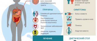
- Oncogastroenterology
What kind of pain occurs with rectal cancer?
- Olga Vladimirovna Khazova
- December 3, 2020
In the first case, diagnosis is not difficult, since the resulting images will clearly show exophytic growth, infiltrative tumor or saucer-shaped ulcerations.
At the beginning of its development, the disease, as a rule, occurs without the manifestation of characteristic clinical signs, which greatly complicates the diagnosis of cancer. The mucous membrane has a slight lesion, which is often mistaken for an inflammatory process.
To increase the effectiveness of the procedure in such situations, doctors use the following techniques:
- study of minimal changes in the mucous membranes of the esophagus;
- performing endosonography;
- the use of chromoendoscopy and endoscopic microscopy;
- use of optical tomography
Thus, thanks to endoscopic examination, it is possible to detect parietal tumor formations, ulcerative lesions and a focus of wall infiltration.