The most common signs of a tumor are:
- Soreness;
- Feeling of heaviness in the right hypochondrium;
- Nausea, feeling of fullness in the stomach, vomiting;
- Jaundice.
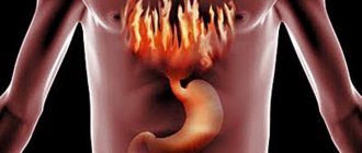
Usually the most characteristic symptoms are pain and a feeling of heaviness in the right hypochondrium, associated with an increase in the size of the liver. The pain can be intermittent, usually it is aching and not intense. When hemangioma vessels rupture or thrombosis, the pain becomes acute, and the patient requires emergency medical care.
If the hemangioma is large and compresses neighboring organs of the abdominal cavity, then there are signs of dysfunction of the stomach or intestines (nausea, vomiting, abdominal pain). Jaundice is possible if the bile ducts are damaged or the evacuation of bile from the gallbladder is impaired. When large vascular trunks are compressed, heart failure and swelling of the lower extremities develop when the inferior vena cava is compressed.
A long asymptomatic course of a hemangioma can result in its rupture and hemorrhage, then the first signs of the presence of a tumor will be acute abdominal pain and symptoms of shock (a sharp decrease in pressure, impaired consciousness and function of vital organs). Massive blood loss and irritation of the peritoneum from the gushing blood pose a threat to the patient’s life and require immediate medical attention.
In rare cases, with diffuse tumor growth, liver failure may develop, and giant nodes, in which a significant amount of blood accumulates, can provoke a blood clotting disorder, combined with thrombocytopenia, and DIC syndrome with its characteristic thrombosis and bleeding (Kasabach-Merritt syndrome) .
Why is a vascular tumor dangerous?
The liver has an abundant blood supply, blood flow per minute is 1.5 liters. The altered section of the vessel may not withstand such a load and rupture.
Factors contributing to hemangioma rupture:
- Increased systemic blood pressure (hypertension).
- Increased pressure in the portal vein system (liver cirrhosis).
The portal vein carries the bulk of blood to the liver (70-75%), the rest through the hepatic arteries.
- Increased intra-abdominal pressure (during heavy physical activity, coughing, constipation).
- Blunt abdominal trauma.
The larger the tumor, the higher the risk of rupture and intra-abdominal bleeding. Death is possible in 75% of cases.
Hemangioma can thrombose, which can result in:
- sclerosis;
- inflammation;
- suppuration of a tumor (liver abscess).
Causes of occurrence in adults
The causes of liver hemangioma are still unknown. Some believe that they are the result of a genetic mutation, which occurs due to hereditary predisposition. Others say that the reason for the growth of formation is a difference in the secretion of sex hormones, which is confirmed by the fact that their formation often occurs during pregnancy. In women who are pregnant once, the tumor is diagnosed more often.
It is believed that the cause of hemangioma in the liver is intrauterine development.
Often, growths on the liver vessels are a hidden congenital pathology.
The most popular theory is innate. She explains the appearance of hemangiomas during pregnancy (a disease, especially of an infectious nature, in the mother at this time) and childbirth. If premature labor occurs, the baby’s liver vessels do not have time to fully form.
There are factors that can provoke the growth of tumors in adults:
- hepatitis;
- injuries;
- smoking;
- infectious diseases;
- venereal diseases;
- high cholesterol;
- viruses;
- intoxication due to poisoning;
- vitamin deficiency (especially vitamin K);
- hypertonic disease;
- alcoholism, etc.
Classification of liver hemangiomas
- According to the structure of the tumor:
There are two most common forms:
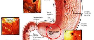
a) Capillary - represents chaotically located tangles of liver capillaries, between the loops of which fine-mesh spaces are formed. Microscopically, these areas have the usual structure of the capillary of the liver tissue, only the course of the vessel is changed. On ultrasound it is determined in the form of a formation with a round shape. This type of hemangioma can be multiple. Rarely exceeds 3 cm in size.
b) Cavernous - a new formation of the liver, the cells of which are larger. It has a dough-like consistency; ultrasound shows the formation of uneven outlines; in the center of such hemangiomas, a cyst filled with liquid contents often forms. The risk of cancerous degeneration and rupture in such tumors is higher.
c) Lymphangiomas - grow from lymphatic vessels.
- According to the course of the disease:
a) Asymptomatic
b) There are clinical manifestations, but no complications.
c) Complicated form.
d) Atypical course of the disease, against the background of concomitant pathology.
Forms
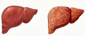
Hemangiomas of the liver are capillary and cavernous. The first of the formations is located in the middle of the liver. There is a shell around it. If the hemangioma enlarges, it can lead to rupture of the membrane and bleeding. A capillary tumor is located on the surface of the liver and is similar in structure to the walls of blood vessels.
Cavernous hemangioma
The cavernous form of liver hemangioma is a single neoplasm that is located inside the liver. It can reach very large sizes, which will lead to the appearance of symptoms. In 10 percent of cases, neoplasms are multiple in nature. The size of tumors can be 2-40 millimeters. Visual examination of the neoplasm can determine the presence of restrictions from the liver parenchyma. New growths are characterized by a lumpy, smooth surface and a reddish-blue color.
Capillary hemangioma
But the capillary form of liver hemangioma is diagnosed in patients in rare cases. They are small in size (up to 7 centimeters). With this type of neoplasm, small cavities are formed, to which a vein approaches. Capillary hemangiomas can grow, especially in the fairer sex during pregnancy. This type of neoplasm does not pose a threat to life.
Multiple liver hemangiomas
Cavernous hemangiomas are rarely multiple. This is typical for capillary type of neoplasms. Multiple liver hemangiomas are neoplasms that are located on the liver. There are more than 2 of them.
Hemangioma of the right lobe of the liver
This is the name for a neoplasm that is an exception. This is a capillary hemangioma, which is located on the right side of the organ and is life-threatening. When this type of neoplasm appears, there is a restriction of liver function, which requires constant and careful monitoring by doctors. Hemangioma of the right lobe of the liver can lead to serious complications or death.
Atypical liver hemangioma
A tumor is formed from mesenchymal cells. Hemangioma is a conglomerate of vessels that have an atypical structure. Atypical liver hemangioma is a type of capillary neoplasm.
what type of liver hemangioma it is and what it is after a detailed examination of the patient.
Symptoms of liver hemangioma
Clinical manifestations depend on:
- tumor size;
- localization;
- stages of development;
- degree of destruction of liver tissue;
- addition of complications.
In 70% of cases, the disease occurs in a latent (asymptomatic) form. As the size of the tumor increases, it can put pressure on adjacent organs, causing a number of symptoms:
- feeling of heaviness, discomfort and pain in the right hypochondrium;
- nausea and vomiting;
- liver enlargement;
- flatulence;
- disorders of appetite, stool and sleep;
- change in skin color - yellowness (observed when the bile ducts are compressed);
- increased temperature due to inflammation in the liver.
When a hemangioma ruptures, signs of intra-abdominal bleeding come to the fore:
- sharp pain in the abdomen;
- decreased blood pressure, increased heart rate;
- pale skin;
- weakness.
The occurrence of such symptoms requires emergency hospitalization of a person in a surgical hospital.
Disease prognosis and prevention
In 2/3 of cases of detection of a vascular defect in liver tissue, the prognosis will be favorable. The pathology progresses slowly, and people do not experience any unpleasant symptoms. Sometimes the tumor lesion disappears completely on its own.
Less commonly, a rapid increase in vascular proliferation is observed - with the capture of increasingly larger areas of tissue, compression of the bile ducts and neighboring organs, and a sharp deterioration in a person’s well-being. In these cases, the prognosis directly depends on the timeliness of medical care. If the hemangioma is quickly removed, then it does not recur, and the person’s health is gradually restored.
Transformation into a malignant neoplasm occurs no more often than in 0.5–1% of cases of hepatic hemangiomas. Following a number of preventive measures helps people prevent such an outcome:
- diet correction - minimizing fatty, fried foods, fast food, preservatives, vessels and stabilizers with dyes in the diet;
- correct drinking regimen - at least 1–1.5 liters of purified water per day;
- absence of abdominal wall injuries;
- prevention of constipation;
- a good night's rest - at least 7-8 hours in a cool, quiet room;
- increasing protective barriers - hardening, taking vitamin and mineral complexes.
Specific measures to prevent the development of hemangiomas have not been developed, since the causes of its occurrence have not been definitively established.
Diagnosis of liver hemangioma
The diagnosis is confirmed using instrumental studies:
- Ultrasound of the liver
The examination shows round formations with heterogeneous contents.
- MRI
Allows you to detect even small hemangiomas. Often performed using contrast enhancement of the liver vessels. It very well determines the structure of the tumor and its exact location.
- Liver scintigraphy
Isotopes are injected intravenously and a two-dimensional image of the liver is obtained by measuring the radiation they emit.
- Angiography of the vessels of the celiac trunk (celiacography)
The contrast agent is injected through the femoral artery, it reaches the aorta, then enters the celiac trunk and along its branches begins to fill the main arteries of the organs, including the liver.
Thanks to this method, it is possible to determine whether the tumor is vascular or parenchymal, malignant or benign (pharmaceuticals that act on blood vessels are additionally administered).
Treatment
There is no clear answer on how to treat hemangioma and whether it is worth doing it at all. The tumor is benign and is asymptomatic in most patients, and the risk of any liver surgery is quite high.
Treatment of hemangioma is not required if there are no symptoms of the tumor, the risk of complications and malignancy is minimal, and if there is absolute confidence that the tumor is benign.
Indications for treatment may include:
- The appearance of tumor symptoms;
- Fast growth;
- Complications;
- It is impossible to completely exclude the malignancy of the neoplasm.
The most serious complication of liver hemanigoma is its rupture and bleeding. In such cases, emergency surgery may be required, but it is quite dangerous and the mortality rate with such resections is high, so it is recommended to first ligate or embolize the hepatic artery, and when the patient’s condition has stabilized, resection of the tumor-affected area of the liver will become possible.
The issue of the need to remove giant hemangiomas is still not resolved. Some surgeons are of the opinion that surgery is necessary due to the likelihood of tumor rupture, but the risk of surgical complications and death reaches 7%, which is unacceptable for benign tumors. In addition, various studies show that the risk of complications with giant hemangiomas is minimal even in the absence of any treatment, so the size of the tumor should not be a reason for surgical treatment. Most experts agree that observation of even large hemangiomas that are asymptomatic is quite safe for the patient. Observation is possible only when there is not the slightest doubt about the correctness of the diagnosis of hemangioma.
There is no conservative therapy to get rid of hemangioma, and the main and most effective method of treatment remains its surgical removal. You can get rid of the tumor by enucleation of the tumor node or liver resection.
Enucleation means the removal of tumor tissue from the liver parenchyma. Such removal is possible due to the fact that a pseudocapsule of compacted liver tissue is formed around the hemangioma, and there are no bile ducts along the periphery of the tumor. When enucleating a hemangioma, it is possible to preserve the existing parenchyma of the organ as much as possible, which is considered an advantage compared to resection. Of course, centrally located tumors are more difficult to remove than nodes on the periphery of the organ, the operation will take longer, and the patient may lose more blood, but in general, such an intervention is well tolerated by patients and produces a minimum of complications.
Resection involves removing a section of the liver along with the tumor. This operation is preferable for large hemangiomas and their deep location. If the doctor doubts the benignity of the tumor, then resection is also indicated for the patient.
examples of liver resection
In some cases, radical treatment is impossible due to the serious condition of the patient, the multiplicity of liver damage by hemangioma, and the location of the tumor near large vessels. The doctor can come to the aid of embolization of the arteries feeding the tumor, which becomes the method of choice for such patients.
Embolization involves the introduction of a sclerosing solution (polyvinyl alcohol) into the tumor vessels, which are “sealed”, leading to a decrease in the size of the tumor. For giant hemangiomas, embolization can be a preparatory step before a planned operation, when reducing the size of the tumor will make the upcoming intervention easier.
RF destruction of liver tumor
The search for gentle methods of treating hemangioma continues. Thus, radiofrequency tumor destruction has been tried, which can be performed through the skin or laparoscopically. The procedure has already shown good results. Ligation of the vessels feeding the tumor can also be very effective.
For tumors that cannot be technically removed, radiation therapy can be prescribed for several weeks, which reduces the size of the tumor, symptoms and, accordingly, the risk of complications.
Liver transplantation is considered the most radical way to treat inoperable hemangiomas, but due to the complexity of donation and the operation itself, it is performed very rarely.
There are no preventive measures for liver hemangioma. It is important to detect a tumor in time, and patients with such pathology require dynamic monitoring.
For newly diagnosed tumors, an ultrasound scan is performed every three months for a year. Patients receiving hormonal medications and pregnant women, who are likely to have further growth of the hemangioma, deserve special attention. In this case, liver ultrasound is performed every three months. For other patients, if the tumor does not grow, annual ultrasound monitoring is sufficient.
Diet for liver hemangioma
A specific nutritional system has not been developed, so the principles of diet for diseases of the liver and biliary tract are used.
- Fractional meals 5-6 times a day in small portions
- Eliminate alcohol, carbonated drinks, coffee
- Limit consumption of fatty, smoked foods.
- Minimize salt and hot spices
- Drink more water (30ml/kg body weight)
- Eat more fiber in the form of vegetables and fruits
- Give preference to low-fat dairy products
- From animal products, it is better to choose low-fat fish, white poultry, and chicken liver.
Phytotherapy
- Oat seed decoction
Pour 1 liter of boiling water over the seeds and leave for 10 hours. Boil for 30 minutes, filter, add 1 liter of boiling water. The course of treatment is 45 days. Half a glass 3 times a day 15 minutes before meals.
- Lime tea
It is recommended to drink every day.
- Infusion of bitter wormwood
Sold in a pharmacy. Take 12 drops 3 times a day. The course of treatment is 2 months. You must complete 3 courses per year.
- Raw potatoes
Eat 50 g daily 30 minutes before meals.