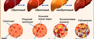
Liver fibrosis (AF) is one of the most common complications resulting from liver diseases. AF manifests itself in the form of sequential proliferation and compaction of connective tissue, which leads to inflammation. In this way, the body tries to create a barrier between the affected area of the body and the healthy one, preventing the infection from spreading. Unfortunately, many people do not know what fibrosis is.
Fibrosis may be a complication of liver disease
Pathogenesis
When factors affecting the liver occur, biologically active substances are formed that trigger the work of macrophages, which secrete substances that aggravate the anti-inflammatory response. The balance is disturbed, which leads to the predominance of fibrotic protein production. The normal exchange between blood and liver cells is disrupted, which ultimately leads to liver necrosis. Sometimes the work of mediators of focal inflammation can stop and then processes are activated aimed at stopping pathological fibrosis formation and restoring the normal functioning of the body.
Complications
In the absence of timely medical intervention, pathology leads to very sad consequences, including:
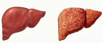
- The development of hepatic cirrhosis, occurring at the final stage of the pathology;
- Ascites and peritonitis;
- Varicose veins of the esophageal veins, accompanied by heavy bleeding;
- Hepatic encephalopathy and confusion;
- The development of hepatopulmonary syndrome, when there is an acute lack of oxygen in the patient’s blood;
- Cancerous tumor in the liver - hepatocellular carcinoma, a difficult to treat tumor disease that develops against the background of viral or alcoholic liver damage;
- Gastropathy of a hepatic nature, which is a gastric lesion due to impaired liver function and changes in blood circulation;
- Hepatorenal syndrome, which is a severe form of liver failure due to cirrhosis;
- Colopathy develops against the background of blood microcirculation disorders and dysfunctional changes in the liver.
Causes of liver fibrosis
A number of factors lead to this complication:
- viral lesions: cytomegaloviruses, viral hepatitis and infectious mononucleosis;
- immune system failure;
- damage to the bile ducts;
- portal hypertension;
- Bud-Chiarri syndrome (retention of venous blood in the liver);
- abuse of alcoholic beverages for 7 years or more (alcoholic liver fibrosis develops);
- genetic conditioning (heredity);
- medicinal substances: anticancer drugs, antirheumatic drugs, vitamin A.
Congenital fibrosis
Congenital liver fibrosis (fibrocholangiocystosis, cystic fibrosis or cystic fibrosis) is a genetically determined pathology, expressed by a morphological disorder of the intrahepatic branches of the portal vein and bile tracts. The clinic is characterized by portal hypertension with bleeding in the digestive system.
Fibrocholangiocystic disease is expressed by fibrotic changes in the liver and the growth of cysts in the intrahepatic bile ducts. This disease is quite rare, but can occur against the background of nephronophthisis, polycystic disease or dysplasia. Pathology is not confined to gender. The prognosis of the disease depends on its form and can be either favorable or fatal.
Symptoms and first signs
The disease often develops asymptomatically at first. Symptoms begin to appear already at a long stage. There are no clinical signs in the early stages; the proliferation of connective tissues can be detected by analyzing a piece of organ tissue. The early manifestations of this pathology include the following nonspecific symptoms:
- decreased performance;
- increased fatigue;
- the appearance of difficulties during physical and psychological stress.
The listed signs may indicate other complications. With fibrosis, the following specific symptoms will further appear:
- anemia;
- deterioration of immunity;
- the appearance of bruises over the entire area of the body;
- bleeding in enlarged veins of the digestive system;
- formation of vascular webs.
Symptoms of liver fibrosis in children are the same as in adults.
Symptoms
At the initial stages, fibrotic liver processes do not express themselves in any way; usually the first symptoms begin to appear 5 years after the onset of development, or even later. Only a comprehensive and comprehensive diagnosis allows a patient to be diagnosed correctly.
Quite often the spleen is involved in the pathological process, which only complicates a reliable diagnosis.
The first pathological manifestations are considered to be signs of fatigue, lack of resistance to physical and psychological stress, and decreased performance.
Examination of the patient may show some enlargement of the spleen tissue, and blood tests will reveal a deficiency of all blood components. Scar tissue can only be detected by ultrasound examination or liver biopsy.
Over time, the patient notices a pathological decrease in immune forces, strange bruises and spider veins form on the surface of the body, and hematemesis may appear, which indicates hemorrhage of the esophageal veins. The liver begins to irreversibly enlarge, and without treatment, cirrhosis develops.
Signs of hepatitis C, B
Usually, with fibrotic lesions against the background of hepatitis, the progression of the pathology is permanent and irreversible.
During the development of pathological processes, changes in the liver structures occur, the extreme stage of which is cirrhosis.
The natural course of hepatitis directly depends on the rate of development of fibrotic pathology. The virus significantly affects the progression of pathological processes, which can manifest itself differently in each patient; for some, the development of irreversible changes slows down, and for others, on the contrary, it accelerates.
But in most cases, the pathological clinical picture rapidly increases, cirrhosis quickly develops, which further complicates the course of hepatitis.
Patients experience painful discomfort in the right hypochondrium, constant headaches, hyperthermia is often present, weight decreases, the sclera and skin become yellow, and fluid accumulation increasingly appears in the abdomen and legs.
In men, against the background of fibrosis and hepatitis, libido decreases and gynecomastia develops. Behavioral changes occur, the liver continues to steadily increase in size, varicose veins are observed on the anterior wall of the peritoneum, and the urine takes on a dark tint.
Mental disorders manifest themselves in a constant pessimistic mood, excessive irritability and anxiety. Patients experience inexplicable fear, they have reduced self-esteem and concentration, they often experience unreasonable guilt, and are in a lethargic state.
Metabolic products constantly formed in the body are no longer detoxified by the liver, as they should be normally, which leads to their neurotoxic effect on brain structures. This explains the occurrence of mental disorders against the background of the development of fibrotic processes.
Forms
The classification of the pathology in question is divided into two types. The first is based on the causes of the disease. Experts distinguish three forms:
- cardiac fibrosis of the liver;
- periportal liver fibrosis (develops due to parasitic infection);
- hereditary form (this is the result of a metabolic disorder caused by excessive stagnation of chemicals in tissues and organs, develops against the background of renal polycystic disease).
Another classification is based on the distribution and localization of tissue growths:
There are different forms of liver fibrosis
- periductal form (concentration of fibrous segments around the bile ducts);
- pericellular form (damage to the surface of hepatocytes, formation of impenetrable membranes around them);
- mixed form (combines several characteristics of different forms);
- venular and perivenular form (damage to the median tissue of the hepatic lobules);
- septal form (appearance of extensive necrosis).
Forms of fibrosis
There are several forms of the disease.
Focal
Focal fibrosis is evidence of focal liver damage in the past, a local process. At the site of the granuloma, small intratubular scars appear. This form occurs when there is a local disturbance of blood circulation in the liver due to local stagnation or a sharp increase in blood pressure in the vessels.
This may be the initial stage of any fibrosis, when the foci are single. The process is accompanied by the development of focal fibrosis of the liver stroma.
Congenital
Congenital fibrosis (fibrocholangiocystic disease) is a hereditary autosomal dominant disease. There are familial and sporadic forms. With this pathology, children develop fibrous lesions and bile duct cysts. Belongs to the group of rare diseases.
Manifestations of this disease begin in children aged three to thirteen years. The pathogenesis of the disease is due to the pathology of the formation of the bile duct plate between the liver lobules. With this form, there is no formation of false lobules. This lesion may be accompanied by pathologies of other organs in the child.
Portal or periportal fibrosis
Portal liver fibrosis is characterized by the formation of pathological tissue in the center of the liver lobules. Where the central vein of the lobule is located. This process develops in various chronic hepatitis. Over time, fibrous tissue grows to surround the portal tracts. They expand, and the liver lobules become smaller. Portal and periportal forms of the lesion are usually combined.
Pericellular
Pericellular fibrosis is characterized by the proliferation of fibrotic tissue around major organ cells. This form of the disease occurs with alcohol damage to the organ and hepatitis (C, B, D).
Venular or perivenular
Venular fibrosis - it is characterized by the proliferation of fibrous tissue in the center of the classical organ lobules, where the main vein of the lobule is located.
Perivenular - it is characterized by the growth of pathological tissue between hepatocytes and the central vein. Next, the main vein of the lobule grows with pathological tissue.
Both forms occur with cardiovascular insufficiency and alcoholism.
Periductal
Periductal fibrosis of the liver - with this disease, pathological tissue grows near the bile ducts. This form occurs with inflammatory diseases of the liver ducts.
Periductal fibrosis is characteristic of:
- congenital defects of the bile drainage tract;
- biliary cirrhosis;
- sclerosing cholangitis.
Fibrous changes in this form lead to even greater disruption of the outflow of bile.
This is interesting: How to determine liver fibrosis by ultrasound? Is fibrosis visible on ultrasound?
Septal fibrosis
Septal fibrosis is characterized by the formation of septa of fibrous tissue (septa).
They grow at the site of necrosis of a large number of liver cells. These septa completely penetrate the parenchyma of the hepatic lobule. A large number of false lobules are formed.
There are 2 types of septa:
Porto-portal – connect adjacent portal tracts.
Portocentral - connects the main hepatic vein and the portal complex. Due to the proliferation of septa, the blood supply to the organ is disrupted. Pathological fistulas are formed. An arteriovenous discharge of blood is formed. Liver cells receive nutrients from the bloodstream and die. This form of fibrosis is characteristic of chronic inflammatory processes.
Mixed fibrosis
This form is the most common. Here there are combinations of all the forms that were described earlier. The form is found in all liver diseases.
Stages of liver fibrosis
To establish the stage of organ destruction, liver biopsy is used as an instrumental method or fibrotest. The results are interpreted according to the METAVIR method.
In accordance with these methods, experts distinguish 4 stages of AF. To determine the degree of liver fibrosis, a scale with positions from 1 to 4 is used.
1st degree
With liver fibrosis of the 1st degree, the organ has lost its full functioning, and the exchange between hepatocytes and blood cells is disrupted. With quick diagnosis and quality treatment, the disease recedes.
2nd degree
Liver fibrosis of the 2nd degree is established with an increase in the area of changes and damage to the organ. Therapy becomes more complicated, the disease is difficult to control with medications at this stage, and then stage 2 fibrosis progresses to stage 3 AF.
3rd degree
Liver fibrosis of the 3rd degree consists in the formation of compactions in the structures of the organ. Without careful treatment, the prognosis becomes negative - the pathology enters the final stage.
4th degree
Stage 4 liver fibrosis is diagnosed when cirrhosis occurs. It is possible to defeat it only with an organ transplant.
Causes of the disease
Fibroblasts play a major role in the development of this process. Increased fibrinogenesis occurs when hepatocytes are damaged. Fibrous tissue contains ten times more collagen than normal connective tissue. The pathological process begins to develop when the formation of collagen becomes greater than its breakdown. There must be signs of inflammation and proliferation of small intrahepatic ducts. Fibrous changes in the liver can trigger elements that appear when liver cells are damaged.
These include:
- peptides;
- macromolecules;
- lysosome fragments;
- sinusoidal surface of a damaged liver cell;
- formation of a basement membrane between hepatocytes and sinusoidal cells;
- stromal fibrosis;
- reduction of hepatic cell villi;
- fibroblasts.
A vicious circle effect appears. Fibroblasts are stimulated due to damage to hepatocytes. There is a malnutrition and damage to liver cells due to the proliferation of fibroblasts.
There are primary and secondary fibrosis.
Primary liver fibrosis is a congenital disease. It is genetic in nature and occurs before birth. This is a serious disease. It affects hepatocytes, liver vessels, and most bile ducts.
Secondary – formed against the background of long-term pathological processes. The outbreaks do not show themselves for a long time. It is very difficult to suspect these diseases.
Causes of secondary fibrosis:
- Alcohol.
- Abuse of fatty foods.
- The predominance of fried foods in the diet.
- Long-term use of certain potent medications.
- Poisoning by chemical agents.
- Chronic poisoning by toxins.
- Amyloidosis.
- Brucellosis.
- Viral hepatitis C.
- Other viral hepatitis.
- Diabetes.
- Schistostomiasis.
- Fat disease.
- Autoimmune hepatitis.
- Galactosemia.
- Hepatoma.
- Primary biliary cirrhosis.
- Hemochromatosis.
- Liver toxoplasmosis.
- Wilson-Konovalov disease.
- Hypothyroidism.
- Echinococcosis.
- Budd-Chiari syndrome.
- Cholelithiasis.
- Weakening of the immune system.
This is interesting: Stage 4 liver fibrosis: what is it? Life expectancy and prognosis for hepatitis C
METAVIR scale
The degree of liver health in medicine is determined using the Metavir scale. It is optimal and helps to interpret the results of a fibrotest, an analysis for alt and ast, or a biopsy, which is the most reliable option for diagnosing various diseases. When examining a tissue fragment, METAVIR assigns two base numbers: the first to determine the degree of inflammatory process, the second to represent the degree of fibrosis, which is set on a scale from 0 to 4. The level of necrotic and inflammatory processes is assessed on a 4-point scale.
Legend
The indicator of the intensity and activity of pathology is indicated in the form of a two-letter and two-digit system. For example, “F2-A3”, “F3-A1”, that is, almost any combination from the list of correspondences on the metavir scale is possible:
- f0 (no fibrosis) – a0 (no activity);
- f1 (portal proliferation of tissue without septa) – a1 (moderate activity);
- f2 (portal proliferation of tissue with several septa) – a2 (medium activity);
- f3 (portal proliferation of tissue with many partitions) – a3 (high activity);
- f4 (cirrhosis).
Other scales
In addition to the METAVIR index, the stages of liver fibrosis can be determined using the Knodel and Ishak scales, which were originally invented to determine the degree of liver fibrosis in hepatitis.
The level of intensity of hepatitis is assessed by the Knodel system. It is more complex than METAVIR, but is characterized by higher accuracy for determining the severity of the inflammatory process that accompanies liver fibrosis in hepatitis C. The method is based on the use of four different scores, which together make up an overall index. The first block of the index (periportal necrosis) varies from zero to ten. The next two blocks (lobular necrosis and portal inflammation) vary from zero to four. The combination of these three components determines the level of the inflammatory process:
- 0 (no inflammation);
- 1-4 (beginning of inflammation);
- 5-8 (low inflammation);
- 9-12 (moderate inflammation);
- 13-18 (severe inflammation).
The fourth block indicates the level of organ compaction and varies from 0 (no scars) to 4 (cirrhosis).
Stages of liver fibrosis according to the Ishak scale:
- 0 – no fibrosis;
- 1 – fibrosis grows into several portal tracts;
- 2 – fibrosis expands into most portal tracts;
- 3 – fibrosis grows into many portal tracts and some portoportal septa;
- 4 – fibrosis covers the portal tracts and many portoportal and portocentral septa;
- 5 – incomplete cirrhosis;
- 6 – full-fledged cirrhosis.
Diagnostic methods
At the initial stage, the disease is difficult to identify due to the fact that the signs of liver fibrosis are nonspecific. For a long period of time, fibrotic changes occur without visible symptoms.
There are several methods for diagnosing liver fibrosis. The most common is laboratory, based on the following tests: general blood test, analysis of alt and ast, blood biochemistry, fibromax, general analysis of urine and feces.
The second group of methods is instrumental. Based on the following procedures:
One of the methods for diagnosing fibrosis is liver biopsy.
- Ultrasound.
- Fine needle biopsy.
- Esophagogastroduodenoscopy.
- CT scan.
- Biopsy.
- Fibrotest.
- Fiber scanning.
- Indirect elastometry.
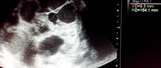
Many people are interested in whether AF is visible on liver ultrasound. Experts say that it is visible, and therefore this method is the fastest and easiest for initial diagnosis, moreover, everyone knows what it is, unlike biopsy or elastometry.
Diagnostics
When examining a patient, the doctor may see:
- Yellow skin.
- Enlarged veins on the abdomen.
- Increased liver size.
- Gynecomastia.
- Darkening of urine.
- Enlarged spleen.
- Ascites.
- Xanthomas.
- Spider veins.
- Xanthelasmas.
- Signs of mental disorder.
- Erythema of the palms.
- Swelling of the legs.
- Kaiser-Fleischner ring.
Laboratory diagnostics
Examination methods:
- Complete blood count (typical: increased ESR, decreased red blood cells, hemoglobin, leukopenia, thrombocytopenia).
- Blood biochemistry (Increase: activity of transaminases, cholesterol, bilirubin. Decrease: albumin, urea, fibrinogen, prothrombin).
- General urine analysis (proteinuria, bilirubinuria, high levels of urobelinogen, cylindruria).
- Immunogram.
Instrumental research methods
- Ultrasound (determines changes in the structure of the liver, detects cysts).
- CT scan (finds changes in the structure of the organ, determines the stage of the disease).
- MRI (assessment of the degree of organ enlargement and changes in its tissue).
- Laparoscopy (examine the appearance and take a piece of tissue for biopsy).
- Biopsy (to determine the degree of fibrosis).
- Isotope scanning (determines the functional state of the organ).
- Elastography (degree of fibrotic changes).
- Fibrotest, Fibromax, Firomet V and Fibrospect II (determine the degree of fibrotic changes).
Treatment
Before treatment, it is necessary to eliminate other problems of the body, if any, (destroy viruses, bacteria and parasites in other organs). Next, you need to get rid of exposure to household and industrial toxic substances. Avoid the use of certain drugs that have an adverse effect on hepatocytes (sedatives, contraceptives and steroids).
Initial treatment of liver fibrosis once diagnosed is with medications. Drugs containing ursodeoxycholic acid help reduce inflammation and maintain liver function. The attending physician also prescribes the latest medications that inhibit the proliferation of connective tissue.
Compliance with a certain diet, the diet of which is based on nutrition with a maximum reduction in the load on the liver, has a significant impact on the healing process. The diet for liver fibrosis is prescribed by the doctor and the patient must eat strictly according to it.
Symptoms and prognosis
Signs of hepatic fibrosis may appear five years after the start of the pathological process. Often the disease already at stage 2 of the disease is accompanied by:
- increase in organ size;
- thrombocytopenia;
- bleeding from the esophagus;
- enlarged spleen.
Fibrosis, which limits the normal functioning of the liver, often causes cirrhosis, as well as liver failure and portal hypertension. This stage cannot be treated and requires a liver transplant. The first and second stages often occur without pronounced symptoms. Therefore, at this stage the disease is difficult to diagnose. The second stage may be accompanied by inflammation and enlargement of the spleen. At the same time, the number of blood cells (leukocytes, platelets) decreases, which causes anemia and anemia. Liver tissue changes significantly.
Important! If therapy is started in a timely manner at the second stage, the prognosis for recovery is favorable.
The rate of progression directly depends on the severity of inflammatory processes in the liver. Further development of the disease (stages 3 and 4) can cause cirrhosis, the occurrence of varicose veins of this organ, hemorrhage, and scar formation.
The prognosis for fibrosis of the second degree depends not only on the individual susceptibility of the body to drug treatment, but also on the normalization of lifestyle and diet.
Based on the degree of prevalence and localization of the pathological process, several main forms of the disease can be distinguished:
- venular – the focus is located in the central part of the organ;
- pericellular – inflammation is mainly concentrated around hepatocytes;
- septal – the presence of large necrotic areas, the formation of a large number of fibrous septa;
- periductal - characterized by constantly growing connective tissue located around the bile canaliculi;
- mixed form is the most common type of fibrosis, which includes all the signs described above.
The rate of development of fibrosis from the initial to the last stage is determined mainly by the type of pathology.
- The non-cirrhotic form can develop in various severe infectious diseases. In this case, sclerotic changes and thrombosis in the hepatic vessels may develop. This pathology is often the result of excessive alcohol consumption, the presence of hepatitis of various natures, exposure to toxic substances, and uncontrolled use of medications.
- The periportal form is complemented by potal hypertension and is characterized by an increased severity of the course. The disease begins with parasites, which occurs through contaminated water. This form is dangerous due to severe complications when the helminth enters the human body.
Traditional medicines
Milk thistle is a folk remedy for treating fibrosis
Many people are wondering: is it possible to cure such a serious disease with folk remedies? Experts have established how to treat AF using alternative medicine. The pathology in question requires a complex effect, and the organic substances of many medicinal plants can have a beneficial effect on the functioning of hepatocytes and reduce the intensity of the inflammatory reaction.
Milk thistle, white cinquefoil, chicken eggs, eryngium, birch leaves, half-palm, dandelion, infusions of buckthorn, prunes, rose hips, apple cider vinegar and honey will help treat liver fibrosis, reducing the unfavorable course of the disease.
Progression of fibrosis
The intensity of fibrosis progression varies significantly among different patients. It occurs most quickly in adult male patients with weak immune systems who often drink alcohol. Diabetes and excess weight also contribute to its development.
At the moment, there is no accurate method for predicting the rate of progression of necrosis in patients. A large amount of alanine aminotransferase in the blood can serve as a marker. Analysis for alt and ast, fibrotest, ultrasound fibroscan examination and biopsy can also help in predicting the development of events.
Life expectancy with fibrosis
It is important to know how long people with AF live. Before cirrhosis, the disease does not threaten a person’s life with proper treatment. After the onset of the fifth stage, many factors influence life expectancy. Sometimes the patient lives longer than ten years, and in other cases he may not last even a year, dying from cirrhosis.
The situation is influenced by: the degree of compensation for liver work, etiology, blood biochemistry, complications of cirrhosis, quality of therapy, associated problems, nutrition, gender and age of the patient.
With timely diagnosis, you can significantly reduce the rate of development of the fifth stage and live with it for ten years.