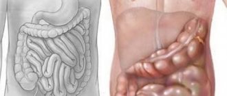
Symptoms and causes
The danger of infection lies in the fact that in some cases the symptoms are not clearly expressed - the patient may simply not attach any significance to them or confuse them with the manifestation of an existing chronic disease (for example, with gastritis, heartburn is a common occurrence).
Signs of esophageal candidiasis often resemble the picture with damage to the gastrointestinal tract:
- decreased appetite;
- weight loss;
- nausea (less often vomiting);
- heartburn;
- pain in the upper abdomen and behind the sternum;
- loose stool.
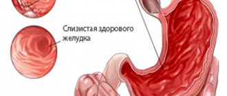
Esophageal candidiasis is also expressed by odynophagia (painful swallowing) and dysphagia (impaired process or inability to swallow food). With such disorders, food or liquid may enter the trachea, nose, or larynx. There may be white films in the vomit, and mucus and bloody discharge in the stool.
Damage to the esophageal mucosa initially appears as separate white areas. They then expand, forming a dense plaque. Underneath, the fungi spread deeper and deeper, reaching blood vessels and muscles.
Esophageal candidiasis occurs for various reasons:
- intestinal candidiasis (high probability);
- contact with an infected person or carrier;
- infected product;
- infection through household items or hygiene products;
- oral candidiasis.
The causes of esophageal candidiasis may be hidden in an imbalance of calcium-phosphorus metabolism. This happens when the functionality of the adrenal glands and thyroid gland is impaired.
There are certain risk factors for the development of esophageal candidiasis:
- reduced immunity;
- narrowed lumen of the esophagus (due to tumor, injury);
- diabetes;
- pathology of the digestive organs;
- intravenous (parenteral) nutrition (the gastrointestinal tract is not involved);
- allergy;
- chronic infections (often HIV, tuberculosis);
- smoking;
- alcoholism;
- long-term use of antibiotics or hormonal drugs;
- malnutrition;
- low acidity;
- taking corticosteroids;
- intoxication;
- taking antacids;
- organ transplantation.
This is not a complete list of factors influencing the development of fungal infection. The significance of each is determined by the state of the human immune system.
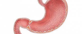
What is esophageal candidiasis?
An infection of the esophageal tube due to the active action of a fungus of the genus Candida, where the origin of the diagnosis comes from, is called candidiasis. According to the International Classification of Diseases, 10th edition, pathology refers to an unspecified disease of the esophagus.
The disease code according to the international classifier is K22.9.
Photo of esophageal candidiasis
The disease has a long treatment period; if diet, daily routine and treatment regimen are not followed sufficiently, the success of recovery is in question. Patients suffering from pathologies of the digestive system, in the amount of 1.5% of the total number of cases, are diagnosed with candidiasis of the digestive system.
The occurrence of the disease is facilitated by a failure of immune reactions, activation of the human immunodeficiency virus, and impaired administration of antibacterial agents.
Diagnostics
A doctor's suspicion of esophageal candidiasis arises from studying the outpatient chart and the presence of risk factors.
The diagnosis is confirmed after several studies:
- X-ray of the esophagus;
- endoscopic examination (esophagoscopy);
- mycological culture analysis;
- cytological and histological analyzes of scrapings;
- biopsy (rare).
Esophagoscopy is a standard procedure if the patient complains of pain and difficulty swallowing. During endoscopy, the disease is expressed by redness, fibrinous plaque and surface vulnerability (on contact).
How to diagnose
If candidomycosis is suspected, an endoscopic examination is recommended in a medical facility. To do this, special equipment is used to examine in detail the affected mucous membrane of the patient's esophagus. Characteristic signs of pathology include increased sensitivity in the area under study, hyperemia, and the formation of fungal plaque. Plaque can have a fibrous appearance with different structures, configurations, sizes and different locations. During diagnosis, three types of candidiasis can be identified depending on the characteristic symptoms.
Catarrhal endophagitis. In this case, swelling is fixed on the mucous membrane, and the inflammatory process is of the diffuse type. Possible bleeding upon contact with the surface of the shell itself. Plaque can be a cobweb-like layer of whitish color without foreign erosions on the tissues.
Fibrous esophagitis. With this type, you can find plaques of a loose type, round in shape. Contact vulnerability and distinct tissue hyperemia are observed.
Fibrous-erosive esophalitis. The plaque becomes gray in color in the form of velvety ribbons on the surface of the esophageal mucosa. Erosion takes on a rounded shape. The mucous membrane is inflamed, has severe swelling, and candidal changes in the esophagus prevent normal examination of the entire organ with an endoscope. Severe pain, stenosis and bleeding are observed.
After the examination, the doctor may prescribe an additional examination using x-rays using contrast agents. Also, for diagnosis, a tissue sample can be taken for histological examination in the laboratory. Doctors can take a culture from the esophagus to determine the type of nutrient medium. After this, the sensitivity of pathogens to antifungal drugs can be determined. A method for eliminating the symptoms of the development of Candida fungus in the esophagus is prescribed after the patient has been diagnosed. The doctor determines the course of therapy based on the test results, the patient’s condition and his individual characteristics of the body, as well as the susceptibility of the drugs to the causative agent of the disease.
Complications
There are several degrees of esophageal candidiasis:
- Stage I infection means that the fungus has not yet penetrated deeply, and there is plaque only in certain areas of the esophagus.
- In stage II of the disease, the fungus penetrates the mucous membrane, and plaque spots begin to merge into large areas.
- Grade III characterizes the penetration of the fungus into the muscles, the development of a bacterial infection and tissue necrosis is possible.
If treatment is not done in a timely manner, complications such as:
- ulcer;
- bleeding;
- phlegmon (purulent inflammation);
- stricture (reduction of the lumen of the esophagus);
- rupture of the esophageal tube;
- tissue death;
- death.
Therefore, it is very important to start therapy in a timely manner and not only act on the pathogen, but also eliminate concomitant diseases.
Traditional treatment
Confirmed symptoms of esophageal candidiasis are eliminated with medication. Typically, treatment of esophageal candidiasis means taking drugs with azole - Fluconazole, Clotrimazole, Oronazole, Miconazole. If they do not give the desired result, then polyene antibiotics are prescribed:
- Levorin;
- Amphotericin B;
- Nystatin;
- Mycoheptin.
In case of severe infection, drugs are administered intravenously with the simultaneous administration of enterosorbent agents (activated carbon, enterosgel, polysorb, etc.).
When esophageal candidiasis manifests itself against the background of reduced immunity, a course of immunostimulants is included in its treatment.
After taking the necessary medications, it is important to restore the gastrointestinal microflora. To do this, you need dental medications - Linex or Baktisubtil.
How dangerous is this disease?
Thrush in the mouth, throat, or esophagus is not often seen in healthy adults.
People at increased risk of candidiasis in the mouth and throat are mainly children, especially infants under one month old, and people who:
- wear dentures;
- diabetics;
- have cancer;
- have HIV/AIDS;
- are taking antibiotics or corticosteroids, including inhaled corticosteroids, such as for asthma;
- take medications that cause dry mouth or have medical conditions that cause dry mouth;
- smoke.
Most people with candidiasis in the esophagus have a weakened immune system. This includes people living with HIV/AIDS and people suspected of having blood cancers (leukemia and lymphoma).
www.cdc.gov
Traditional treatment
It is possible to treat esophageal candidiasis with folk remedies, but it is better to combine it with traditional medicine. Herbal infusions of chamomile, rose hips, calendula, juniper, St. John's wort, yarrow, eucalyptus, and oak bark are effective. You should only combine these medications with medications after consulting your doctor.
One of the traditional methods of treatment is a strained decoction of oats. It must be filled with water in a ratio of 1:3, brought to a boil and after 3 minutes remove from heat. Drink this decoction at night and before meals (3 times a day).
Kombucha tincture also helps - take it 4 times a day (1 glass per day).
After eating, rinsing your mouth with soda solution is effective.
New therapies
Experts have proven the effectiveness of autohemotherapy in the treatment of candidiasis. What it is? With this method of treatment, a small portion of blood is taken from the patient, irradiated with ultraviolet light and injected back. This helps activate the immune system to fight infection.
Autohemotherapy is usually combined with another new treatment method - endoscopic injection of granulocyte mass. Donor blood cells - granulocytes - are delivered into the esophagus through a special tube. To activate the immune system, it is necessary to carry out several such procedures. Immunomodulatory therapy allows for faster cure.
Diet
Diet for esophageal candidiasis is one of the important components of treatment. It has certain similarities with the diet for intestinal candidiasis. Portions should be significantly reduced, but also the time between doses should be reduced.
The amount of carbohydrates in the diet should be reduced as much as possible, and some foods will have to be eliminated completely:
- honey and chocolate;
- potato;
- alcohol;
- carbonated drinks;
- bread;
- raw mushrooms;
- sugar;
- sweets and confectionery;
- salty;
- yeast;
- spicy.
The diet must include dairy and fermented milk products - milk, cottage cheese, cheese, kefir, yoghurts. You should also include brown rice, buckwheat, cabbage, herbs, onions and garlic in your diet. As for drinks, it is better to limit yourself to tea, rosehip decoction and dried fruit compote.
For esophageal candidiasis, pickled vegetables and kombucha are useful - such products must be of proven quality.
If a severe form of candidiasis is observed, then high-protein mixtures administered through a tube are used for nutrition.
Gastroenterology of St. Petersburg, 2006, No. 1-2, pp. 17-20.Standards and prospects for pharmacotherapy of candidiasis of the digestive organs.
M.A. Shevyakov
St. Petersburg MAPO.
The increase in the number of patients with immunodeficiency conditions of various origins has led to the fact that gastroenterologists and doctors of other specialties are increasingly faced with the problem of choosing an antifungal drug for the treatment of candidiasis of the digestive organs.
In the treatment of candidal infections of the mucous membranes of the upper digestive tract, azole antifungal drugs dominate. These drugs can be used topically or systemically and have been shown to be safe and effective. In the treatment of dysbiotic conditions associated with intestinal overgrowth of Candida spp
Polyene antimycotic drugs in combination with pro- and prebiotics have taken their place. A significant problem associated with candidiasis of the mucous membranes is the tendency of some patients to relapse. In a small number of cases, the explanation for the reasons for such relapses is obvious (for example, relapses of candidiasis in people with manifest and uncontrolled HIV infection, decompensated diabetes mellitus, long-term users of high doses of glucocorticosteroids), but in other patients the cause of relapses remains unclear (1).
When choosing an antifungal drug for the treatment of candidiasis, the clinician has to take into account seven factors: features of pathogenesis, localization of the lesion, sensitivity of the pathogen, safety profile of the antifungal drug, the state of the patient’s liver and kidney function, possible interactions with other drugs, and, finally, pharmacoeconomic aspects.
Pathogenesis and diagnosis of candidiasis of the upper digestive tract.
The pathogenesis of candidiasis of the mucous membranes of the digestive tract is characterized by the sequential passage of the following stages - adhesion, invasion, candidemia and visceral lesions. At the first stage, micromycetes adhere to the epithelial cells of any part of the mucous membrane. Subsequently, defects in the resistance system allow micromycetes to penetrate (invade) the mucous membrane and underlying tissues through transformation into pseudomycelium. Cytopenia is a decisive factor that allows invading fungi to reach the vascular wall, destroy it and circulate in the vascular bed. This stage is called candidemia. In the absence of adequate therapy, candidemia leads to the formation of foci of invasive candidiasis in visceral organs, for example, lungs, endocardium, central nervous system, liver, etc.
At first glance, it is paradoxical, but the invasion of fungi of the genus Candida
is more often observed in areas where the mucous membrane is represented by multilayered epithelium (for example, the oral cavity, esophagus) and much less often - in single-layer epithelium (for example, the stomach, intestines).
The “gold standard” for diagnosing candidiasis of the mucous membranes is the detection of Candida
spp. during morphological examination.
In order to detect pseudomycelium, morphological mycological methods are used: cytological - with staining of smears, for example, according to Romanovsky-Giemsa, and histological - with staining of biopsy specimens, for example, the CHIC reaction. Thus, knowledge of the dimorphism of Candida
spp. is the key to the differential diagnosis between candidiasis and candidiasis. In modern conditions, the clinician has the right to expect from a morphologist an accurate description of the detected morphological structures of the fungus, because the detection of blastomycetes, as a rule, indicates candidiasis, and the detection of pseudomycelium allows us to confirm the diagnosis of candidiasis.
Treatment of oropharyngeal candidiasis and esophageal candidiasis.
The goal of treatment for these forms of candidiasis is to eliminate the symptoms and clinical signs of the disease, as well as to prevent relapses. For some patients with oropharyngeal candidiasis, “local” risk factors are important - wearing dentures and inhaling corticosteroids. Symptoms of oropharyngeal candidiasis and esophageal candidiasis, in addition to subjective discomfort, can impair the swallowing of food and liquids and significantly reduce the quality of life.
To treat oropharyngeal candidiasis, both locally acting and systemically acting antifungal drugs can be used. Abroad, for local therapy, for example, lozenges with clotrimazole or a suspension of nystatin or amphotericin B are used. In our country, such dosage forms are not registered, and local therapy usually involves chewing or applying crushed tablets of nystatin or natamycin to the mucous membrane in doses of 1 to 4 million units per day with a course duration of up to 2-4 weeks (2). The effectiveness of this “improvised” treatment method has not been studied in controlled studies. Local antifungal therapy for esophageal candidiasis is ineffective.
For candidiasis of the mucous membranes of the upper digestive tract, systemic azole antimycotics (ketoconazole, fluconazole, itraconazole or voriconazole), intravenous amphotericin B (especially for azole-refractory infections), as well as echinocandin drugs (caspofungin, micafungin, etc.) can be used. For patients with impaired swallowing, parenteral forms of antimycotic drugs have been developed.
The effectiveness and safety of azole and echinocandin antimycotic drugs have been studied quite well, a large evidence base based on controlled studies has been accumulated, and the results of these studies have formed the basis of therapeutic standards (3-5).
The effectiveness of antimycotic therapy for oropharyngeal candidiasis was assessed in multiple randomized prospective studies in both patients with AIDS and patients with malignant neoplasms. Most patients respond to treatment with topical forms of polyene antimycotics (lozenges and suspension). However, in HIV-infected patients, relapses of oral candidiasis may occur more quickly after local therapy than after treatment with fluconazole at a dose of 100 mg per day, and resistance of the pathogen can develop with the use of any therapy option. It has also been shown that fluconazole is superior in effectiveness to ketoconazole, and itraconazole capsules are equivalent in effectiveness to ketoconazole. Oral itraconazole solution is better absorbed from the gastrointestinal tract than the capsule form and is comparable in effectiveness to fluconazole. A dose of itraconazole solution 2.5 mg/kg twice daily has been recommended for the treatment of oropharyngeal candidiasis in children 5 years of age and older. The effectiveness of itraconazole oral solution may be greater due to the summation of local and systemic effects.
Recurrent candidiasis is usually observed in patients with immunodeficiencies, especially with AIDS. Long-term maintenance therapy with fluconazole has been shown to be effective in preventing the development of oropharyngeal candidiasis in both cancer patients and AIDS patients. Long-term maintenance therapy with fluconazole in HIV-infected patients reduces the incidence of invasive candidiasis, but does not affect overall survival. Continuous maintenance antifungal therapy reduced the incidence of relapse of candidiasis compared with intermittent treatment, but the levels of development of microbiological resistance were similar in both groups.
Oral polyene agents such as amphotericin B or nystatin are less effective in preventing mucosal candidiasis. At the same time, it was found that itraconazole 200 mg per day was effective as anti-relapse therapy for oropharyngeal candidiasis for 6 months. It has been shown that 64-80% of patients with fluconazole-refractory infections are cured of oropharyngeal candidiasis when using itraconazole oral solution. Amphotericin B oral solution has also been used successfully in the treatment of fluconazole-resistant oral candidiasis.
Micafungin or caspofungin intravenously at a dose of 50 mg daily may be a reasonable alternative treatment, especially for the treatment of candidiasis caused by fluconazole-resistant strains, but their use is limited by the high cost of treatment. Intravenous antifungal therapy may sometimes not be necessary if either interferon-gamma or granulocyte-monocyte colony-stimulating factor is used in combination with oral antifungal therapy.
C. albicans remains the most common pathogen for esophageal candidiasis in patients with HIV infection or esophageal cancer
. The presence of oropharyngeal candidiasis together with symptoms of esophagitis (dysphagia or odynophagia) strongly suggests esophageal candidiasis. As a treatment for esophageal candidiasis, fluconazole is superior in effectiveness to ketoconazole, itraconazole capsules and flucytosine, but itraconazole capsules in combination with flucytosine are as effective as fluconazole. Itraconazole oral solution has comparable efficacy to fluconazole. Up to 80% of patients with fluconazole-refractory infections are cured with the use of oral itraconazole solution. Voriconazole (200 mg twice daily for an average of 14 days) is as effective as fluconazole (400 mg loading dose followed by 200 mg daily for an average of 15 days) but has a higher incidence of side effects. Voriconazole has been successfully used to treat cases of esophageal candidiasis refractory to fluconazole. In these cases, intravenous amphotericin B, caspofungin, or micafungin may also be effective (2,6).
In patients with AIDS, relapses of esophageal candidiasis are quite common, and long-term suppressive therapy with fluconazole (100 mg/day) is effective in preventing relapses.
In both oropharyngeal and esophageal candidiasis, the vast majority of infections are due to either C. albicans
, or mixed culture (2.7).
However, infections caused by C.glabrata, C. krusei
and other pathogens that are insensitive to treatment with various antifungal drugs have been described.
In vitro antifungal susceptibility tests generally reliably predict clinical response to fluconazole and itraconazole. The use of antiretroviral therapy in HIV-infected patients has led to both a decrease in the level of carriage of C. albicans
and a decrease in the frequency of relapses of oropharyngeal candidiasis and esophageal candidiasis. However, it must be recalled that repeated courses of treatment with azole antifungals or the use of long-term therapy for recurrent infections are the main factors leading to the development of azole-resistant fungal strains.
To summarize the above, we formulate the following key recommendations:
- The primary episode of oropharyngeal candidiasis can be treated by prescribing topical nystatin 500 thousand units four times a day or natamycin 100 mg 4 times a day (dissolving tablets, applying crushed tablets to the mucous membrane) for 7-14 days, but you need to be prepared that the effectiveness of such treatment is lower than treatment with azole drugs.
- The drugs of choice for the treatment of oropharyngeal candidiasis remain oral fluconazole 100 mg/day for 7-14 days or oral itraconazole solution 200 mg/day for 7-14 days.
- 3. Ketoconazole and itraconazole capsules are less effective for the treatment of oropharyngeal candidiasis than fluconazole due to unstable absorption in the digestive tract.
- In case of fluconazole-resistant oropharyngeal candidiasis, therapy is prescribed with itraconazole 200 mg/day per os, preferably in the form of a solution, or intravenous caspofungin or micafungin (50 mg per day), or intravenous amphotericin B (at least 0.3 mg/kg/day) . The duration of treatment courses is usually 1-2 weeks.
- Oropharyngeal candidiasis associated with dentures requires complete disinfection of the latter for final cure.
- For effective treatment of esophageal candidiasis, systemic antifungal therapy is required for 2-3 weeks with fluconazole (100-200 mg/day) per os or itraconazole oral solution (200 mg/day) per os.
- Cases of esophageal candidiasis resistant to fluconazole therapy should be treated with itraconazole solution (at least 200 mg/day per os), voriconazole (200 mg 2 times a day) or caspofungin (50 mg per day) or micafungin (50 mg per day), or intravenous amphotericin B (0.3-0.7 mg/kg/day) until clinical response is obtained.
- In vitro antimycotic susceptibility testing is not necessary for primary episodes of either oropharyngeal candidiasis or esophageal candidiasis, but may be useful in guiding therapy in patients with refractory or recurrent disease.
- In patients with HIV/AIDS, treatment of the underlying infection with antiretroviral therapy is the most important factor in preventing relapses and treating candidiasis.
Apparently, a significant improvement in the results of treatment of candidiasis in general can be associated with the development of methods of active specific immunotherapy. For example, they are completing a clinical trial of the drug Mycograb, created on the basis of recombinant human antibodies to hsp90 (heat shock protein 90), a molecular fragment necessary for the viability of yeast-like fungi. Initial results show its antifungal activity and synergism with amphotericin B both in vitro and in vivo (8).
Diagnosis and principles of treatment of intestinal candidiasis.
The differential diagnosis of candidiasis and candidiasis in the intestines can present certain difficulties. A fairly common mistake is diagnosing candidiasis only on the basis of identifying the fact of the growth of yeast-like fungi in feces. Meanwhile, this fact may reflect transient candidiasis.
To diagnose candidiasis, as well as for any other opportunistic infection, it is important not only to demonstrate the pathogen, but also to prove its participation in the pathological process. One of the fundamental criteria for the diagnosis of an opportunistic infection is the presence of defects in the system of antimicrobial resistance, and for the form of infection we are considering, this is a deficiency of obligate bacteriobiota - natural competitors of micromycetes. Thus, the diagnosis of intestinal dysbiosis with overgrowth of Candida
set if the following criteria are present (9):
- intestinal dyspepsia syndrome,
- laboratory confirmed intestinal dysbiosis,
- height Candida
spp. over 1000 CFU/g on stool culture.
Endoscopic studies with cytological examination of preparations are not very informative, since most often pathological changes are not detected. Differential diagnosis of intestinal candidiasis is also carried out with infectious colitis, helminthiasis and parasitic intestinal lesions, fermentopathy, tumors, irritable bowel syndrome.
The treatment plan for a patient with intestinal candidiasis should include the following therapeutic directions (10):
- Treatment of the underlying disease according to accepted standards
- Candida fungi
- Prescription of pro- and prebiotics and other drugs that help restore the natural antimicrobial resistance of the intestinal mucosa in order to prevent relapses of dysbiosis
- Symptomatic therapy and diet therapy.
For selective decontamination of intestinal candidiasis, poorly resorbable polyene antimycotics (nystatin and natamycin) are often used. The antifungal activity of some synthetic (for example, quinolone) and biological enteroseptics (for example, including live cultures of Saccharomyces boulardi
). The effectiveness of these drugs as antimycotics still has to be finally assessed in comparative studies, but we can already talk about some advantages and disadvantages of these drugs. Quinolone drugs have a wide spectrum of antibacterial and antiprotozoal activity, which makes them preferable for mixed intestinal infections. At the same time, their pronounced antibacterial activity can have a depressing effect on the resident normobiota and on the effectiveness of simultaneously prescribed probiotic agents.
Preparations containing live strains of Saccharomyces spp
in addition to enteroseptic, they showed an immunomodulatory effect (due to stimulation of IgA production by the intestinal mucosa).
However, recent observations demonstrate the serious danger of using them in groups of patients with severe immunodeficiencies. This describes 92 cases of invasive mycoses caused by Saccharomyces in patients with typical risk factors (patients with cytopenia, as well as those receiving antibiotics and having an intravenous catheter installed), with 40% of patients receiving biological products containing Saccharomyces boulardi
(11).
Nystatin, historically one of the first antimycotic drugs, is still widely used in our country and abroad. As the results of analyzes by the Pharmexpert Center for Medical Research showed, in 2001, in physical terms, that is, in terms of the number of packages (units) of the drug sold on the domestic antimycotic market, nystatin was in first place (12). For selective decontamination of intestinal candidiasis, nystatin is prescribed orally in the form of tablets of 250 and 500 thousand units, the daily dose is usually 3-4 million units, and courses of treatment last 2-3 weeks. However, the instability of the active substance (nystatin largely loses activity after exposure to sunlight and open air) and irritating effect on the mucous membranes (nausea, vomiting and diarrhea are sometimes noted when taking the drug) limit the use of this drug.
The optimal drug for selective decontamination of the intestines against micromycetes of the genus Candida
is natamycin (enteric-soluble tablets Pimafucin) (13,14).
Unlike imidasal antimycotics, natamycin is practically not resorbed in the intestine. This property, on the one hand, makes the treatment absolutely safe and applicable even for pregnant women, lactating women and newborns. On the other hand, the absence of resorption causes a consistently high concentration of natamycin in the intestinal lumen, which is the key to successful treatment of intestinal dysbiosis with overgrowth of Candida
spp. The enteric coating of Pimafucin tablets protects the mucous membranes of the digestive tract from irritation and ensures stability during storage, which distinguishes it from other polyene drugs.
Pimafucin is prescribed for adults 1 tablet containing 100 mg of natamycin four times a day, and for children 1 tablet 2 times a day. Courses of treatment of 5-10 days usually lead to the elimination of yeast-like fungi from the intestines, however, if factors that reduce the effectiveness of local antifungal barriers persist, a recurrence of candidiasis is possible. Pimafucin is also used for the prevention of intestinal candidiasis, for example, during courses of antibiotic therapy.
The successful solution of the main task - selective decontamination - allows us to count on success in the restoration and consolidation of eubiosis - i.e. persistent elimination of intestinal normobiota deficiency. For this purpose, pro- and prebiotics are traditionally used.
Probiotics are preparations containing live cultures of pharmacopoeial strains of normobiota. The main disadvantages of probiotics are significant inactivation as they overcome the acid and enzyme gastrointestinal barriers, as well as as a result of joint use with synthetic antibacterial agents. Recently, the problem of biocompatibility of resident and pharmacopoeial strains of normobiota has also been discussed. For example, according to N.A. Glushanova and A.I. Blinov, in vitro, out of 24 studied strains of resident lactobacilli, only 6 turned out to be biocompatible with the probiotic strain L.acidophilus 317\402
and another 9 – with
L.plantarum 8PA3
.
At the same time, not a single resident strain has been identified that is simultaneously biocompatible with both L.acidophilus 317\402
and
L.plantarum 8PA3
. The bioincompatibility of probiotic and resident lactobacilli was manifested by the development of two types of reactions: “resident strain against probiotic” and “probiotic against resident strain” (15).
Perhaps it is the biological incompatibility of pharmacopoeial and resident strains of normobiota that is the cause of side effects (increased diarrhea and symptoms of intestinal irritation) and ineffective treatment when using probiotics. It must be assumed that when correcting intestinal dysbiosis, it is more correct from a pathogenetic point of view not to try to “instill” a probiotic strain in the intestine, but to stimulate the “remnants” of resident strains with which the individual has been contaminated since the time of the first contacts with the skin and mucous membranes of the mother. Most likely, it is these resident strains of normobiota that are most beneficial for human health, including from the standpoint of immunological compatibility. If we assume that each patient is “normobiotically” individual to a certain extent, then it is more logical to use prebiotics to correct dysbiosis.
Choice of prebiotics, i.e. stimulators of intestinal growth of resident normobiota that do not contain live cultures of normobiota are quite wide today. The active principle of such agents may be lactulose, pectin, some biologically active metabolic products of obligate bacteria and others.
One of the most effective modern domestic products with a pronounced prebiotic effect is Eubicor. The drug contains inactivated non-viable yeast Saccharomyces cerevisiae (vini),
dietary fiber, vitamins, amino acids, macro- and microelements. It is known that dietary fiber, without being altered by enzymes of the digestive tract, easily reaches the colon, where it is metabolized by anaerobic microbiota to short-chain fatty acids. The latter are the main energy sources in the mucous membrane of the colon; they stimulate the proliferation of its cells, mucus formation and blood circulation.
In addition, dietary fiber creates additional area for the adhesion of various intestinal bacteria. Thus, in the lumen of the colon, the number of sites of fixation of obligate microorganisms increases many times, which leads to a significant increase in their number and an increase in the metabolic activity of intestinal contents. At the same time, it is bifidobacteria and lactobacilli that are primarily fixed on dietary fiber. On the other hand, the detoxifying effect of dietary fiber against pathogenic microorganisms and their toxins is well known.
The second active principle of Eubicor is inactivated Saccharomyces cerevisiae. Saccharomyces cerevisiae (vini)
are medicinal yeast micromycetes. When assessing the effectiveness of Eubicor, the results of a microbiological study of the contents of the colon showed that when taking Eubicor, there is a statistically significant increase in the number of bifidobacteria, lactobacilli, and full-fledged Escherichia coli, with a concomitant decrease in the content of opportunistic bacteria until their elimination.
To date, an extensive evidence base has been accumulated regarding the polyvalent clinical effectiveness of Eubicor (16, 17). As a result of increasing the protective and regulatory properties of normal intestinal microflora, Eubicor has an immunomodulatory and antioxidant effect, helps restore the enzymatic function of the intestine, the protective barrier of its mucous membrane. The pronounced sorption properties and metabolic effects of Eubicor determine its characteristic antitoxigenic and desensitizing effects. Eubicor is prescribed 1-2 sachets 3 times a day, added to food, in courses of 3-4 weeks. Eubicor can be used together with drugs for selective intestinal decontamination, as well as for the prevention of intestinal dysbiosis.
During the period of therapy, patients with intestinal candidiasis are recommended to eat a diet with a limit on foods containing simple carbohydrates (sugar, sweet fruits and berries, confectionery, honey, milk, white cabbage, legumes, kvass, beer).
Some patients with intestinal candidiasis report the phenomenon of perianal candidiasis. This disease is manifested by persistently recurrent anal itching and burning. Upon examination, the doctor reveals hyperemia and maceration of the perianal area, sharply limited from the surrounding skin, as well as peeling, superficial cracks and erosions. The diagnosis of perianal candidiasis is confirmed by the detection of pseudomycelium of Candida spp.
microscopy of skin scales in the perianal area. To treat this specific dermatitis, ointments and creams containing antifungal agents, for example, natamycin, terbinafine, clotrimazole or others, are used topically. For example, a good effect is noted with topical application of Pimafucort cream or ointment twice a day for 14 days. However, local therapy alone does not prevent relapses of dermatitis, since it cannot selectively decontaminate the main reservoir of candidal infection - the intestines. Therefore, in case of perianal candidiasis, it is advisable to simultaneously treat intestinal candidiasis according to the principles outlined above.
It remains to add that patients with candidiasis of the digestive tract, difficult from the point of view of diagnosis and treatment, should be examined in specialized mycological clinics.
Literature.
1.
Shevyakov M.A., Kolb Z.K., Borzova Yu.V.
Recurrent esophageal candidiasis in HIV-uninfected patients. (Abstracts of reports of the 7th Kashkin Readings) // Problems of medical mycology, 2004, T.6., N2, P.131. 2.
Barantsevich E.P., Barantsevich N.E., Bogomolova T.S., Kolb Z.K., Lebedeva T.S.
Oropharyngeal candidiasis in patients with hemoblastosis. Problems of medical mycology, 2005, T.7, No. 1, P.19-20. 3.
Pappas PG, Rex JH, Sobel JD, Filler SG, Dismukes WE, Walsh TJ, Edwards JE Guidelines for Treatment of Candidiasis Clinical Infection Diseases – 2004 -Vol.
38-P. 161-189. 4.
Laine L. The natural history of esophageal candidiasis after successful treatment in patients with AIDS.
Gastroenterology, 1994, Vol.107, N3, P.744-746 5.
Parente F., et al., Prevention of symptomatic recurrences of esophageal candidiasis in AIDS patient after the first episode: a prospective open study.
American Journal of Gastroenterology, 1994, Vol. 89, N3, P.416-420. 6.
Suleiman J., et al.
Open label study of micafungin in the treatment of esophageal candidiasis. Abstracts of 42nd ICAAC, September 27-30, 2002, San Diego, USA, (M-892). 7.
Shevyakov M.A., Melekhina Yu.E., Vybornova I.V., Kolb Z.K., Bogomolova T.S., Klimko N.N.. Species and sensitivity to fluconazole of pathogens of recurrent esophageal candidiasis in HIV -negative patients // Abstracts of scientific and practical conference on medical mycology (VIII Kashkin Readings), Problems of medical mycology, 2005, Volume 7, No. 2, P.81.
8.
Mattheus R. Candida immunotherapy.
Trends in Medical Mycology, Joint meeting of the 9th Congress of the ECMM and the 7th TIFI, 2003, S11.04, P.53. 9.
Klimko N.N. et al. List of basic methods and criteria for diagnosing mycoses (methodological recommendations).
SP b MAPO, St. Petersburg, 2001, 24 p. 10.
Shevyakov M.A.
Candidiasis of the digestive organs: clinical picture, diagnosis and treatment: abstract. dis...doctor of medical sciences. – St. Petersburg, 2000. – 41 p. 11.
Enache-Angouvant A., Hennequin C. Invasive Saccharomyces infection: a comprehensive review.
Clinical Infection Diseases – 2005 -Vol. 41-P. 1559-1568. 12.
Pharmacotherapy of mycoses.
Under. ed.: Sergeev Yu.V., Shpigel B.I., Sergeev A.Yu. –M.: Medicine for everyone, 2003, - 200 pp. 13.
Intestinal candidiasis in general therapeutic practice (clinical significance, diagnosis, treatment): Educational and methodological manual.
– St. Petersburg, 2004. – 36 p. 14.
Shevyakov M.A., Kolb Z.K., Savelyeva O.G., Borzova Yu.V.
The nature of intestinal microbiocenosis disorders in patients with irritable bowel syndrome. (Abstracts of reports at the 6th scientific and practical conference on medical mycology (6th Kashkin Readings.) // Problems of medical mycology, 2003, T.5, No. 2, pp. 43-44. 15.
Glushanova N.A., Blinov A. I. Biocompatibility of probiotic and resident lactobacilli. Abstracts of the 7th Slavic-Baltic Scientific Forum "St. Petersburg - Gastro-2005" // Gastroenterology of St. Petersburg, No. 1-2, 2005, M31.
16.
Grinevich V.B. and etc. Theoretical and practical justification for the clinical use of the drug "Eubikor" for diseases of the digestive system (methodological manual), St. Petersburg, 2002, 22 P.
17.
Grinevich V.B. et al. Clinical aspects of diagnosis and treatment of intestinal dysbiosis in general therapeutic practice (educational manual). St. Petersburg, 2003, 37 P.
Prevention
To reduce the risk of developing candidiasis, it is important to follow some preventive measures:
- do not cause any diseases or infections;
- use personal hygiene products (especially a toothbrush!);
- use individual dishes;
- exclude contact with patients with candidiasis;
- strengthen the immune system.
Candidiasis of the esophagus is not a rare disease, but does not have pronounced symptoms (at the initial stage). When symptoms appear, it is important to start diagnosis in a timely manner. Delayed treatment can lead to unnecessary complications. It is also important to always remember basic preventive measures - they can save you from many problems.