Abdominal hernia
is the exit of an internal organ under the skin and its protrusion on the surface of the body as a result of a violation of the integrity or weakening of muscle tissue. This is a fairly common pathology that can occur at any age. Both men and women are susceptible to this disease, however, in men this pathology is more common.
Abdominal hernia
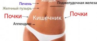
can be internal or external. The external ones, in turn, are umbilical, femoral, and inguinal. They can have different shapes and sizes. The most common is a hernia of the linea alba, since the muscle tissue in this area of the body is the weakest. Internal hernias are a consequence of the penetration of an organ into the cavity formations of the peritoneum inside the abdomen.
Etiology of the disease and ICD-10 code
According to the international classification of diseases, the ICD-10 code for abdominal hernia ranges from K40 to K46. It all depends on the type of education, its location, and problems. There are such hernia codes:
- K40 - inguinal;
- K41 - femoral;
- K42 - umbilical;
- K43 - hernia in the anterior abdominal wall;
- K44 - diaphragmatic;
- K45 - other hernias in the peritoneum;
- K46 - unspecified.
In medical terms, a hernia is the free migration of one of the internal organs into various parts of the peritoneum or under the skin. The problem occurs especially often in areas of weak muscle walls. Outwardly, it looks like a protrusion above the plane of the skin, like a bump. Sometimes the hernia is removed by simply pressing the hand (reduced), but then comes out again.
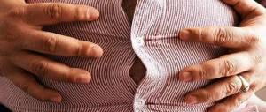
Flabbiness of the abdominal muscles is one of the causes of hernia
The problem occurs for the following reasons:
- Features of the body structure of a particular person. Congenital or acquired defects, abdominal abnormalities.
- Heredity to the formation of protrusions.
- Heavy physical labor with tension on the abdominal muscles.
- Pregnancy.
- Enhanced pumping of the lower abs.
- Eating disorders and stool disorders (constipation).
- Frequent hysterical screaming or crying in infants.
- Severe, continuous cough similar to whooping cough.
- Prostate adenoma and difficulty urinating against this background.
- Flabbiness of the abdominal muscles.
- Unsuccessfully performed surgical intervention with improper suturing of the peritoneal tissue.
A hernia does not occur suddenly. Its formation requires time and a number of listed predisposing factors.
How do internal abdominal hernias manifest?
The main symptoms of any external hernia are periodically appearing visible protrusion and discomfort in its area. Since an internal abdominal hernia cannot be examined with the eye, all its signs boil down to the periodic appearance of discomfort:
- symptoms may be completely absent;
- periodically experience pain in the epigastric region, accompanied by a feeling of heaviness and fullness;
- from time to time, pain may occur in the left iliac or periumbilical region;
- pain varies in intensity - from dull aching to sharp, cramping, unbearable;
- the pain goes away in a certain position of the body, when the intestinal loops return to their place under the influence of gravity or pressure from neighboring organs - for example, lying on your back.
Patients are often examined for a long time and undergo repeated courses of treatment from a therapist, gastroenterologist, or even a neurologist and psychiatrist, but the unpleasant symptoms do not disappear.
Classification of protrusions in adults
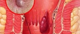
The structure and external manifestation of umbilical hernias
There are different types of abdominal hernias. They are classified as follows:
According to the degree of formation and development:
- initial;
- full;
- channel
By method of origin:
- congenital - is an anomaly of fetal development;
- acquired.
The process of formation and strangulation of a hernia
According to the presence of various complications:
- simple - uncomplicated;
- complex - accompanied by peritonitis, irreducibility, strangulation, inflammation.
Reduce if possible: reducible and not amenable to conservative treatment.
Along the course, primary hernias are distinguished, recurrent (repeating) and postoperative.
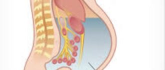
Diaphragmatic hernia
Diaphragmatic hernia is the penetration of part of the stomach, esophagus, or small intestine into the thoracic region through weak spots or defects in the diaphragm.
Photo: diaphragmatic hernia
Causes:
- obesity;
- heavy physical activity;
- pregnancy;
- thinning and weakening of connective tissue with age.
A diaphragmatic hernia often manifests itself as symptoms of reflux esophagitis: belching, heartburn, chest pain, which can be confused with angina.
Complications:
- cicatricial narrowing of the esophagus;
- strangulation of a hernia in the esophageal opening of the diaphragm;
- bleeding from the vessels of the esophagus;
- perforation of the esophagus.
The diagnosis is made by X-ray examination. Conservative treatment uses drugs that reduce gastric secretion. For large hernias and complications that arise, surgical treatment is performed.
Symptoms and signs
Inguinal hernia
The clinical picture of the pathology depends on the type and type of protrusion. There are such signs depending on the problem:
- Inguinal hernia. Localized in the inguinal canal area. It can be observed along the spermatic cord (oblique) or at the posterior wall of the groin (straight). More often, the oblique protrusion has an oval shape and extends into the labia in women or into the scrotum in men. A hernia is painful. The direct inguinal protrusion is less often infringed, but more often recurs, unlike the oblique one.
- Femoral. The size of the lateral protrusion is about 1-2 cm. The problem is localized in the groin closer to the thigh. The lump is soft and mobile to the touch. Therefore, it is difficult to confuse it with an ordinary wen or omentum.
- Postoperative. Located in the area of unsuccessful sutures of the abdominal cavity. Accompanied by pain due to irritation of the vagus nerve, nausea, and vomiting.
- Umbilical. Location: navel area. A hernia in the area of the birth scar ring can be clearly palpated and even reduced. A person with such a protrusion feels nausea and pain.
- Hernia of the white line. It is also called preperitoneal lipoma. Here, fat first leaks through the gaps in the tendon fibers, and then parts of the internal organs protrude.
With various types of abdominal protrusion, you can feel the hernial orifice - the so-called push of the internal organs when the patient coughs.
Increased level of lymphocytes in the blood
To determine this parameter, it is enough to take a general clinical blood test. The test is taken on an empty stomach; before the test, you should not engage in physical activity during the day, do not eat fatty foods, and do not smoke for 2-3 hours. Blood for general analysis is usually taken from a finger, less often from a vein.
A complete blood count allows you to find out how different types of white blood cells relate to each other. This ratio is called the leukocyte formula. Sometimes the number of lymphocytes is directly indicated in the analysis transcript, but often the transcript contains only English abbreviations. Therefore, it is sometimes difficult for an ignorant person to find the necessary data in a blood test. Typically, the required parameter is indicated as LYMPH in the blood test (sometimes also LYM or LY).
In adults
The relative norm for the content of lymphocytes in the blood in this category of the population is considered to be values in the range of 20–34 percent. In absolute values (units), the variation ranges from 1 to 4.5X10⁹/liter.
In children
In children, the content of such cellular elements varies within very wide limits and primarily depends on age.
- Up to one year - from 55 to 75 percent or 4–10.5X10⁹/l.
- From one to four years - from 45 to 65 percent or 2–8X10⁹/l.
- From four to six years - from 35 to 55 percent or 1.5–7X10⁹/l.
- From six to ten years - from 30 to 50 percent or 1.5–6.5X10⁹/l.
- From ten to 21 years - from 30 to 45 percent or 1–4.8X10⁹/l.
https://www.youtube.com/watch?v=ytabout
As can be seen from the above inverse arithmetic progression, with increasing age, the relative and absolute level of lymphocytes gradually decreases.
What does it mean?
In the medical environment, an increased level of lymphocytes relative to the norm is called lymphocytosis. This condition is not a disease - it is a protective reaction of the body and an indicator of developing pathological processes. In this case, both the absolute readings of the content of the basic cellular element in the blood and its relative parameter, expressed as a percentage of the basic immune map of all plasma elements, are analyzed.
An increased level of lymphocytes can be caused not only by diseases, but also by physiological characteristics - for example, in women during the menstrual cycle, tests can give unexpected results, and in a number of people with a reactive immune system, even the slightest malfunction in the body, such as a common cold, often produces a high concentration of this cell type.
READ MORE: Skin with varicose veins
Because elevated lymphocyte levels are not a disease, there is no specific treatment for this condition. In the absence of clear symptoms of a specific disease, except for the results of laboratory tests, a medical specialist can refer the patient for radiography, ultrasound, CT/MRI, prescribe histological/cytological analysis, etc.
In adults and children, specific therapy is prescribed only after receiving an accurate diagnosis. In the vast majority of cases, the specialist prescribes antiviral drugs, antibiotics, antipyretics, anti-allergenic and anti-inflammatory drugs, in some cases - corticosteroids, chemotherapy, bone marrow transplantation and other necessary measures, developed individually based on the patient’s current condition, severity of the disease and other parameters.
Diagnostics and treatment methods
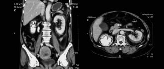
If pathology is suspected, an ultrasound of the abdominal organs is prescribed
To identify and confirm a hernia, you should consult a surgeon. Only he can see and accurately determine the type of problem. The doctor will conduct a standard examination and external palpation of the desired area. If there are doubts or suspicions of other pathologies, the doctor prescribes an ultrasound of the abdominal organs, an X-ray of the barium passage of the small intestine or an X-ray. In rare cases, diagnostic laparoscopy is performed.
Treatment tactics are selected depending on the degree of complexity of the pathology.
More often, the surgeon decides on surgical intervention for a hernia. Conservative therapy is used only in cases of initial uncomplicated protrusion or for elderly patients for whom surgery is contraindicated. In this case, the patient is recommended to wear a bandage, reduce physical activity, and normalize nutrition.
Reduction is carried out in the absence of infringement.
Surgical mesh for umbilical hernia
A standard operation is performed for complex hernias, as well as in the presence of strangulated protrusions. Small hernias are operated on under local anesthesia. For larger ones, a full abdominal operation is performed. The problem area is strengthened with a polypropylene mesh (mesh graft). This helps prevent relapse.
There are times when a patient undergoes an emergency excision. Indications for this are necrosis of strangulated tissues, perforation of the walls of an internal organ, peritonitis. An extended laparotomy is performed to assess the condition of the internal organs of the peritoneum. All necrotic areas of the omentum and intestines must be removed.
After the operation, wearing a bandage is indicated. Physical activity is temporarily contraindicated. It is advisable to follow a soft stool diet. Defecation should take place without straining.
Lymphocytes in the blood are increased. Increased lymphocytes in the blood - causes, treatment
Why does the white blood cell count increase? This symptom can have several causes. First of all, these are infectious diseases. Many infections, especially viral ones, cause the immune system to produce increased numbers of killer T cells and NK cells. This type of lymphocytosis is called reactive.
Viral infections that can cause an increase in lymphocytes in the blood include:
- Flu,
- AIDS,
- Infectious mononucleosis,
- Herpes,
- Viral hepatitis,
- Chickenpox,
- Measles,
- Rubella,
- Whooping cough,
- Adenoviral infection
- Mumps.
Also, increased lymphocytes in the blood can be observed during bacterial and protozoal infections:
- Tuberculosis,
- Syphilis,
- Brucellosis,
- Toxoplasmosis.
However, not every bacterial infection is accompanied by lymphocytosis, since many bacteria are destroyed by other types of leukocytes.
Thus, an increase in lymphocytes in the blood may indicate infection with some viruses, bacteria, fungi, protozoa or multicellular parasites. If the symptoms of the disease by which it could be determined are not obvious, then additional tests are carried out.
An increase in the number of white blood cells can be observed not only during illness, but also some time after recovery. This phenomenon is called post-infectious lymphocytosis.
Another cause of lymphocytosis is diseases of the hematopoietic system (leukemia) and lymphatic tissue (lymphoma). Many of them are malignant. With these diseases, lymphocytosis is observed in the blood, but the immune cells are not complete and cannot perform their functions.
The main diseases of the lymphatic and circulatory systems that can cause lymphocytosis:
- Lymphoblastic leukemia (acute and chronic),
- Lymphogranulomatosis,
- Lymphoma,
- Lymphosarcoma,
- Multiple myeloma.
Other reasons that can cause an increase in the number of immune cells:
- Alcoholism;
- Frequent smoking of tobacco;
- Taking narcotic substances;
- Taking certain medications (levodopa, phenytoin, some analgesics and antibiotics);
- The period before menstruation;
- Prolonged fasting and diets;
- Long-term consumption of foods rich in carbohydrates;
- Hyperthyroidism;
- Allergic reactions;
- Poisoning with toxic substances (lead, arsenic, carbon disulfide);
- Immunity disorders;
- Endocrine disorders (myxedema, ovarian hypofunction, acromegaly);
- Early stages of some cancers;
- Neurasthenia;
- Stress;
- Vitamin B12 deficiency;
- Injuries and wounds;
- Splenectomy;
- Accommodation in high mountains;
- Radiation injuries;
- Taking certain vaccines;
- Excessive physical activity.
Lymphocytosis can also be temporary or permanent. The temporary type of disease is usually caused by infectious diseases, injuries, poisoning, and medications.
Since the spleen is an organ where immune cells break down, its surgical removal for some reason can cause temporary lymphocytosis. However, subsequently the hematopoietic system returns to normal and the number of these cells in the blood stabilizes.
Sometimes a situation opposite to lymphocytosis can be observed - lymphopenia, when lymphocytes are low. For lymphocytes, a decrease is typical in the following cases:
- Severe infections that deplete lymphocytes;
- AIDS;
- Tumors of lymphoid tissue;
- Bone marrow diseases;
- Severe types of heart and kidney failure;
- Taking certain medications, for example, cytostatics, corticosteroids, antipsychotics;
- Radiation exposure;
- Immunodeficiency state;
- Pregnancy.
A situation where the number of immune cells is lower than normal may be a temporary phenomenon. So, if during an infectious disease a lack of lymphocytes is replaced by an excess, this may indicate that the body is close to recovery.
However, it should be remembered that lymphocytosis in children can also be caused by such a serious disease as acute lymphoblastic leukemia. Therefore, it is important to regularly check your baby's white blood cell count through blood tests.
Does lymphocytosis manifest itself in any other way besides changes in blood composition? If it is caused by an infectious disease, the patient will experience symptoms characteristic of this disease, for example, fever, chills, headaches, cough, rash, etc. But these symptoms are not symptoms of lymphocytosis itself. However, in some cases, with an increase in lymphocytes caused by non-infectious causes, an enlargement of the lymph nodes and spleen, the organs where most lymphocytes are located, may be observed.
When the number of lymphocytes increases, the reasons for the increase are not always easy to detect. First of all, it is recommended to consult a general practitioner. Most likely, he will give directions for several additional tests - blood for HIV, hepatitis and syphilis. In addition, additional studies may be prescribed - ultrasound, computed tomography or magnetic tomography, radiography.
An additional blood test may be required to rule out the error. To clarify the diagnosis, an operation such as a lymph node or bone marrow puncture may be necessary.
When determining the cause of an increase in lymphocytes, an important role is played by determining the number of typical and atypical types of cells.
Atypical lymphocytes are blood cells that have different properties and sizes compared to normal ones.
Most often, atypical cells are observed in the blood in the following diseases:
- Lymphocytic leukemia,
- Toxoplasmosis,
- Pneumonia,
- Chicken pox,
- Hepatitis,
- Herpes,
- Infectious mononucleosis.
On the other hand, in many diseases a large number of atypical cells are not observed:
- Measles,
- Mumps,
- Rubella,
- Flu,
- AIDS,
- Adenoviral infection
- Malaria,
- Autoimmune diseases.
A factor such as erythrocyte sedimentation rate (ESR) should also be taken into account. In many diseases this parameter increases. The dynamics of other blood components are also taken into account:
- Total white blood cell count (may remain unchanged, decrease or increase),
- Dynamics of platelet count (increase or decrease),
- Dynamics of the number of red blood cells (increase or decrease).
An increase in the total number of leukocytes with a simultaneous increase in lymphocytes may indicate lymphoproliferative diseases:
- Lymphocytic leukemia,
- Lyphogranulomatosis,
- Lymphoma.
READ MORE: Bruise due to varicose veins
This condition may also be characteristic of:
- acute viral infections
- hepatitis,
- endocrine diseases,
- tuberculosis,
- bronchial asthma,
- removal of the spleen,
- cytomegalovirus infection,
- whooping cough
- toxoplasmosis,
- brucellosis.
Relative lymphocytosis (in which the total number of white blood cells remains approximately constant) is usually characteristic of severe bacterial infections such as typhoid fever.
In addition, it occurs in the case of:
- Rheumatic diseases,
- Hyperthyroidism,
- Addison's disease
- Splenomegaly (enlarged spleen).
A decrease in the total number of leukocytes against the background of an increase in the number of lymphocytes is possible after severe viral infections or against their background. This phenomenon is explained by the depletion of the reserve of rapid immunity cells, primarily neutrophils, and the increase in long-term immunity cells - lymphocytes. If this is the case, then, as a rule, this situation is temporary, and the number of white blood cells should soon return to normal. Also, a similar state of affairs is typical for taking certain medications and poisoning.
A decrease in the number of red blood cells due to lymphocytosis is usually characteristic of leukemia and bone marrow diseases. In addition, cancer of the bone marrow is usually accompanied by a very large increase in lymphocytes - approximately 5-6 times higher than normal.
A simultaneous increase in the number of red blood cells and lymphocytes can be observed in heavy smokers. The ratio of different types of lymphocytes can also be of diagnostic value. For example, with myeloma, first of all, the number of type B cells increases, with infectious mononucleosis - types T and B.
- Drug-related reactions
- Serum sickness
- Injury
- After removal of the spleen
- Heavy smokers
- Lymphocytosis of giant granular lymphocytes
- B cell lymphoproliferative disease
- malignant thymoma
- chronic lymphocytic leukemia
- acute lymphoblastic leukemia
- malignant non-Hodgkin's lymphoma
Below are typical causes of elevated lymphocyte levels.
In adults
- During the menstrual cycle of women - a physiological reason for the increase immediately before menstruation.
- The “reactive” type of immunity is a physiological cause in the absence of serious diseases, an extremely strong immunological response to any malfunction in the body or the forced functioning of a number of organs.
- Prolonged fasting.
- Viral diseases of the liver with enlargement of the latter and spleen.
- Tuberculosis of any type, even apparently asymptomatic.
- A variety of bacterial infections, including syphilis, brucellosis.
- Infectious mononucleosis.
- Allergic manifestations.
- Hypertrophied thyroid function.
- Lymphocytosis of smokers and alcohol addicts, developing against the background of stress.
- Pathogenic autoimmune processes, including rheumatoid arthritis, systemic lupus erythematosus, scleroderma, dermatomyositis.
- Lymphocytic leukemia of chronic benign type.
- Progressive lymphosarcoma.
- Direct poisoning with a number of chemicals, in particular arsenic, chlorine, lead.
- Crohn's disease.
- Multiple myelomas.
- Endocrine diseases.
- Adverse reactions to a number of medications.
- Broad spectrum neurasthenia.
- The turning point of acute diseases with the start of the recovery period, as well as the transition from relapse to remission of chronic forms of diseases.
In children
- Anemia, especially acute lack of vitamin B12.
- Classic infectious diseases, in particular rubella, measles, encephalitis, chickenpox, whooping cough, smallpox, mumps, malaria.
- Malignant tumors and oncology.
- Lymphocytosis of the infectious type, also known as Smith's disease.
- Bronchial asthma and other types of pulmonary diseases.
- Endocrinological problems.
- Physiological lymphocytosis in children under four years of age in the absence of manifestations of other diseases and normal health.
https://www.youtube.com/watch?v=ytpress
To understand why lymphocytes in the blood are elevated, you should know that there are two types of lymphocytosis - absolute and relative. The first pathological condition is characterized by an increase not only in the number of leukocytes in the blood, but also in the total number of lymphocytes. With a relative deviation, the number of leukocytes remains at the same level. This occurs due to the reduction of granular species, namely neutrophils, eosinophils and basophils.
- Relative lymphocytosis. This deviation usually manifests itself during viral infections (during influenza, brucellosis, typhoid fever, etc.) and purulent-inflammatory processes.
- Absolute lymphocytosis. This deviation can also be detected in children after they take a general blood test. Lymphocytes are elevated in a child against the background of absolute lymphocytosis in diseases such as rubella, mumps, chickenpox, secondary syphilis, measles, hyperthyroidism of the thyroid gland, mumps, relapsing fever, whooping cough, infectious mononucleosis, tuberculosis, malaria, lymphosarcoma, scarlet fever, leishmaniasis, toxoplasmosis, viral hepatitis, etc.
- Infectious lymphocytosis. This syndrome most often occurs in young children aged 2 to 7 years. If we talk about the causes of the disease in question, they have not been identified at the moment. However, there is an opinion that this pathological condition is associated with viral infections. After all, this disease is most often observed in schools, kindergartens, sanatoriums and summer camps. The incubation period of this disease lasts about 2-4 weeks, and its peak occurs in autumn and spring.
Possible complications and consequences
Types of strangulated hernias
The most terrible and dangerous complication of any protrusion is its infringement. Part of the organ is compressed in the hernial orifice. This causes tissue necrosis, death and decay. Doctors distinguish these types of infringements:
- Strangulation or elastic. The vessels of the mesentery of the intestine are compressed.
- Obstructive. There is a bend in the intestine. In this case, the movement of feces stops. Constipation may occur, which will lead to further poisoning of the body with waste products.
- Regional. A small section of the intestine is pinched, not the entire loop.
Do not under any circumstances try to treat a hernia using traditional methods. This can cause serious pathologies and complications, including death.
Causes
At the initial stage of development, such formations do not cause pain and are almost invisible. But under the influence of physical activity, illness or other factors, the symptoms of an abdominal hernia begin to manifest themselves more strongly.
The main localization methods are the following:
- umbilical ring;
- groin area;
- midline (white) line of the peritoneum.
Often the formation is observed on postoperative scars.
As a rule, abdominal hernia manifests itself in the same way in men and women. The cause is disturbances in the anatomical position of organs and malfunctions in their functioning.
There are external and internal protrusions. In the first case, the pathology is visually determined, and in the second it is detected using ultrasound or x-ray examination. With external formations, the organs emerge together with the parietal abdominal layer. Internal pathologies are localized inside the peritoneum, or protrusion occurs into the chest cavity through natural or artificially formed openings.
Indications for physical therapy
The ligamentous apparatus of the knee is a rather complex system that includes different anatomical elements. Carrying out therapeutic exercises will be advisable for damage to different types of knee joints:
- If there is a rupture of the anterior cruciate ligament of the knee joint, rehabilitation will be more effective and faster when performing exercise therapy. Tears of this nature are most often found in athletes;
- in case of damage to the lateral lateral ligament, which is quite often caused by walking in high heels;
- when the lateral internal ligament is torn. In most cases, such ruptures are accompanied by dislocations of the limb;
- in case of damage to the cruciate posterior ligaments caused by a sharp bend of the knee;
- with the help of gymnastic exercises, recovery from ligament rupture near the meniscus occurs much faster;
- a torn quadriceps muscle will heal faster with regular exercise therapy.
Each of these types of injuries requires serious and proper treatment, since the further functioning of the knee and the fullness of its mobility depend on it. And one of the extremely important stages in restoring the functionality of the knee is physical exercise when the ligaments are torn.
How is the disease diagnosed?
At an early stage it is difficult to determine, since the protrusion is almost invisible. But if there is any suspicion of an inguinal hernia in a woman and symptoms characteristic of the disease, you should consult a therapist or gynecologist. Using palpation, the doctor will be able to detect the presence of a hernia, but a surgeon and gastroenterologist will be able to finally confirm the diagnosis.
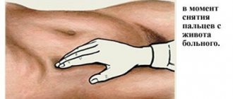
To determine the presence of a disease, the doctor places his fingers on the relevant area and asks the patient to cough. A cough provokes an increase in pressure in the abdominal area, which creates jerking movements of the hernia, and this can be clearly felt with the fingers.
Sometimes additional examination will be required to determine an inguinal hernia.
Additional diagnostics include:
- Ultrasound of the abdominal region, pelvis, hernial bulge;
- irrigoscopy, which allows you to determine various developmental anomalies and intestinal diseases;
- herniorgafia - X-ray examination of a hernia.
Exercise therapy for sprains and bruises of ligaments
Even minor injuries to the knee can lead to partial or even complete limitation of motor activity. If you do not respond to an injury in a timely manner, recovery from a sprained ankle or knee joint will take a long period and require great effort.
What to do first after a ligament rupture, how to restore ligaments in case of sprains or bruises{q} Regardless of which element of the ligament joint is injured and what is the severity of the injury, its restoration is only possible with multi-stage rehabilitation.
Moderate or severe pain occurs in the knee almost instantly after a bruise or sprain. Typically, swelling also occurs at the site of injury. The first thing they do in such cases is to numb the limb and temporarily immobilize it.
Recovery of the knee joint after a sprain will be complete if special physical therapy is carried out during the rehabilitation period.
The exercises are quite simple in terms of technique, but the benefits from them will be enormous:
- treatment by position. The injured leg must be placed on a functional splint, the knee bend angle should be approximately 30-45 degrees. Such exercises are best performed during rest after physical activity;
- performing flexion and extension movements by the ankle joint, while trying to achieve the greatest possible load angle;
- sit on a chair, straighten your back, bend your knees. Perform swings with both limbs in different directions like a pendulum;
- Stretch your arms forward, straighten your torso and perform half squats.