In the famous Soviet-era comedy “Pokrovsky Gate” there is a wonderful episode in which Rimma Markova (surgeon), smoking a cigarette on a clip, answers her friend on the phone that she should cut without waiting for peritonitis (we were talking about appendicitis). Indeed, this condition poses a serious threat to the patient’s life, and delaying the operation is literally like death.
According to statistics, the disease is diagnosed in 15-20% of patients with an “acute abdomen”, and in 11-43% it causes emergency laparotomy (revision of the abdominal organs). Despite significant advances in medicine, the mortality rate for this pathology is quite high and ranges from 5 to 60 percent or more. The wide range of numbers is explained by many factors: the cause and stage of the process, its prevalence, the age of the patient, concomitant pathology, and others.
Peritonitis: definition
Peritonitis is called aseptic inflammation or bacterial infection of the peritoneum, and, accordingly, develops in the abdominal cavity. This process is a serious complication of inflammatory diseases of the abdominal organs and is included in the group of acute surgical pathologies referred to as “acute abdomen”. According to statistics, this disease develops in 15–20% of cases in patients with acute surgical diseases, and the need for emergency laparotomy for this reason reaches 43%. Mortality with such a complication is observed in 4.5–58% of cases. The huge range of numbers is explained by many factors (the cause and stage of the process, its prevalence, the age of the patient, and others).
The high mortality rate for this condition is explained by two factors:
- failure of patients to seek specialized care in a timely manner;
- an increase in the number of elderly patients (the process is not so acute, which leads to late consultation with a doctor);
- an increase in the number of patients with cancer;
- errors and difficulties in diagnosing the process, inappropriate treatment;
- severe course of the process if it spreads (spread peritonitis).
Mechanism of peritonitis
Let's consider the mechanism of peritonitis using the example of appendicitis (inflammation of the vermiform appendix of the cecum).
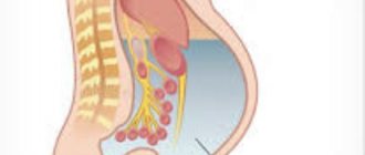
The peritoneum is a thin connective tissue membrane lining the organs and walls of the abdominal cavity. Constantly produces a small amount of fluid that facilitates the sliding of internal organs, contains a large number of blood vessels and nerves. Normally, the peritoneum and abdominal cavity are sterile.
The cecum, like any other part of the large intestine, contains a huge number of bacteria in its lumen. Under normal conditions, this microflora performs useful functions, participating in digestion, synthesis of vitamins, and supports the immune system. However, when these same bacteria enter the sterile abdominal cavity, they begin to exhibit their pathogenic properties, causing inflammation and intoxication. The situation is complicated by the large area of the peritoneum, through which bacterial waste products and toxins are absorbed.
In the first hours after the onset of appendicitis, inflammation of the appendix is limited to its walls. The pain that occurs during this period is caused by reactive irritation of the nerve endings located in the peritoneum, covering the appendix on all sides. As the disease progresses, inflammation leads to swelling and increased permeability of the walls of the appendix (phlegmonous appendicitis). Peritonitis, which begins at this stage, involves the peritoneum of the appendix, which provokes intense pain in the right iliac region. Surgical removal of the appendix at this stage prevents possible complications in the form of diffuse peritonitis with the spread of inflammation to other areas of the abdominal cavity.
Late seeking medical help provokes gangrenous appendicitis, with necrosis and perforation of the wall of the appendix. The contents of the cecum and transudate enter directly into the abdominal cavity, seeding it with intestinal flora. A change in body position provokes the flow of infected transudate into the subhepatic region and other parts of the abdomen, which leads to the spread of infection. 24 hours after perforation of the appendix, we can talk about diffuse peritonitis, with intestinal paresis (paralysis).
The infectious process leads to activation of the immune system, massive intoxication of the body and other complications. Without treatment, peritonitis leads to sepsis - blood poisoning that occurs with multiple organ failure, ending in death.
The body's response to infection depends on the state of immunity and health of the patient, the aggressiveness of the infection, and the volume of intestinal contents leaked into the abdominal cavity.
A little anatomy
The abdominal cavity is lined from the inside with a serous membrane called the peritoneum. The area of this shell reaches 210 meters and is equal to the area of the skin. The peritoneum has 2 layers: parietal and visceral. The visceral peritoneum covers the internal organs of the abdomen and pelvis and is their third layer, for example, the uterus has the endometrium (inner layer), myometrium and serosa.
The parietal layer covers the abdominal walls from the inside. Both layers of the peritoneum are represented by a single continuous membrane and are contiguous over the entire area, but form a closed sac - the abdominal cavity, which contains about 20 ml of aseptic fluid. If in men the abdominal cavity is closed, then in women it communicates with the external genitalia through the fallopian tubes. Visually, the peritoneum looks like a shiny and smooth membrane.
The peritoneum performs a number of important functions. Due to the secretory-resorptive and absorption functions, the serous membrane produces and absorbs up to 70 liters of fluid. The protective function is ensured by the content of lysozyme, immunoglobulins and other immune factors in the abdominal fluid, which ensures the elimination of microorganisms from the abdominal cavity. In addition, the peritoneum forms ligaments and folds that secure the organs. Due to the plastic function of the peritoneum, the focus of inflammation is delimited, which prevents further spread of the inflammatory process.
Diagnosis of peritonitis
First of all, the doctor performs a palpation examination of the abdomen. With its help, the tone of the muscles of the anterior wall of the peritoneum is established, the areas that react most acutely to inspection are identified, and the intensity of muscle resistance to palpation is determined.
Next, the doctor identifies the presence of peritoneal symptoms.
- Conducts the Shchetkin-Blumberg test (increased pain when you stop pressing on the patient’s stomach).
Shchetkin-Blumberg symptom - Determines the presence of Voskresensky's symptom (increased pain upon completion of a sliding movement with the fingertips towards the patient's iliac region while pulling the patient's shirt by its lower edge).
- Uses the Medel method (tapping the anterior peritoneal wall with fingertips).
- Identification of Bernstein's symptom in men (the testicles and penis are reflexively pulled towards the anterior abdominal wall as a result of contraction of the abdominal muscles).
Abdominal puncture
The doctor also prescribes a blood test, a rectal examination for men and a vaginal examination for women. In conjunction with the examination, the patient may be sent for an X-ray examination and ultrasound of the internal organs of the abdominal cavity in order to identify areas of fluid accumulation. If the above diagnostic methods are insufficient, the doctor may resort to laparocentnesis - taking a puncture to determine the composition of excess fluid in the abdominal cavity.
Causes of the disease
The leading cause of this complication is bacteria that penetrate the abdominal cavity. Depending on the route of entry of microorganisms, there are 3 types of inflammation of the peritoneum:
Primary peritonitis
The inflammatory process in this case occurs against the background of preserved integrity of the internal organs of the abdomen and is a consequence of spontaneous blood dissemination of bacteria into the peritoneum. Primary inflammation of the peritoneum is in turn divided into:
- spontaneous peritonitis in children;
- spontaneous inflammation of the peritoneum in adults;
- tuberculous inflammation of the peritoneum.
Pathogenic pathogens represent one type of infection or monoinfection. The most common type is streptococcus pneumoniae. In sexually active women, inflammation of the peritoneum is usually caused by gonococci and chlamydia. In the case of peritoneal dialysis, gram-positive bacteria (eubacteria, peptococci and clostridia) are detected.
In children, spontaneous inflammation of the peritoneum, as a rule, occurs in the neonatal period or at 4–5 years. At four to five years of age, systemic diseases (scleroderma, lupus erythematosus) or kidney damage with nephrotic syndrome are a risk factor for the development of this complication.
Spontaneous inflammation of the peritoneum in adults often occurs after emptying (drainage) of ascites, which is caused by cirrhosis of the liver or after long-term peritoneal dialysis.
Tuberculous damage to the peritoneum occurs with tuberculous damage to the intestines, fallopian tubes (salpingitis) and kidneys (nephritis). Mycobacterium tuberculosis enters the serous tissue of the abdominal cavity through the bloodstream from the primary source of infection.
Secondary peritonitis
Secondary inflammation of the peritoneum is the most common type of described complication and includes several varieties:
- inflammation of the peritoneum caused by impaired integrity of internal organs (as a result of their perforation or destruction);
- postoperative;
- post-traumatic inflammation of the peritoneum as a result of blunt trauma to the abdominal area or penetrating injury to the abdominal cavity.
The causes of the first group of inflammation of the peritoneum are the following types of pathologies:
- inflammation of the appendix (appendicitis), including perforation of the appendix (gangrenous and perforated appendicitis);
- inflammation of the internal genital organs in women (salpingitis and oophoritis, endometritis), as well as ruptures of an ovarian cyst or fallopian tube during ectopic pregnancy or in the case of pyosalpinx;
- intestinal pathology (intestinal obstruction, intestinal diverticula, Crohn's disease with perforation of ulcers, perforation of duodenal ulcers, perforation of intestinal ulcers of other etiologies: tuberculosis, syphilis, etc., malignant intestinal tumors and their perforation);
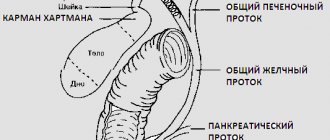
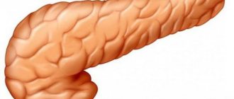
- diseases of the liver, pancreas and biliary tract (gangrenous cholecystitis with perforation of the gallbladder, suppuration and rupture of various hepatic and pancreatic cysts, rupture of parapancreatic cysts, cholelithiasis).
Peritonitis after surgery is classified as a separate group, despite the fact that this type of disease is caused by abdominal trauma. But it should be taken into account that the injury caused by the operation is inflicted on the patient under certain conditions, in compliance with the rules of asepsis, and the negative response of the body to the surgical injury is associated with complex anesthetic management.
Post-traumatic inflammation of the peritoneum occurs as a result of a closed abdominal injury or due to a penetrating injury to the abdomen. Penetrating wounds can be caused by a gunshot wound, stab objects (knife, sharpening) or due to iatrogenic factors (endoscopic procedures accompanied by damage to internal organs, abortion, uterine curettage, hysteroscopy).
Tertiary peritonitis
This type of inflammation of the peritoneum is the most difficult to diagnose and treat. Essentially, this is a relapse of previous inflammation of the peritoneum, and, as a rule, occurs after surgery in those patients who have experienced emergency situations, as a result of which their body’s defenses are significantly suppressed. The course of this process is characterized by an erased clinical picture, with the development of multiple organ failure and significant intoxication. Risk factors for tertiary peritoneal inflammation include:
- significant exhaustion of the patient;
- a sharp decrease in plasma albumin levels;
- identification of microorganisms resistant to multiple antibiotics;
- progressive multiple organ failure.
Tertiary inflammation of the peritoneum is often fatal.
Types of peritonitis
Depending on the etiological factor, the following types of peritonitis are distinguished:
- idiopathic (or primary) - occur when pathogenic microorganisms enter along with the flow of lymph, blood or through the fallopian tubes in diseases such as enterocolitis, salpingitis, tuberculosis of the genitourinary organs;
- secondary - develop due to injuries or inflammatory-destructive diseases of organs and are usually detected with perforated, gangrenous or phlegmonous appendicitis, perforation of a stomach or duodenal ulcer, rupture of ovarian cysts, pancreatic necrosis, Crohn's disease, phlegmonous-gangrenous cholecystitis and pancreatitis, occlusion of mesenteric vessels, diverticulitis ah and other pathologies.
Depending on the microbial factor, peritonitis is:
- bacterial – provoked by inflammation caused by microorganisms;
- aseptic - caused by substances that are aggressive towards the peritoneum and provoke inflammation.
In the clinical practice of surgeons, secondary peritonitis occurs more often than idiopathic peritonitis, which occurs in only 1-1.5% of patients.
Traumatic peritonitis is classified into a separate group:
- arising as a result of closed or open injuries causing damage to the abdominal organs;
- arising as a result of surgical interventions, accompanied by anastomotic failure, suture defects, accidental mechanical trauma to the peritoneum and the development of hemoperitoneum (blood accumulation).
Special types of peritonitis include the following:
- carcinomatous (cancerous);
- parasitic;
- rheumatoid;
- granulomatous.
Depending on the nature of the fluid accumulating in the abdominal cavity, the following types of peritonitis are distinguished:
- serous;
- fibrinous;
- purulent;
- hemorrhagic.
The nature of the peritoneal lesion may be as follows:
- limited - an abscess or infiltrate occurs on the peritoneum;
- unlimited – the area of inflammation does not have clear boundaries and is diffuse.
The prevalence of peritoneal lesions may be as follows:
- local - only one anatomical zone of the peritoneum is affected;
- common – inflamed from 2 to 5 zones;
- total (or general) – 6 or more zones are affected.
According to the clinical course, in most cases, peritonitis is acute. However, sometimes the inflammation takes a protracted course, in such situations the pathological process is considered chronic.
The above characteristics of peritonitis are difficult to remember for people who do not have a medical education, so in practice the more simplified formulation “acute” is often used. The remaining classifications are usually omitted and used only for maintaining medical records.
Development mechanism
How quickly this complication will develop and how severe it will be is largely determined by the state of the body, the virulence of microorganisms, and the presence of provoking factors. The mechanism of development of peritoneal inflammation includes the following points:
- intestinal paresis (lack of peristalsis), which leads to disruption of the absorption function of the peritoneum, as a result of which the body becomes dehydrated and loses electrolytes;
- dehydration leads to a decrease in blood pressure, which results in rapid heartbeat and shortness of breath;
- the rate of development of the inflammatory process and its prevalence are directly proportional to the number of pathogenic microbes and the severity of intoxication;
- microbial intoxication is complemented by autointoxication.
Classification
There are many classifications of inflammation of the peritoneum. Today the classification recommended by WHO is used:
Depending on the current:
- acute peritonitis;
- chronic inflammation of the peritoneum.
Depending on the etiological factor:
- aseptic inflammation of the peritoneum;
- microbial (infectious) peritonitis.
Origin of the complication:
- inflammatory;
- perforated (perforation of internal organs);
- traumatic;
- after operation;
- hematogenous;
- lymphogenous;
- cryptogenic.
Depending on the exudate:
- serous peritonitis;
- hemorrhagic;
- fibrinous;
- purulent peritonitis;
- putrid or ichorous.
Depending on the spread of inflammation:
- delimited (appendicular, subphrenic, subhepatic and others);
- widespread: diffuse - damage to the peritoneum covers 2 floors of the abdominal cavity;
- diffuse - inflammation of the peritoneum in more than two areas of the abdominal cavity;
- general - the inflammatory process is widespread over the entire area of the peritoneum.
Viral peritonitis does not develop in humans; it is diagnosed only in animals (cats, dogs).
Why does peritonitis occur in children?
Depending on the cause, the child may be diagnosed with primary or secondary intestinal peritonitis. Peritonitis often occurs against the background of other pathologies:
- complication of acute appendicitis;
- parasitic infection;
- inflammation in the abdominal cavity;
- umbilical sepsis.
In children, peritonitis is manifested by a significant deterioration in well-being, an increase in temperature to 38°C, the child becomes nervous, capricious, cries for no reason and does not want to eat
. Primary peritonitis is less common, the causes of which may be:
- hemorrhages in the abdominal cavity;
- internal organ injuries;
- intestinal obstruction;
- diplococcal infection.
With peritonitis, the child’s general well-being deteriorates significantly. He becomes nervous, capricious, cries for no reason and does not want to eat. The temperature rises to 38°C, vomiting begins, and the skin becomes dry and pale.
Symptoms
With peritonitis, the symptoms are very diverse, but have a number of similar signs. The clinical picture of this disease depends on its stage and primary pathology, the age of the patient, previous treatment and the presence of severe concomitant processes. Elderly patients, in whom inflammation of the peritoneum is mild and atypical, requires special attention. Signs of peritonitis are combined into a number of characteristic syndromes.
Pain syndrome
This syndrome is inherent in every form of inflammation of the peritoneum. The localization of pain, its irradiation and nature depend on the primary disease. For example, if a stomach or duodenal ulcer is perforated, a very sharp pain occurs, like a stab with a knife (dagger pain), and the patient may lose consciousness. In this case, the pain syndrome is localized in the epigastric region. In case of perforation of the appendix, the patient indicates the localization of pain in the iliac region on the right.
As a rule, sudden sharp pain and rapid development of the disease up to a shock-like state are observed in such acute surgical pathologies as strangulation intestinal obstruction, pancreatic necrosis, perforation of the intestinal tumor, thrombosis of the mesenteric veins. In the case of an inflammatory disease, the clinical picture increases gradually. The intensity of pain depends on the duration of peritonitis.
The maximum severity of the pain syndrome is at the beginning of the disease, and the pain intensifies with the slightest movement of the patient, changing body position, sneezing or coughing, and even when breathing. The sick person takes a forced position (on the sore side or on the back), with his legs brought to his stomach and bent at the knees, tries not to move, cough and holds his breath. If the primary focus is located in the upper abdomen, the pain radiates to the scapula or back, supraclavicular region or behind the sternum.
Dyspeptic syndrome
With peritonitis, intestinal and stomach disorders manifest themselves in the form of nausea and vomiting, retention of stool and gas, loss of appetite, false urge to defecate (tenesmus), and diarrhea. At the onset of the disease, nausea and vomiting occur reflexively, due to irritation of the peritoneum.
With further progression of inflammation of the peritoneum, intestinal failure increases, which leads to disruption of motor-evacuation function (weakening and then complete absence of peristalsis), and is manifested by retention of stool and gases. If the inflammatory focus is localized in the pelvis, tenesmus, repeated loose stools and urination disorders occur. Similar symptoms are characteristic of retrocecal phlegmonous or gangrenous appendicitis.
Case Study
At night (as usual), a young woman of 30 years old was delivered by ambulance. Complaints of very severe pain in the lower abdomen for 5 - 6 hours. The pain becomes more intense over time, pulling, sometimes cutting. The temperature is 38 degrees, there is nausea, vomiting several times, frequent and painful urination. First of all, they called the gynecologist on duty. On examination, the abdomen is tense, painful in the lower sections, the Shchetkin-Blumberg sign is positive, more in the iliac region on the right. During a gynecological examination, the uterus is not enlarged, elastic, displacement behind the cervix is sharply painful. The area of the appendages is sharply painful; it is not possible to palpate possible inflammatory formations. The posterior fornix bulges, sharply painful on palpation. When performing a puncture through the posterior vaginal fornix, a large amount of turbid peritoneal fluid (more than 50 ml) was obtained. Preliminary diagnosis: Pelvioperitonitis (inflammation of the peritoneum in the pelvis) Acute right-sided adnexitis? I called a surgeon for a consultation. The surgeon is very experienced, palpated the abdomen and with the words: “Not mine,” retired to his room. The patient received infusion therapy for two hours. After 2 hours, the patient’s condition did not improve, the pain syndrome persists. She decided on exploratory laparotomy. The surgeon refused to assist. After dissecting the abdominal wall and examining the appendages (slight hyperemia of the fallopian tube on the right - mild salpingitis), a surgeon appears in the operating room (apparently, something suggested that maybe “it’s his”) and stands at the table. He inspects the intestines, primarily the cecum, and discovers gangrenous retrocecal appendicitis. An appendectomy is performed and the abdominal cavity is drained. The postoperative period was uneventful.
I cited this case as an example: it is easy to miss peritonitis even with such a banal disease as appendicitis. The vermiform appendix is not always located in a typical manner; it is not without reason that surgeons say that appendicitis is the monkey of all diseases.
Intoxication-inflammatory syndrome
Typical signs of this syndrome are temperature that rises to 38 degrees and above, fever alternating with chills, an increase in leukocytes in the peripheral blood and an acceleration of ESR. Breathing becomes more frequent, its frequency exceeds 20 respiratory movements per minute, the pulse increases (fastens) to 120 - 140 per minute. It is typical that the heart rate does not correspond to the increasing temperature (the pulse is ahead of the temperature).
Peritoneal syndrome
This syndrome is caused by many signs detected during examination of the patient, palpation and auscultation of the abdomen, determination of pulse, blood pressure and respiratory rate:
- Face of Hippocrates
Hippocrates was the first to describe a suffering face, characteristic of widespread inflammation of the peritoneum. The patient’s facial features become sharpened due to dehydration (dehydration), and there is a pained expression on the face. The skin is pale, sometimes with an earthy or gray tint, dry mucous membranes, yellowness of the sclera. As the disease progresses, a cyanotic skin color appears. Drops of sweat appear on the forehead, especially after each painful attack.
- Abdominal examination
The mobility of the abdominal wall during breathing is assessed by examining the abdomen. The abdomen either participates in breathing to a limited extent or does not participate at all. There may be a change in the shape of the abdomen (asymmetry or retraction - tension in the abdominal muscles).
- Auscultation and percussion
When listening to the intestines, weakened peristalsis or its complete absence (deafening silence) and the appearance of pathological intestinal sounds are determined. Percussion (percussion of the abdominal cavity): liver dullness disappears, tympanitis (drumming sound) is detected in all areas of the abdomen. In some cases, it is possible to identify accumulated fluid.
- Palpation
When palpating the anterior wall of the abdomen, its pain is determined, usually sharp, the abdomen is tense - board-shaped in the case of perforation of a hollow organ, the Shchetkin-Blumberg symptom (a sign of irritation of the peritoneum) is determined. There may be a lack of abdominal muscle tension, which is observed in elderly patients, with exhaustion, in case of severe intoxication, or the retroperitoneal or pelvic location of the primary focus.
A characteristic sign of peritoneal irritation is the Shchetkin-Blumberg symptom. During palpation of the abdomen, the patient feels pain, and after applying pressure at the site of greatest pain and the doctor abruptly removing the hand, the pain intensifies significantly.
When performing a rectal and vaginal examination, you can palpate an infiltrate, an abscess (abscess) or an accumulation of inflammatory fluid in the pelvis. In women, pain, flattening or bulging of the posterior vaginal fornix is determined.
Clinical picture of nonspecific peritonitis
Chronic nonspecific peritonitis has a blurred clinical picture. The smooth development of the pathology is explained by the absence of typical symptoms: acute abdominal pain, nausea and vomiting. In the initial stages, the patient’s body compensatoryly copes with the effects of toxins released by the pathogen. Severe intoxication can lead to the development of asthenic syndrome. This specific disease has the following symptoms:
- Significant reduction in body weight;
- Increase in body temperature to thirty-eight degrees;
- General weakness of the body;
- Increased fatigue;
- Drowsiness;
- Episodic pain syndrome;
- Increased sweating;
- Shortness of breath when performing physical exercises;
- Abnormal stool.
The main causes of chronic nonspecific peritonitis are tubercle bacilli and pneumococci.
Diagnostics
Diagnosis of abdominal peritonitis includes a thorough history taking and assessment of the patient's complaints. The chronic pathology of the digestive organs, how the disease began, its course, the severity of pain and intoxication syndromes, the duration of the disease (up to 24 hours, two days or 72 hours or more) are clarified. During a clinical examination, the pulse (up to 120), blood pressure (a decrease is noted), respiratory rate and abdomen are assessed. The abdominal wall is palpated, the abdominal cavity is auscultated, and signs of peritoneal irritation are determined. Laboratory research methods are used:
- general blood test (increase in leukocytes to 12,000 and above or decrease in leukocytes to 4,000 and below, shift of the formula to the left, acceleration of ESR);
- biochemical blood test (albumin, liver enzymes, sugar, pancreatic enzymes, etc.);
- general urine analysis;
- the acid-base state is determined.
Instrumental examination methods:
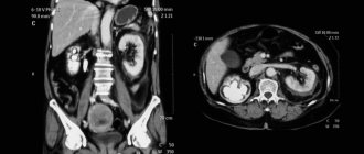
- Ultrasound of the abdominal organs (if indicated and pelvis);
- X-ray of the abdominal cavity (in case of perforation of the ulcer - the presence of free gas, in case of intestinal obstruction - Kloiber cups);
- laparocentesis (puncture of the abdominal cavity - obtaining a massive effusion);
- puncture through the posterior vaginal fornix (for pelvic inflammatory processes);
- diagnostic laparoscopy.
Types of peritonitis
According to the nature of occurrence and its specificity, peritonitis is divided into three types.
Types of peritonitis
Primary . It is the result of harmful viruses or infection through the lymph or blood. In turn, it is divided into:
- spontaneous child (preschool girls are most often at risk);
- spontaneous adult (as a result of hemodialysis for renal failure);
- primary peritonitis as a consequence of active tuberculosis.
Secondary . Caused by damage or inflammation of the internal organs of the abdominal cavity. This includes:
- peritonitis as a consequence of a violation of the integrity of the membrane of the internal organs of the peritoneum;
- peritonitis caused by abdominal trauma;
- peritonitis in the postoperative period as a consequence of surgery.
Peritonitis may result from trauma
Tertiary . Such peritonitis is rare and is a relapse of an existing disease. Most often, its occurrence is accompanied by a failure of the internal organs of the abdominal cavity. The body loses its protective properties, treatment does not work, and the course of the disease leads to death.
There are other classifications of peritonitis. Depending on the pathogen, the disease is divided into two types.
- Bacterial . Caused by aerobic and anaerobic microorganisms, for example, E. coli, staphylococci, clostridia, etc. The most common cause of peritonitis is the introduction of several harmful microorganisms into the body at once.
- Aseptic . It develops in the process of contact of the peritoneum with the contents of the stomach, intestines, blood, bile or pancreatic juice. As a result, within a few hours pancreatitis passes from the aseptic type to the bacterial type.
Main pathogenetic syndromes in peritonitis
According to the degree of spread of the inflammatory process, they are distinguished:
- local peritonitis (one part of the peritoneal cavity is affected);
- widespread (covers from two to five departments);
- total (managed to affect six or more parts of the abdominal cavity).
Types and stages of peritonitis
Treatment
Treatment of this complication requires immediate hospitalization and, as a rule, emergency surgery. Under no circumstances should the disease be treated on an outpatient basis, since the course of this disease is unpredictable and, in addition to surgical intervention, requires observation of the patient both before and after surgery.
Treatment of peritonitis must be timely and comprehensive and consists of several stages:
- preoperative preparation;
- surgical intervention;
- intensive care and monitoring after surgery.
Preoperative preparation
Preparation for surgery should be complete and last no more than 2, maximum 3 hours. Preoperative preparation includes:
- catheterization of the central vein (installation of a subclavian catheter);
- urinary catheterization;
- gastric emptying (removal of gastric contents using a gastric tube);
- massive infusion therapy of colloids and crystalloids of at least 1.5 liters (replacement of circulating blood volume, normalization of microcirculation disorders, fight against metabolic acidosis);
- preparation for anesthesia (premedication);
- administration of antibiotics (drugs are selected empirically before surgery);
- antienzyme therapy;
- normalization of the cardiovascular system;
- maintaining liver and kidney function.
Surgery
Surgical intervention has the following goals:
- eliminate the primary focus that caused inflammation of the peritoneum;
- cleansing the abdominal cavity;
- intestinal decompression;
- effective drainage of the abdominal cavity.
Operation stages:
- Anesthesia
Anesthesia for surgery is carried out in several stages. Endotracheal anesthesia is preferable; in extreme cases, spinal anesthesia (SMA) is performed. When performing SMA, a catheter is placed in the subdural space through which local anesthetics (lidocaine) are administered in the postoperative period, which reduces the need to use narcotic drugs.
- Access
In case of inflammation of the peritoneum, a median laparotomy is performed (an incision from the pubis to the navel and above, to the sternum), which provides good access to all floors of the abdominal cavity.
- Eliminating the source of the complication
After an incision in the anterior abdominal wall, an inspection of the abdominal organs is carried out and the original source of the disease is established. Further surgical intervention is carried out depending on the situation. In case of perforation or rupture of an organ, the wound is sutured; in case of inflammation (appendicitis, pyovar, etc.), the organ is removed. In case of intestinal obstruction, intestinal resection with anastomosis is performed, and in the case of purulent inflammation of the peritoneum, enterostomies are formed.
- Sanitation of the abdominal cavity
The effusion is removed from the abdominal cavity; after its elimination, the abdominal cavity is repeatedly washed with antiseptic solutions (chlorhexidine, dioxidine, furacillin) and dried.
- Bowel decompression
A tube with numerous side holes is inserted into the small intestine. Administration is carried out through the nose, rectum or enterostomy (necessary for removing gases from the intestines).
- Drainage
Drainage of the abdominal cavity is carried out with silicone or rubber tubes (exited to the anterior abdominal wall), which should ensure the removal of effusion from all parts of the abdomen.
- Suturing the wound
The operation ends with suturing the postoperative wound or applying a laparostomy. During laparostomy, the abdominal wall is not sutured; only the edges of the wound are brought together with special sutures.
Postoperative therapy
Management of the postoperative period should be carried out under monitoring, be complete and adequate, with a quick change of prescriptions and tactics in the absence of positive dynamics.
Postoperative patient management includes:
- adequate pain relief;
- carrying out intensive infusion therapy (up to 10 liters per day);
- carrying out detoxification therapy (hemodialysis and lymphosorption, administration of diuretics, hemosorption, lavage of the abdominal cavity through drains or sanitation through laparostomy);
- prescription of antibiotics in maximum doses, intravenous route of administration (combination of cephalosporins with aminoglycosides and metronidazole);
- immunocorrective therapy;
- prevention of intestinal paresis (administration of proserin) and intestinal failure syndrome (administration of atropine, potassium preparations);
- normalization of the functioning of all organs and systems;
- prevention of complications.
Treatment of peritonitis
Treatment of peritonitis is carried out in the emergency surgery department. If you suspect acute peritonitis, you should not take food, water or painkillers, use heating pads or give enemas, the patient should maintain a supine position. The treatment of peritonitis, with the exception of rare cases (limited peritonitis, state of agony, etc.), is surgery.
Before the operation, preparations are carried out aimed at at least partially stabilizing the patient’s condition. Preparation consists of replenishing fluid balance, relieving pain shock and normalizing blood pressure.
Surgical intervention for the treatment of peritonitis is performed under general anesthesia. During the operation, the primary infectious focus is eliminated, the inflammatory effusion is removed, the abdominal cavity is washed with antiseptics and drainage is installed. Then the intestinal obstruction that developed as a result of sepsis is restored, and intestinal compression is eliminated. After the operation, it is time for drug treatment of peritonitis, for which active antibacterial therapy is used, as well as therapy aimed at maintaining the vital functions of the body.
Video from YouTube on the topic of the article:
Postoperative care in the early phase
The patient is transported on a gurney to the intensive care ward, where he is carefully transferred to a special functional bed with clean linens. The patient is provided with warmth and comfort. A warm heating pad is placed on the legs, on a blanket, and an ice pack is placed on the postoperative wound (for no more than half an hour), which will prevent bleeding from the wound and somewhat reduce the pain.
The patient is placed in a Fowler's position in bed - the head end is raised 45 degrees, and the legs are slightly bent at the knee and hip joints. If the patient is unconscious (under anesthesia), he is laid horizontally, removing the pillow from under his head. To avoid retraction of the tongue, the head is tilted back slightly and the lower jaw is brought out. In the first 2–3 days after surgery, the patient is prescribed fasting and strict bed rest. If necessary, artificial ventilation of the lungs is continued, and if the patient’s condition is satisfactory, he is periodically given inhalations of humidified oxygen.
The first dressing change is carried out on day 2, under the supervision of a doctor. If the bandage has become loose or bleeding from the wound has increased, dressing should be done earlier. Honey. The nurse monitors not only the pulse, respiratory rate, blood pressure (every hour) and temperature, but also controls urine output (the urinary catheter is left in place for another 2 to 3 days after surgery) and the amount and nature of discharge through the drains. The drains are periodically washed, and the dressings on the drains are changed by a doctor.
The patient’s nutrition after surgery begins on the 2nd day and parenterally (infusion therapy). Basically, parenteral nutrition includes the administration of 10% glucose and amino acid salts. The volume of infusions is calculated according to the formula: 50 – 60 ml/kg of patient’s body weight.
On the first day after surgery, the patient is not given anything to drink, and to relieve thirst, the lips are wiped with a damp cloth. As soon as peristalsis is established (usually on day 2), the patient is allowed to drink (1 teaspoon of water every hour) and proceeds to enteral nutrition (administration of liquid food and mixtures through a nasogastric tube).
It is undesirable for the patient to remain in bed for a long time (physical inactivity provokes postoperative complications). Taking into account the patient's condition, early activation is started.
By the end of the first day, the patient should begin to actively behave in bed (turn over, bend, straighten limbs). On the 2nd - 3rd postoperative day, the patient first sits down in bed, then, after several deep breaths and exhalations and clearing his throat, he must get up and walk around the room, after which the patient is put to bed. Honey helps to lift the patient. sister. As the condition improves and pain decreases, the patient expands the regimen according to the doctor’s instructions.
Late phase
As soon as the patient establishes constant peristalsis, the passage of gases is established and stool appears, he is transferred to independent feeding. Food is taken at room temperature, in small portions, up to 6 times a day.
- During the first week, food should be liquid (broths: water after boiling is drained and replaced with new one, soft-boiled eggs, jelly and jelly, pureed vegetables with a small amount of butter).
- On days 3–4, the patient’s menu includes pureed cottage cheese, boiled beef, lamb, pureed chicken and fish, slimy porridges and soups (rice, oatmeal). Coarse fiber and foods that are difficult to digest and irritate the digestive tract (legumes, cabbage, radishes and radishes, stringy meat, skin and cartilage of poultry and fish, cold drinks) are excluded. The intake of fats should come from vegetable oils, sour cream and cream, and a small amount of butter. Easily digestible carbohydrates (marmalade and honey, jam, marshmallows, chocolate, etc.) are limited. Dried bread or yesterday’s baked bread is included in the menu for 5–7 days.
- Free mode (walks around the department and on the hospital grounds) is prescribed for 6–7 days. If the postoperative period is favorable, the sutures are removed on days 8–9, and the drains are removed on days 3–4. The patient is usually discharged on the day the sutures are removed.
Distant phase
After discharge, the patient must follow a number of medical recommendations:
- limiting heavy lifting (no more than 3 kg) and heavy physical activity for 3 months;
- sexual rest for up to 1.5 months;
- performing therapeutic exercises (training the respiratory and cardiovascular systems, strengthening the abdominal muscles and preventing the development of hernias, restoring working capacity).
Rehabilitation of the patient is facilitated by skiing, hiking, hiking, and swimming. The patient is also recommended for sanatorium treatment.
The patient should eat sparingly (up to 5 times a day), not overeat, but also not starve. It is recommended to boil, steam, stew or bake food (without crust). Limit the consumption of foods that irritate the gastrointestinal tract (spices, peppers, marinades and pickles, bitter and sour vegetables: sorrel, radish, garlic, onions, radishes). You should avoid refractory fats (margarine, lard, smoked foods) and limit your consumption of sugar (sweets, jam) and baked goods.
Treatment in the postoperative period
After surgery, it is necessary to continue taking medications to avoid the development of complications. On the second day after surgery, the patient will be prescribed parenteral nutrition. Nutrients enter the body through intravenous infusion, bypassing the gastrointestinal tract. Enteral nutrition is intended to restore intestinal motility. Nutrient mixtures are administered using a tube through the mouth and nose. The composition of the mixtures and the duration of treatment are selected by the doctor individually.
Note! If the patient shows positive dynamics, then for normal intestinal functioning one should switch to natural nutrition. This usually occurs 5 days after surgery.
It is recommended to follow a low-calorie diet, including low-fat meat broths, vegetable purees, compotes and jelly in the diet. Gradually, eggs, meat and dairy products can be added to the menu. When eating on a diet, you should avoid smoked products, spices, legumes, sweets and chocolate. It is prohibited to drink carbonated drinks and coffee.
The patient should check the postoperative wound several times a day, paying attention to the cleanliness of the dressing and the degree to which it is wet. The dressing should be constantly changed, while observing the rules of antiseptics and preventing displacement of the drainage tube.
Consequences and complications
Early complications of peritonitis that can occur in the acute period in the absence of timely treatment include life-threatening conditions:
- infectious-toxic shock;
- acute vascular insufficiency and collapse;
- bleeding;
- development of sepsis;
- acute renal failure;
- intestinal gangrene;
- cerebral edema;
- dehydration;
- pulmonary edema;
- DIC syndrome;
- death of the patient.
Long-term consequences of peritonitis (after surgical treatment):
- formation of intra-abdominal adhesions;
- infertility (in women);
- interintestinal abscess;
- intestinal eventration;
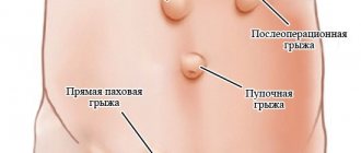
- ventral hernia;
- intestinal paresis and obstruction.
Diagnosis and treatment
Correct diagnosis on the first day of disease development is the key to successful treatment. Diagnosis is based on examination of the patient by a doctor, ultrasound and x-ray examinations, and laboratory blood tests.
Often the patient’s condition does not allow for extensive diagnostics.
The only treatment for acute peritonitis is surgery. During the operation, the doctor removes pus from the abdominal cavity, performs general sanitation of the peritoneum, excises abscesses, seals and stitches ruptures. Typically, the patient is given a drain to drain the newly formed pus. Additionally, antibacterial therapy, detoxification of the body, and immune support are used. A few days after surgical treatment, the patient is prescribed intestinal stimulation to activate its work.
Forecast
The prognosis after peritonitis largely depends on the duration of the clinical picture before medical care, the extent of peritoneal damage, the age of the patient and concomitant pathology. The mortality rate for this complication still remains at a high level, for example, with diffuse inflammation of the peritoneum it reaches 40%. But with timely and adequate therapy, early surgical intervention in compliance with all the requirements for performing an operation for this complication, a favorable outcome is observed in 90% of cases or more.
Author:
Sozinova Anna Vladimirovna obstetrician-gynecologist