The human digestive system is a complex of hollow organs through which food passes, covering a distance of 4-5 meters from the mouth to the rectum. The stomach is part of this route, the first stop of ingested food, where it mixes with gastric juices before moving on. The organ also performs immunological functions and is connected to a network of specialized nerves that are not found in any other part of the digestive tract.
Where is the stomach
This hollow muscular organ is located in the upper left abdominal region under the ribs and is controlled by the autonomic nervous system (responsible for regulating functions associated with normal life, such as breathing, digestion, blood circulation). It is also important to know that acid sensitivity is controlled by molecules called acetylcholine, histamine and gastrin.
The human stomach is located just below the diaphragm, just below the lungs and heart, and is in contact with the pancreas, liver, spleen and colon. Its upper part is protected by the chest. The organ's shape resembles a J-shaped empty leather pouch, consisting of several layers of muscle that move with force to thoroughly mix the food before entering the duodenum.
Structure of the stomach
The stomach is a sac shaped like a bean or bean grain. The outer layer of the stomach, separating it from the abdominal cavity, is the serosa. Three other layers behind it:
- Muscular, responsible for stomach contractions;
- Axillary - connective tissue that protects the stomach wall from rupture;
- Mucous – a membrane that prevents rapid erosion of the walls of the stomach.
Currently reading: What is gastroscopy of the stomach - types, prices in Moscow and how to prepare
The mucous membrane has the largest size - due to the muscular layer it is all in small folds, which speeds up the process of digesting food.
Anatomical structure
The stomach has two openings: the inlet - cardia or cardiac, and the outlet - pylorus or pylorus. They are both circular muscles called sphincters that open and close as needed. The cardia connects the esophagus to the stomach, regulates the flow of chyme and prevents reflux. The pylorus connects the organ with the duodenum.
The body of the stomach is its most powerful part, which is anatomically called the lesser and greater curvature. Its walls consist of different layers: mucous membrane, submucosal, muscle and serous tissue. Muscular – the one that helps form a bolus. The fundus of the stomach, its highest part, is located on the left under the diaphragm. This is where gases accumulate. And the antrum or antrum is a narrow area that is located in front of the pylorus.
The stomach is smooth on the outside, wrinkled on the inside. Its inner membrane has small pores, which are the openings of the glands. They secrete gastric juice - a liquid containing hydrochloric acid, pepsin, gastrin and lipase. All these compounds are necessary for the high-quality digestion of carbohydrates, proteins and fats, the absorption of nutrients from them and their absorption into the blood.
Anatomical characteristics
Human stomach, illustration from the book "Grey's Anatomy of the Human Body"
Topography of the stomach
The length of the stomach goes from top to bottom, from left to right, from back to front.
Skeletotopy of the stomach
- cardiac section, lat. pars cardiaca - at the level of the VII rib on the left;
- fundus of the stomach (vault), lat. fundus (fornix) ventricul - at the level of the cartilage of the 5th rib on the left;
- body of the stomach, lat. corpus ventriculi - between the fundus of the stomach and the pyloric region, respectively;
- pyloric section (pyloric), lat. pars pylorica - at the level of the Th12-L1 vertebra on the right.
The fundus of the stomach is adjacent to the ribs, the pyloric region is adjacent to the spinal column.
Syntopy of the stomach
Adjacent to the stomach:
- in front - the left lobe of the liver;
- behind, on the left, above - the spleen;
- behind - the pancreas;
- below - loops of the small (jejunal) intestine.
Holotopy of the stomach
Covered with peritoneum on all sides (intraperitoneal), projected into the epigastrium (regio epigastrica).
Structure of the stomach
- anterior wall of the stomach, lat. paries anterior
- posterior wall of the stomach, lat. paries posterior
- lesser curvature of the stomach, lat. curvatura ventriculi minor
- greater curvature of the stomach, lat. curvatura ventriculi major
Functions of the organ
The main purpose of the stomach is to digest ingested food, and saliva, pancreatic juice and other secretions from the digestive tract contribute to this. After chewing, which occurs in the mouth, it goes through the esophagus to the stomach, where, under the influence of mechanical (peristaltic) muscle movement and the chemical action of hydrochloric acid, bile and other enzymes, it is converted into chyme. This mushy mass rises through the digestive tract to continue assimilation of essential nutrients and the resulting waste that cannot be used by the human body.
Topography and structure
The stomach is located in the epigastrium between the esophagus and duodenum (duodenum), under the diaphragm and liver. The volume of an organ in an adult is 1–3 liters, the length of an empty organ is 18–20 cm, and the length of a filled organ is 22–26 cm.
The stomach consists of the following parts:
- The cardiac part, which is adjacent to the area where the esophagus enters the stomach;
- Bottom (vault);
- Body;
- The pyloric part consists of the vestibule and the canal (pylorus);
- Minor and major curvature (walls).
The wall of the stomach consists of the following layers: the muscular layer, the serous layer and the mucous layer.
Muscularis , which includes:
- The outer layer is the rectus muscles (lesser and greater curvature);
- Middle - circular muscles (sphincter - a valve that prevents the exit of the food bolus);
- Internal - oblique muscles (give the stomach its shape).
The muscular layer is responsible for the activity of contractions (peristalsis) of the organ and the advancement of the food bolus.
Serous layer , which is separated from the muscular layer by a thin subserosal layer, it is responsible for nutrition and innervation (supply of nerve endings) of the organ. This layer covers the stomach completely, provides shape and fixes the organ. The layer contains lymphatic, blood vessels and Meissner's nerve plexuses.
Mucosal layer – forms folds that increase the surface area of the stomach for more efficient digestion. In addition to the folds, the layer contains gastric fields (round elevations), on their surface the ducts of the endocrine glands open, which produce gastric juice.
The organ is supplied with blood through the celiac trunk, the left and right omental arteries of the stomach and small intragastric arteries. Lymphatic drainage occurs through the hepatic lymph node, the innervation of the organ is carried out by the submucosal, subserosal and intermuscular plexuses (intramural nerve plexuses), and the vagus and sympathetic nerves are also involved.
Gastrointestinal system
This organ is part of the gastrointestinal tract. The general goal of this system is to process food at its entire stage, digestion and absorption of elements necessary for the body. The entire gastrointestinal tract consists of the following sections:
- oral cavity – food is crushed here;
- esophagus - crushed food passes through it to other sections;
- stomach – primary digestion of food occurs here;
- duodenum - here food continues to be digested and transported to the intestines;
- small intestine - a number of chemical elements are absorbed here, which may or may not be needed by the body.
- large intestine - here the necessary components of food are finally absorbed and feces are formed;
- rectum - transports feces directly to the anus.
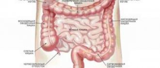
The system is also equipped with a number of necessary glands: salivary, sublingual, pancreas, etc. Often the liver and gallbladder are part of this same system, although they also have other functions. A diagram of the gastrointestinal tract is shown below (Figure 2).
The nature of the discomfort
The patient should pay attention when abdominal pain begins. Time plays an important role in diagnosing major diseases; pain can be:
1. Early. If your stomach hurts 15-30 minutes after eating, most likely the cardiac region, fundus, or body are affected.
2. Late ones appear 1-1.5 hours after eating, indicating an ulcer, gastritis, pancreatitis, cancer.
3. Hunger pains occur between meals, most often occurring with ulcerative lesions of the stomach and duodenum.
In most cases, a person’s stomach is located in the left hypochondrium and epigastrium, because these areas are the center of unpleasant sensations.
The nature of the pain is:
- Spicy.
- Dumb.
- Aching.
- Bursting.
- Compressive.
- Pulling.
- Stabbing.
- Cutting.
- Dagger pain.
A patient never has only one symptom; as a rule, there are accompanying complaints, which allow the doctor to make a preliminary diagnosis.
Possible diseases
1. Gastritis.
There are acute and chronic forms. In the first case, the pain in the stomach area is dull and aching. The patient does not pay attention to them for a long time. The acute process is characterized by more pronounced changes. Unpleasant sensations in the abdomen are associated with food; the condition worsens 30 minutes after eating. The patient may notice a certain pattern: some foods are well tolerated, others cause aggravation. Additional complaints:
- Nausea, vomiting.
- Feeling of heaviness, fullness in the stomach.
- Belching sour, rotten.
- Heartburn.
- General weakness, pallor, dry skin.
- Decreased appetite.
2. Ulcer.
The disease is characterized by the formation of defects in the mucous membrane. The dangerous fact is that the symptoms do not always correspond to the severity of the changes. Deep lesions may not be clinically apparent. The patient does not seek help or take medications, since the complaints are minimal. In this case, the risk of complications is high. Perforation and bleeding are life-threatening. If your stomach hurts even slightly, it is better to consult a doctor. Features of the ulcer:
- When there is a breakthrough, there are pains in the stomach, the pain is dagger-like in nature.
- The person’s condition is close to shock: pale skin, sticky sweat, increased breathing and pulse.
- It is characterized by a cyclical course: alternating periods of remission with exacerbations (autumn, spring).
3. Cancer.
The pain in the stomach area is dull, rather the person feels some discomfort. This condition is not related to food. It is recommended to consult a doctor if the following symptoms are present:
- Aversion to meat food.
- Rapid weight loss in a short period of time without changing your diet or physical activity.
- Pale skin.
- Decreased appetite, feeling of fullness, heaviness even with a minimal amount of food.
- The stomach hurts constantly, physical activity and stress do not affect the severity of the discomfort.
4. Infections.
In case of poisoning or consumption of low-quality products, nausea, vomiting, and stool disorders come to the fore. The above complaints are accompanied by stomach pain. Often the patient may point to a specific product, which raises suspicion. Additionally, there is an increase in temperature, general weakness, and dehydration.
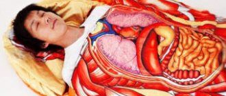
Where is the human stomach located in an anatomical photo
The human stomach is located under the diaphragm on the left. An elastic plate separates it from the chest organs. The localization of the organ is on the anterior wall of the abdomen - to the left of the lower part of the sternum. Below is the gastrointestinal tract. An enlarged abdomen, when bloated, closes the gastric wall. To the right is the liver. It closes the upper gastric corner. With the accumulation of gases in the abdomen, it is difficult to palpate the organ in the right hypochondrium. Only clinical and instrumental methods help to study its structure and properties in pathology of the gastrointestinal tract.
Clinical studies show that the volume of hydrochloric acid per day is about 2.5 liters. Approximately this amount is enough to ensure complete processing of food. When eating portioned meals in small volumes, acidity does not decrease, since the body “learns” to produce pepsinogen at certain times of the day according to daily rhythms. Try to maintain consistent meal times.
The location of the stomach and other abdominal organs changes with cancer, intestinal flexures, adhesions, and anasarca. It is difficult to palpate hollow organs with your hands, so clinical and instrumental methods should not be neglected.