Location
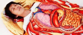
Schematic representation of the esophagus (in the center) and the organs located around it.
Among specialists, it is customary to correlate the beginning and end of the esophagus with the visible and permanent bone formations of the human skeleton:
- it begins at the VI cervical level (in front, this is the area of the lower edge of the cricoid cartilage of the larynx);
- ends in the area of the X-XI thoracic vertebra.
Traditionally, there are 3 sections of the esophagus:
- cervical,
- chest,
- abdominal.
Cervical region
Its boundaries:
- above – the lower edge of the cricoid cartilage (level of the VI cervical vertebra);
- below – the jugular notch of the sternum (level of the I-II thoracic vertebra).
The length of this part of the esophagus is small and is only 5-6 cm in an adult.
Heading down, the esophagus passes behind the trachea, and on either side of it are the common carotid arteries and recurrent nerves.
Thoracic region
It starts from the jugular notch of the sternum and ends at the level of the X-XI thoracic vertebra in the place where the esophagus leaves the chest cavity through the hole in the diaphragm. This is the longest part, its length is 15-18 cm.
In the chest area, the esophagus is closely surrounded by other organs:
- in front of it are the trachea, aortic arch, tracheal bifurcation, left bronchus, pericardium with the heart located in it;
- behind – the thoracic lymphatic duct, vertebral column, aorta, azygos vein;
- on the sides - mediastinal pleura, vagus nerve.
Abdominal
This is the shortest part, its length is 1-3 cm. It starts from the esophageal opening of the diaphragm and ends at the junction with the stomach. Here the esophagus comes into contact with:
- liver;
- vault of the stomach;
- often with the spleen.
Nervous system
The innervation of the esophagus occurs due to the vagus nerves and the trunks of the sympathetic nerves bordering them. The neurons of these nerves are located in the motor nuclei of the brain stem. Efferent fibers transmitting nerve impulses form plexuses that penetrate the wall of the organ. The straight and circular muscle layers form a plexus with neurons that have a specific autonomous function; a short neural arc can close at their level.
The cervical and thoracic sections of the organ supply branches with nerves that ensure their connection with the central nervous system, which form strong plexuses, which in turn stimulate the heart and trachea. In the thoracic section of the organ, in its middle part, in the nerve plexuses there are incoming branches of the sympathetic trunk and splanchnic nerves. In the lower part of the thoracic region, the plexuses again form trunks.
In the part of the esophagus above the diaphragm, the vagal trunks are closely adjacent to the walls of the esophagus and branch in a spiral state. The left trunk goes to the anterior surface of the stomach, the right – to the back. Centripetal nerve fibers from the esophagus enter the spinal cord.
Part of the organ’s autonomic nervous system, which is connected to the sympathetic system, but is functionally opposed to it, helps reflexively regulate the motor function of the esophagus. The mucous membrane of the organ is sensitive to heat, light, pain and tactile effects. The areas of the pharyngeal-esophageal and esophageal-gastric boundaries are particularly sensitive.
Structure
There are 3 layers in the wall of the esophagus, which go from the inside to the outside as follows:
- The mucous membrane is the innermost layer, is easily renewed, has a folded structure, contains cells that produce slightly alkaline mucus, and numerous receptors that carry information to regulatory centers regarding the process of swallowing and the movement of food through the esophagus.
- The submucosal layer is quite loose; there are rich arterial, venous, nervous and lymphatic plexuses.
- The muscle layer is represented by two types of fibers, in the upper third there are striated muscles, and below there are smooth muscle fibers, also located in 2 layers. Circular fibers run almost in a spiral inside, and longitudinal fibers run outside.
- Adventitia is the outer lining of the esophagus, where nerve fibers and blood vessels of the esophagus pass.
Esophageal sphincters
Circular muscle fibers form small thickenings (sphincters), the prolonged contraction of which contributes to the normal functioning of the upper gastrointestinal tract. The most important of them are:
- upper (pharyngeal-esophageal) - protects against the throwing of food from the esophagus back into the pharynx;
- lower - prevents the reflux of gastric contents into the esophagus.
Narrowing of the esophagus
Esophageal narrowings are divided into 2 groups:
- physiological,
- anatomical.
Anatomical narrowings are always present, but physiological ones are present only in a living person. In places of narrowing, it may be difficult for a bolus to pass through, and foreign objects accidentally swallowed by small children stop here, which can be seen on an x-ray.
The following narrowings of the esophagus are distinguished:
- pharyngeal (cricopharyngeal, cricopharyngeal) - the area formed by the cricoid cartilage and the inferior pharyngeal constrictor;
- aortic – in the area of the aortic arch;
- bronchial - at the point of contact of the esophagus and the left bronchus;
- diaphragmatic - in the area where the esophagus passes through the diaphragmatic ring;
- cardiac - at the entrance of the esophagus directly into the stomach.
In this case, the cardiac and aortic are considered to be physiological narrowings, and the diaphragmatic, bronchial and pharyngeal - anatomical.
Diseases caused by disruption of the esophagus
Diseases of the esophagus contribute to impaired motility of the organ and can affect the development of the oncological process. Pathologies may be accompanied by the following symptoms:
- Belching, heartburn with an unpleasant odor;
- Difficult passage of food through the esophagus;
- The occurrence of pain during direct consumption of food;
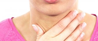
- Constant feeling of a lump in the throat;
- Nausea and vomiting;
- The appearance of hiccups;
- Severe pain in the epigastric region.
Most diseases of the esophagus at the initial stage are asymptomatic and begin to manifest themselves in an acute form. In the absence of proper and timely treatment, irreversible complications develop in the body.
Emerging diseases of the esophagus are classified into two separate types:
- Congenital pathologies;
- Acquired diseases.
Congenital pathologies are defects that are diagnosed a few days after birth. The most common organ diseases are discussed below.
Spasm and cardiospasm of the esophagus
If there is severe discomfort during swallowing and the passage of food through the esophagus, a spasm of the organ can be diagnosed, which often occurs in young people with strong excitability and instability of the central nervous system.
Currently reading: Technique and algorithm for gastric lavage
This condition often occurs under severe stress, rapid absorption of food, or mechanical damage to the esophagus. Against the background of such factors, a reflex spasm develops, which most often occurs at the junction of the esophagus and stomach.
Cardiospasm is accompanied by expansion of the esophagus and the formation of an enlarged cavity with a morphological change and the appearance of a sharp narrowing in the cardiac part. This pathology can develop against the background of the influence of external and internal provoking factors, dysfunction of a neurogenic nature leading to atony.
Causes of the disease:
- Traumatic injury, ulcer or tumor in the esophagus;
- Exposure to toxic substances (vapors from hazardous industries, cigarettes, alcoholic beverages);
- Development of esophageal stenosis against the background of previous diseases (syphilis, tuberculosis, scarlet fever);
- The presence of pathologies of the diaphragm (sclerosis of the opening, aerophagia, gastroptosis, gastritis, splenomegaly, hepatomegaly, peritonitis);
- The appearance of supradiaphragmatic processes (aortic aneurysm, aortitis, mediastinitis, pleurisy);
- Neurogenic factors against the background of the development of infectious pathologies (typhoid fever, measles, diphtheria, poliomyelitis, influenza, meningitis);
- Poisoning with toxic substances (lead, alcohol, arsenic, nicotine);
- Changes in the organ of congenital pathology that develop during embryonic development.
Achalasia
Achalasia of the esophagus is neurogenic in origin and is marked by identified functions of esophageal disorders. A characteristic symptom is gastric perilstatic disorder. The lower esophageal sphincter, which is responsible for the automatic closing function between the organ and the stomach, loses the ability to relax, which leads to the development of pathology. The main provoking factors are considered to be psychogenic, genetic and infectious predisposition. The average age of diagnosis of the disease is from 20 to 40 years.
Burns of the esophagus
A burn to the esophagus caused by exposure to strong chemicals or extremely hot liquids. Statistics show that the age of diagnosed pathology is children under 10 years old. Such data indicate an oversight in children who taste everything due to excessive curiosity. Burns of the esophagus in adults are most often diagnosed when caustic soda or a concentrated acid solution is ingested. Damage to the mucosa under the influence of phenol and Lysol also occurs.
The following degrees of burn are distinguished:
- In the first degree, damage to the esophageal mucosa is noted;
- The second degree is characterized by pathological changes in muscle tissue;
- The third stage of a burn involves damage to all the membranes of the organ (in this case, the victim experiences pain shock and loss of consciousness).
A characteristic symptom of a burn is a strong burning sensation in the sternum, inability to swallow naturally, vomiting, and swelling of the mucous membranes.
Presence of a foreign body
In medical practice, it is quite common to encounter the presence of foreign bodies in the esophagus, which entered during food consumption (poorly chewed pieces) or due to other circumstances. The most common cause of discomfort is fish or chicken bones.
In some cases, a person accidentally swallows sharp objects, which can cause an acute inflammatory process in the body. After removing the foreign body, it is necessary to follow a special diet to restore the normal functionality of the organ.
Currently reading: Diagram of the human stomach with pictures
Ulcer development
This disease often develops when gastric juice enters the esophagus. In this case, the pathology is accompanied by damage to other organs (stomach, duodenum). During diagnosis, single or multiple ulcers are detected on the walls of the organ.
An ulcer can develop against the background of provoking factors (consequence of surgery, perilstatic disorder). The main symptoms: heartburn, pain in the sternum, belching with a characteristic sour taste.
Atresia
This pathology is a rather complex defect, which represents the blind end of the upper part of the organ, and the lower segment is connected to the trachea. Against the background of a progressive syndrome, additional diseases develop in the body.
The cause of the pathology is considered to be abnormal deviations during the embryonic development of the fetus. Most often, such symptoms are observed if at 4–5 weeks of pregnancy a woman was under the influence of negative factors.
Esophagitis
The reason for the development of the disease is the consumption of very hot and cold foods, which have a strong irritation on the mucous membranes of the organ. Pathology can develop due to trauma or infection of the body.
The presence of the disease implies the occurrence of increased pain and salivation. Treatment is based on following a diet, drinking warm milk, and soups with vegetables. In some cases, temporary fasting is recommended.
Diverticulum
With this disease, severe protrusion of the walls of the esophagus occurs. The pathology is formed due to a strong accumulation of food, which is detected by belching when the body position changes. The main signs of the disease are constant nausea, vomiting, sore throat, increased salivation, and bad breath. The treatment method is based on the prescription of an individual therapy regimen.
Functions of the esophagus
The main function of the esophagus is to carry food from the mouth to the stomach. Once in the lumen of the esophagus, the food bolus causes the walls of the esophagus to expand in front of itself and close behind it for 5-6 cm. The contraction of the longitudinal muscles pushes food towards the stomach. In this case, the lower sphincter opens a few seconds earlier than the food bolus reaches it. Such coordinated work occurs due to complex regulatory processes on the part of various parts of the nervous system and the action of local hormones.
Various mental factors, including stress, as well as diseases of the chest and abdominal cavity, can lead to motor dysfunction of the esophagus when they occur:
- difficulty swallowing (feeling of a lump in the throat);
- the appearance of antiperistaltic waves directed from the stomach to the pharynx, etc.
On the other hand, when the mucous membrane is irritated, reflex disorders in the functioning of other organs can occur - increased heart rate, respiratory rate, increased salivation or lacrimation.
Another important function of the esophagus is to prevent the reflux of stomach contents into the airways, pharynx and oral cavity.
Alimentary canal
This is a very special part of the organs. The total length of this channel is approximately 8-10 meters! This part of the organs includes the human digestive system. The structure and functions of the channel are also special.
Its first component is the oral cavity. Everyone knows what it is. The cavity consists of the salivary glands, tongue and teeth. This is where the food is ground. Also, thanks to the tongue receptors, a person senses the taste and temperature of the food or drink consumed. Thanks to the tongue and saliva, so-called food lumps are formed, which are then sent into the pharynx.
The esophagus also includes the digestive system. Its structure and functions are very specific. The esophagus is a long 25 cm tube, the upper part of which consists of striated muscle tissue. The lower one is smooth. And most importantly, the esophagus is the place through which processed food enters the stomach.
Anomalies in the structure of the esophagus
If for some reason the development of the esophagus is disrupted, then various anomalies of this organ may occur, which are treated primarily surgically. The most famous among them are:
- absence of the esophagus (aplasia);
- obstruction (atresia);
- doubling;
- extension;
- abnormal narrowing;
- the presence of fistulas connecting the esophagus to the trachea;
- shortened esophagus;
- the presence of stomach cells on the mucous membrane that produce hydrochloric acid and gastric juice.
Developmental defects
The structure and function of the human esophagus can be disrupted as a result of abnormal development. This is often facilitated by the negative impact of a polluted environment, ionizing radiation, and harmful working conditions that involve prolonged contact with toxic compounds (heavy metal salts, mercury vapor, arsenic). The most common types of pathologies are:
- Atresia. This type of pathology is characterized by complete occlusion of the lumen of the esophagus. It is noticed immediately after birth, since after feeding the child regurgitates food, foam and mucus are released from the nasal passages and mouth. The danger of this condition is that aspiration pneumonia may develop. In this case, the baby can only be helped through surgical therapy.
- Stenosis. Typically, hypertrophy of the muscular tissue of the lining of the esophagus also leads to pathology. In this case, dysphagia leads to a pathological expansion of the food canal, where the accumulation of food masses stagnates, which causes the process of putrefactive fermentation.
- Tracheoesophageal fistula. A congenital pathology that results in abnormal communication between the trachea and esophagus. Because of this, there is no possibility of feeding the newborn through the mouth. Any intake of food causes a paroxysmal cough with severe cyanosis of the skin. Treatment is possible only surgically.
- Short esophagus. A disease in which dysphagia in a child causes a gag reflex with the presence of blood streaks. To eliminate this pathology, not only surgical intervention is used. Sometimes providing an elevated position of the body after eating, or during a night's rest helps relieve negative symptoms.
Remember!
The alimentary canal is an important part of the digestive system. Its proper digestion in the stomach depends on how correctly the food bolus is formed.
Its detailed study allows us to understand the pathogenesis of the development of diseases such as esophagitis, esophagospasm, reflux gastritis. And also choose preventive measures that will prevent the development of these pathological processes.
How to take care of your esophagus
The habit of drinking very hot tea increases the risk of developing esophageal cancer.
To avoid causing a burn to the esophagus, do not consume any chemically active substances:
- In everyday life, burns with subsequent scarring of the mucous membrane are most often caused by banal vinegar essence, which in appearance is confused with water or vodka.
- Always keep corrosive liquids in labeled containers.
- The habit of drinking tea that is too hot increases the risk of esophageal cancer.
Try to eat food in a peaceful state. Remember that strong negative emotions and stress can lead to dysfunction of the esophagus and cause difficulty in moving the food bolus towards the stomach.
Enzymes
This topic should be focused on. The human digestive system (functions, structure were discussed above) is a very complex and multifaceted part of our body. It was previously mentioned in passing that it contains enzymes that affect the breakdown of food. Now let's talk about these substances in more detail and list them all.
Amylase breaks down glycogen and starch, from which maltose is formed. It, in turn, is processed by maltase. And in the end you get two glucose molecules. The listed enzymes are salivary.
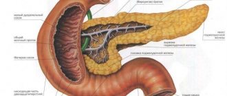
Pepsin and chymosin are found in the stomach. Proteins are broken down and peptides are formed. The pancreas contains trypsin, which processes these same peptides. As a result, amino acids are obtained. Amylase and lipase break down fats and starch.
The gallbladder and liver contain salts, which activate digestive enzymes and emulsify fats. Finally, a few words should be said about small intestinal enzymes. There are a lot of them: maltase, lactase, phosphatase, sucrase... They break down a lot of different substances, resulting in the formation of elements vital for the body. These are glucose, fructose and free phosphate.