What is a biopsy, features of taking a biopsy from the stomach
This study consists of obtaining a certain amount of abnormally changed tissue for further examination under a microscope in order to confirm or exclude an oncological lesion. Thanks to this diagnostic technique, specialists in most cases make the correct diagnosis and prescribe an adequate course of therapy. In modern oncological practice, it is possible to obtain a biopsy from almost any part of the body, but each organ has its own methods and features of taking biopsy material.
A gastric biopsy is usually performed as follows:
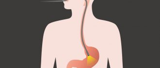
- the patient lies on his side, and an endoscope is inserted into his esophagus, which is carefully advanced to the stomach;
- Once this apparatus reaches the main digestive organ, the doctor makes a visual inspection of its walls and, having discovered suspicious areas, cuts off small pieces of altered tissue from them.
Such a biopsy is called targeted, i.e. the specialist does not act blindly, but takes visible pathological structures. After the biological material is obtained, the patient can go home. But this is only the case when anesthesia was not used during the biopsy. If general anesthesia is used during this diagnostic test, the patient will have to stay in the hospital for some time to recover.
Additional modern research
How is tissue testing performed? First, it is crushed, then it is degreased before a histological examination.
To make the fabric strong and flexible, it is filled with paraffin and then cut into thin plates. In this form it is placed on a glass slide.
A conclusion based on the results of the analysis can be given by a histomorphologist who conducts histology using an electron microscope, which allows a detailed examination of all elements.
During the procedure, microtrauma to the mucous membrane of the esophagus is possible. They are not dangerous and do not lead to complications.
The instruments used during the procedure are miniature; they cannot affect muscle tissue.
The procedure does not cause severe pain; the patient may experience mild discomfort.
Immediately after the examination, the patient’s tongue sensitivity is restored, the swallowing reflex is normalized, and he can go home. After the endoscopy, you are not allowed to eat for another 2 hours.
A tissue sample taken during gastroscopy is placed in a container with a preservative, labeled, numbered and sent to the histology laboratory.
The study is carried out by a pathologist. The tissue sample must be cut into thin sections suitable for examination under a microscope (that is, almost transparent). To do this, the material must be compacted and cut with a special cutting device.
Paraffin is used for compaction (for routine research) or the sample is frozen (for urgent analysis).
Next, microscopic sections are made from the frozen dense sample. A microtome is used for this.
The sections are placed on glass and stained. The finished preparations are examined under a microscope.
The pathologist, when examining the biopsy specimen, states in his conclusion:
- Thickness of the mucous membrane.
- The nature of the epithelium with clarification of the degree of secretion (atrophy, hypertrophy or normal secretion).
- The presence of dysplasia and metaplasia of the epithelium.
- The presence of inflammatory infiltration, the depth of its spread, the degree of inflammation activity. It is assessed by the number of lymphocytes, plasma cells, eosinophils infiltrating the mucosa.
- Signs of atrophy or hyperplasia.
- Presence of Helicobacter pylory and degree of contamination.
The main signs of malignant cell atypia:
- Other cell sizes (tumor cells tend to be much larger than normal tissue cells).
- Cell shape. Polymorphism is noted, the cells are completely different in shape, which is not typical for normal tissue.
- Increase in nuclear size, polymorphism, fragmentation of nuclei.
- A large number of dividing cells in smears.
- Disruption of normal communication between cells: indistinguishability of cell boundaries or, conversely, separation of cells.
- Inclusions in the cytoplasm, vacuolization of the cytoplasm.
There are reliable morphological changes that are classified as precancerous conditions, that is, in the presence of such changes, the risk of developing stomach cancer is several times higher:
- Adenomatous polyps. These are benign neoplasms originating from glandular cells. They have a very high probability of cancerous degeneration.
- Intestinal metaplasia of the gastric mucosa. This is a situation where part of the gastric epithelium is replaced by villous epithelium of the intestine.
- Chronic atrophic gastritis. With this gastritis, a sharp decrease in the number of glands is revealed in a biopsy of the mucous membrane.
- Chronic gastritis type B. This is chronic antral gastritis associated with Helicobacter pylori infection.
- Xanthomas of the stomach. These are accumulations of fat cells in the stomach lining.
- Menetrier's disease. A disease in which overdevelopment of the gastric mucosa occurs with the development of adenomas and cysts in it.
Examination of a tissue sample under a conventional light microscope is usually sufficient. An experienced doctor is able to quickly assess the morphological picture and see cell atypia. But sometimes other methods are used to clarify:
- Electron microscopy. Examination under an electron microscope allows you to examine all cell organelles. Images can be photographed and stored in computer memory for later comparison. The disadvantage of electron microscopy is that only a few cells are visible in the field of view.
- Immunohistochemical methods. The method is based on the principle of antigen-antibody interaction. In some doubtful cases, special sera are used that contain antibodies to certain molecules that are unique to certain tumor cells.
Equipment for taking biopsies from the digestive organs
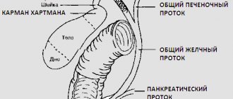
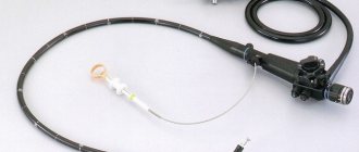
A biopsy sample is taken from the stomach using special instruments. The main instrument is a fibrogastroscope, which is a rigid but flexible tube (probe), the distal (introduced into the cavity of the digestive organ) end of which contains the opening of the instrumental channel. Through it, instruments are brought out to carry out manipulations to collect biopsy material, and water and air are supplied. In addition, the distal section of the fibrogastroscope is equipped with a microscopic lens and a lighting device. The control unit of the device and the eyepiece for monitoring the manipulations being carried out are located on its handle.
Microscopic instruments are used to collect samples for examination through the instrumental channel of the fibrogastroscope:
- Biopsy forceps, which can be used to pinch off a small piece from the surface of a suspicious area.
- An excision loop is used when a biopsy of a gastric polyp is necessary. This instrument allows you to remove a tumor growing on the walls of the stomach entirely.
Also in the operating room there are containers with special solutions into which the resulting biopsy is placed to be sent to the laboratory.
Indications and contraindications for the procedure
A study based on taking a biopsy from the walls of the digestive organ and further studying it, in cases where the use of other diagnostic methods is impossible or does not provide sufficient information to make a diagnosis.
The main indications for a patient to undergo a gastric biopsy are:
- studying the nature of tumor structures to differentiate cancer from polyps and cysts;
- examination of ulcers that do not heal for a long time and tumors of unknown etiology;
- the appearance of signs of dyspepsia in patients at risk for cancer;
- analysis of the degree of injury to the mucosa and the level of malignant degeneration of cells;
- preoperative clarification of the regenerative capabilities of the stomach walls to clarify the possible scope of the operation;
- control of therapeutic measures (resection of the main digestive organ, antitumor drug therapy and radiation).
But even with these indications, a gastric biopsy is not always performed. It can only be prescribed by the attending physician, provided that the sick person has no contraindications to this procedure.
It is absolutely unacceptable to take a biopsy from the walls of the digestive organ in several cases:
- the onset of an asthma attack;
- acute form of heart attack or stroke;
- stenosis of the esophagus, making it impossible to insert a probe.
There are also relative contraindications, i.e. those conditions when conducting such a study is highly undesirable, but in case of urgent medical need it is permissible with the permission of a specialist. These include: fever, inflammatory process in the nasopharynx, accompanied by severe nasal congestion, heart failure, physiologically determined possibility of narrowing the lumen of the esophagus, mental illnesses, epilepsy.
What can a specialist tell you?
After undergoing such a procedure, it is important to listen to your inner state. If any deterioration is observed, or vomiting appears, and the temperature rises, consult a doctor. Deviations are rarely observed.
The results will be known to the patient after 3 days. They will be incomplete if there is not enough material, so the procedure may be repeated.
The results will be benign. The specialist indicates which benign formation is located in the patient’s stomach.
Here, too, repeated testing cannot be ruled out. If the results indicate a malignant formation, then the type of cancer, tumor size, margins and location are indicated.
Possible findings on endoscopic biopsy:
- Polyps;
- Ulcers;
- Benign education;
- Malignant tumor.
Based on the results obtained, treatment is prescribed.
Analysis can occur through targeted or search techniques.
The first is necessary when the pathology has already been detected, the specialist needs to take a sample on the border between healthy and affected tissues. The search is carried out so that the doctor, as a result of the study, can find crushed areas, which will be polyps, ulcers or some kind of compaction.
The first method is the most effective for detecting ulcers, polyps, and other diseases. It is used in 95% of cases.
Preparation for collecting biopsy material
To obtain accurate results that allow a correct diagnosis to be made without repeated examination, the patient must properly prepare for the procedure and follow all the instructions of the attending physician.
The main preparatory measures for patients scheduled for gastric biopsy are:
- visiting the treatment room or operating room on an empty stomach (at least 6-8 hours must have passed since the last meal);
- exclusion of alcohol, chocolate, and fatty foods from the diet at least 2 days before the scheduled event;
- You can drink water on the day of the biopsy, but it is better to refrain from quenching your thirst for the last 4 hours.
The specialist who will carry out manipulations with taking the biopsy should be warned in advance about medications taken for life-saving reasons, an increased gag reflex and the presence of allergic reactions.
Important! All preparatory activities should be taken very seriously. The accuracy of the diagnosis and the accuracy of prescribing a treatment course depend on the accuracy of their implementation. In addition, following your doctor’s recommendations for preparing for a biopsy will help you avoid repeating a not entirely pleasant procedure.
Patient reviews about biopsy
Almost all patients focus on discomfort at the time of insertion of the endoscope; they emphasize that there is no pain because anesthesia was previously performed.
According to the general opinion, the process of plucking the material itself is painless, the damaged area heals quickly and easily, but the tests as a result of the biopsy give a clear picture of the state of health, which leads to quality treatment.
All patients advise those who are planning to undergo a biopsy that they should not stock up on negative impressions from the Internet before the event, because the moral and psychological attitude plays a big role, especially for impressionable people.
Doctor:
Shishkina Olga ✓ Article checked by doctor
Surgical and endoscopic types of examination, what is the difference?
This procedure has several varieties, which are directly dependent on the technique used.
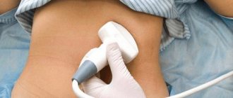
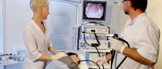
Endoscopic research method
Most often, gastric biopsy is performed using endoscopy. This technique is considered the main one and is carried out using a special device - a gastroscope, which is also called an endoscope, under the constant visual supervision of a gastroenterologist. A biopsy is obtained through endoscopic examination by pinching off small pieces from the gastric mucosa.
Endoscopic research method
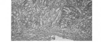
The second method of taking a biopsy sample is surgical
Surgical research method
This biopsy has several varieties:
- Open. Material for further histological examination is obtained during surgery for resection of the entire digestive organ or part of it.
- Loop. The biopsy is cut with a special loop during surgery.
- Brush biopsy. Biomaterial is also collected during abdominal surgery, but to obtain it, a special brush with hard villi is used, which makes it possible to remove pathologically altered cells from the deeper layers of the stomach walls.
In what cases is a biopsy prescribed?
A biopsy is necessary in cases where other methods of examining the stomach do not provide accurate results.
Very often, a biopsy is performed if the patient has a tumor or ulcer, after which the doctor will be able to accurately say whether the tumors in the organ are malignant or whether it is a polyp that does not pose a threat to the patient’s life.
In addition, an organ biopsy can be prescribed for acute and chronic gastritis - with its help, the doctor will be able to accurately determine at what stage the disease is, whether there is a risk of it developing into an ulcer, and also how badly the organ tissue is damaged.
Sometimes a biopsy is prescribed to identify the cause of gastritis, in particular the detection of Helicobacter pylori bacteria on the mucous membrane, which often cause inflammation of the mucous membrane of the organ.
If a patient has an ulcer, a biopsy is mandatory and is prescribed to almost all patients, since an ulcer is very often a precancerous condition.
In an advanced stage of a stomach ulcer, its manifestations can be almost identical to a malignant tumor, and a biopsy in this case will help the doctor determine exactly what pathology the patient is suffering from.
A biopsy is prescribed not only for ulcers, but also for established damage to the patient’s gastric mucosa. In this case, not only an examination is carried out, but also the removal of the detected polyp.
READ How long can you live with stomach cancer?
Very often, this procedure can be repeated after removal of a polyp, as well as after more serious operations on the stomach.
In this case, a detailed examination is necessary to assess the rate of restoration of the stomach wall, as well as to notice in time the complications that have begun.
Progress of the procedure with the collection of biopsy material
The technique of conducting this diagnostic study has some features that distinguish it from a similar procedure performed on another organ.
A gastric biopsy is performed as follows:
- The patient is placed on the couch on his left side.
- The oral cavity, upper part of the esophagus and respiratory tract are treated with a local anesthetic (usually lidocaine), which stops the gag reflex.
- A mouthpiece is placed in the patient's mouth, through which the endoscope is carefully inserted. The sick person helps push the endoscope tube, making swallowing movements to do this.
- Once the device reaches the main digestive organ, the doctor conducts a visual inspection of its walls and, if a pathologically altered area is detected, plucks several (at least 5) pieces from its surface. To obtain a more accurate picture of the pathological process, they must be taken from different places in the abnormal structure.
Possible complications
As a rule, biopsies performed during EGD rarely cause serious complications. Typically, patients may feel slight discomfort in the stomach area in the first hours after undergoing the test. In addition, minor bleeding from the resulting lesions in the area where samples are taken is possible, and this will go away on its own.
However, if the following signs appear, you should definitely see a doctor or call an ambulance:
- brown vomit, similar in color to coffee grounds;
- nausea, pain in the stomach;
- stomach ache;
- increased temperature, fever;
- severe weakness, increased fatigue;
- sharp deterioration in general condition;
- inflammation of the mucous membranes of the oral cavity, nasopharynx;
- difficulty breathing, chest pain.
Biopsy examination, how is it carried out?
Tissue samples taken from a suspicious area of the stomach wall are placed in a special container and sent to the laboratory, where a pathologist performs the following manipulations with them:
- the fabric is filled with a special preservative or paraffin to give it density;
- The thinnest sections are made from the hardened biological material and stained with a special substance;
- Samples prepared in this way for histological examination are placed on glass slides and transferred under a microscope.
Worth knowing! Sometimes in clinical practice there is a need for urgent histological examination, for example, during an operation, it is necessary to urgently obtain the result to determine the extent of surgical manipulations. In this case, they resort to accelerated preparation of the biopsy, i.e., a piece of the taken tissue is simply frozen.
Results of the study, what can be revealed during the procedure?
The interpretation of the result is directly dependent on the medical center where the FGDS biopsy was performed. This is due to the fact that they all use different methods of obtaining information.
But basically, deciphering the answer received from the laboratory will come down to the following parameters:
- Norm. There are no abnormal changes in the examined material, the diagnosis was not confirmed.
- Benign neoplasm. There are pathological changes in the cells, but the process of malignancy is absent. Regular monitoring with repeat testing after a certain time is necessary.
- Malignant structure. A tumor contains cancer cells. It is necessary to carry out clarifying diagnostics to select treatment tactics.
- Incomplete analysis. To obtain an informative picture, the biopsy specimen sent to the laboratory was not enough, so the gastric biopsy must be repeated.
Treatment of the disease depending on the results of the examination
A biopsy is considered a diagnostic procedure, after which the treatment tactics will be correct. It all depends on the test results.
If a biopsy is performed if a tumor is suspected, then using this method it is possible to determine the type of formation and composition. The result will be final. If the conclusion is negative, then the specialist may prescribe the test again.
Surgical treatment is carried out only if the atypical cellular composition is confirmed. Treatment of the disease depends on the results shown by the biopsy.
Consequences and care after the procedure
If all manipulations during biopsy sampling are carried out correctly, taking into account possible contraindications and the general physiological condition of the patient, a biopsy performed on the stomach during the FGDS procedure will not cause complications. However, one should also keep in mind the fact that a person may have latent (secretly occurring) pathological processes.
In this case, a number of negative consequences may occur:
- poor blood clotting or a collapsing vessel, absent in the anamnesis, provoke heavy internal bleeding;
- an undetected ulcerative defect contributes to perforation (rupture) of the stomach wall at the site of biopsy material collection.
Also serious complications of this diagnostic study are anaphylactic shock in the presence of an allergy to the anesthetic used and collapse. But all these consequences occur extremely rarely, so gastric biopsy is rightfully considered a safe procedure.
After such an examination, if it was performed under local anesthesia, no special care is required.
The patient can go home immediately, but he is advised to adhere to several instructions:
- refrain from eating food for 5-6 hours;
- For 2-3 days, exclude hot, salty and spicy foods from the menu.
These dietary restrictions are enough for the gastric mucosa to return to normal.
Any recommendations after the event?
Gastrobiopsy is a surgical procedure that leads to certain complications, so the specialist, taking into account the characteristics of the patient’s condition, after taking the biopsy, will familiarize him with the necessary rules of behavior after the intervention. Following these recommendations will allow the patient to quickly recover at home and return to everyday life.
After completing the procedure, the subject continues to be under the supervision of a doctor for another 2 hours, during which time the active effect of the drugs necessary to create the effect of numbness of the root of the tongue and pharynx continues. During this period, the specialist recommends that the patient not eat or drink until the sensitivity of the tongue is completely restored and the swallowing reflex appears, which will happen in a couple of hours.
The residual cessation of the effect of sedatives will occur after 12 hours, during which time you should not drive.