Home Diseases and treatment Gastroenterology
2 Mar 2020, 20:05 Anna 3 474 No
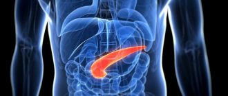
The pancreas is one of the most important organs of the gastrointestinal tract. The gland has exocrine and intrasecretory functions. For digestion, the most significant external secretion is the synthesis of digestive enzymes.
If the secretory function is impaired, a person has problems with digestion and processing of food. In order to understand that it is the pancreas that is affected, a common study such as ultrasound is performed.
Before performing an ultrasound examination, preparation is required on the part of both the doctor and the patient. The accuracy of the research result and, consequently, the diagnosis depend on the thoroughness of preparation.
Indications and contraindications
An ultrasound examination is completely painless and does not take much time. Using a special sensor and a gel-like guide, the doctor examines the position of the pancreas, its size, possible deviations and pathologies.
Also, using an ultrasound sensor, you can take a puncture of the organ for further and more detailed study.
It should be noted that ultrasound is usually performed in combination to assess the condition of the liver, gallbladder, ducts and kidneys. The activities of all these organs are closely interconnected, so pathologies extend to other “participants” of the digestive process.
Indications:
- Pain in the upper abdomen, most often occurring after eating.
- Diagnosed pancreatitis in the acute or chronic stage.
- Suspicion of neoplasms of this organ.
- Changed sizes of internal organs during previous examinations.
- Unfavorable laboratory tests.
- Decreased appetite and sudden weight loss for no apparent reason.
- Digestive disorders that do not go away for a long time.
- Suspicion of jaundice in a patient.
- A sharp increase in blood sugar levels.
The study provides a lot of information about the condition of the organ, and therefore is in demand in diagnosing many diseases.
There are currently no contraindications for ultrasound, since this examination is performed on pregnant women and even newborns.
Structure and functions of the pancreas
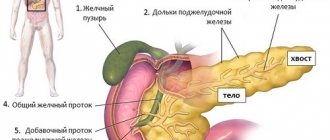
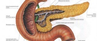
This is a digestive organ located in the upper abdominal cavity, behind the stomach. It has 3 sections: head, body, tail. The head is localized in the right hypochondrium next to the duodenum, the body is in the epigastric region behind the stomach, and the tail extends to the left hypochondrium and is adjacent to the spleen.
The pancreas has two main functions: it produces digestive enzymes and insulin. Pancreatic enzymes are needed to digest proteins, carbohydrates and fats. Insulin regulates carbohydrate metabolism, increasing tissue consumption of glucose.
The duct of Wirsung runs through the center of the organ, through which pancreatic enzymes enter the cavity of the small intestine. The bile and pancreatic ducts have a single mouth, so often the pathology of one organ leads to disruption of the other.
The hormone insulin enters directly into the blood. It is produced by the islets of Langerhans. These are clusters of glandular cells, most of which are located in the tail of the gland.
Preparation
High-quality diagnostics is impossible without preliminary preparation. To do this, you need to consult with a specialist who will perform an ultrasound.
Usually the patient is prescribed a special diet that excludes fatty and stomach-heavy foods and alcohol.
It is advisable to exclude foods from your diet that can cause bloating. There should be a light dinner the day before, but not earlier than 12 hours before the examination. You can take a laxative, but only on the recommendation of a doctor. Before the examination, you should not drink or eat, or smoke.
If you are taking any medications, you must notify the specialist before the examination.
Often, the results of ultrasound are influenced by proper preparation. If you strictly adhere to your diet and follow your doctor’s recommendations, you can count on more reliable information.
Moments of preparation for research
Many diseases of the digestive system are often hidden, and symptoms and signs are not noticed by humans. Even with a thorough examination of the abdominal space with an ultrasound machine, it may be impossible to determine the presence of the disease. Therefore, additional tests are performed in adults to confirm the diagnosis.
To perform an ultrasound scan of the pancreas, preparation must be done as accurately as possible. Even small deviations in the recommendations will blur the picture of the conclusion. Correctly and consistently performed actions affect the accuracy of results up to 60%.
The main and fundamental point in preparing for an ultrasound diagnosis is diet. The use of certain medications is taken into account in accordance with the doctor's recommendation.
Preparation for an ultrasound scan of the pancreas begins 3 days before the examination. It is necessary to completely exclude proteins from the diet: various types of meat, fish, nuts, mushrooms.
The diet in preparation for an ultrasound excludes the use of:
- Legumes: peas, soybeans, beans (quite heavy products, difficult to remove from the body);
- Whole milk;
- Black bread;
- Fresh vegetables and fruits (the presence of a large amount of fiber in them irritates the mucous membranes).
All consumed products undergo heat treatment. Boiled or baked vegetables, porridges, and semi-liquid soups are recommended. There are no restrictions on liquids, but do not drink sweet or carbonated water.
It is also advisable to take activated carbon (espumisan, smecta) to eliminate possible gas formation. This measure especially applies to people with a chronic course of the disease, during which there is flatulence and bloating. If there is a problem with constipation, then laxatives and bowel emollients are taken.
You should adhere to your diet especially strictly the day before the examination. How to prepare for an ultrasound of the pancreas a day in advance:
Ultrasound diagnostics are performed on an empty stomach. You should not drink or smoke before the examination. During the examination, there are cases when the pancreas extends beyond the stomach and is poorly visualized. In this case, the diagnostician asks the patient to drink water in small sips or through a straw. The organ reacts to the incoming fluid and secretes digestive secretions.
Preparing for an ultrasound for babies and children is significantly different from preparing for adults.
Children will not be able to adhere to such recommendations so clearly. Infants and newborns are examined for the pancreas and all organs of the digestive tract, also including the kidneys and pelvic organs. Before diagnosis, it is recommended to wait 2.5-3 hours after feeding. And if the child is kept on artificial nutrition, then the time increases to 3-3.5 hours. Older children are examined on an empty stomach, but if you do not have the patience to withstand hunger, then give the baby water to drink. Do not give tea, juices or sweet water under any circumstances.
The pancreas is involved in the digestive tract and is located in the abdominal cavity of the stomach. It produces enzymes that help break down and absorb nutrients and minerals. If an organ malfunctions, the consequences can be negative for the entire organism. Ultrasound examination of the pancreas helps to quickly identify pathologies.
Methodology
The procedure itself is carried out in a clinic or hospital using special equipment. Duration ranges from 10 to 20 minutes, depending on the qualifications of the specialist.
For convenience, the patient lies down on the couch, the doctor applies a lubricating gel and runs the sensor over the abdominal area. When clarifying controversial indications, additional examination may be required in the lateral, standing, and prone positions.
Normal values in adults and children:
- Homogeneous structure.
- Dimensions are within normal limits.
- Average reflectivity.
- No deformation of the vascular pattern.
- The Wirsung duct should not be dilated (within 1.5 - 2.5 cm).
Some indicators can be considered conditionally normal if the patient is diagnosed with certain problems with the gastrointestinal tract. Also during the examination it is necessary to take into account the weight, age and gender of the patient, on which the parameters of the organ may depend.
What does an abdominal ultrasound show?
Interpretation of abdominal ultrasound is a series of numbers and characteristics of reflected ultrasound, which you can see in the protocol of your own study.
To understand them at least a little before you go to the doctor, we suggest you read the following information.
The content of the article
What will the transcript of an abdominal ultrasound show?
First, let's look at what this ultrasound shows.
Behind the anterior wall of the abdomen there is a large space - the abdominal cavity. There are quite a few organs located in it, which an ultrasound of the abdominal cavity will show. This:
- stomach
- intestines
- pancreas
- liver
- bile ducts: intra- and extrahepatic
- spleen
- gallbladder
- kidneys
- adrenal glands
- abdominal part of the aorta and its branches
- lymph nodes
- lymphatic trunks and vessels
- division of the autonomic nervous system
- nerve plexuses.
The abdominal cavity is lined with two layers of a thin membrane - the peritoneum. It is this inflammation that is called peritonitis and is a life-threatening condition. The organs are covered in different ways by the peritoneum: some are wrapped in it, some do not even touch it, but are located within the boundaries outlined by it. Conventionally, the cavity is divided into the abdominal cavity itself and the retroperitoneal space. The latter includes the bottom of the list of organs, starting with the kidneys.
All these organs - both the abdominal cavity and the space behind the peritoneum - are examined during an ultrasound examination of the abdominal cavity. This study can detect the presence of structural damage, inflammation, pathological formations, enlargement or reduction of the organ, and disruption of its blood supply. Ultrasound does not see how a sick or healthy organ copes with its functional responsibilities.
What does ultrasound give? The study helps to find the cause of the disease in such cases:
- pain or discomfort in the abdomen
- bitterness in the mouth
- feeling of a full stomach
- intolerance to fatty foods
- increased formation of gases
- frequent bouts of hiccups
- feeling of heaviness in the right or left hypochondrium
- jaundice
- high blood pressure
- lower back pain
- fever not due to a cold
- weight loss not related to diets
- belly enlargement
- as control over the effectiveness of treatment of pathologies of the digestive system
- and also as a routine examination, including for existing anomalies of organ development, cholelithiasis.
Pathology determined by ultrasound
What does abdominal ultrasound diagnose? Using this study, the following diseases can be identified:
1. From the gallbladder side:
- acute and chronic cholecystitis
- empyema of the bladder
- gallstone pathology
- during a choleretic breakfast, the motor function of the bladder can be assessed
- developmental anomalies (kinks, septa).
2. From the liver:
- cirrhosis
- hepatitis
- abscesses
- tumors, including metastases
- hepatosis
- "stagnation" in the liver due to cardiopulmonary diseases
- fatty liver change.
3. From the kidneys and urinary system:
- kidney tumors
- "wrinkled bud"
- pyelonephritis
- narrowing of the ureters
- stones and “sand” in the kidneys.
4. From the side of the spleen, ultrasound of the abdominal cavity reveals:
- cysts
- tumors
- abscesses
- heart attacks
- organ enlargement in infectious and parasitic diseases
5. From the pancreas:
- cysts
- tumors
- abscesses
- stones in the ducts
- signs of acute and chronic pancreatitis.
6. Ultrasound reveals free fluid in the abdominal cavity
7. From the abdominal part of the aorta or its branches, an aneurysm and its dissection, vasoconstriction may be visible
8. From the side of the retroperitoneal lymph nodes, their enlargement and homogeneity of structure are visible
How to understand the research results
To do this, consider the ultrasound form (protocol). It indicates points that relate to each organ separately.
Liver
Interpretation of abdominal ultrasound in relation to this organ includes:
Share sizes:
ParameterWhat is written on the formNormal ultrasound indicators in adults
| Dimensions of the entire organ | Normal, decreased, increased (underline as appropriate) | Norm |
| right | The numbers are indicated in cm for each item | Up to 12.5 |
| left | Up to 7 | |
| caudate | 30-35 | |
| Oblique-vertical dimension (OVR) of the right lobe | Numbers in mm | Up to 150 mm |
| Outlines | It is emphasized whether they are even or not | Smooth |
| Capsule | It is emphasized whether it is differentiated or not, thickened or not | Differentiated, not thickened |
| Left lobe thickness | Number in mm | 50-60 |
| Right lobe thickness | 120-125 | |
| Echostructure of parenchyma | Emphasized, normal, increased or decreased | Norm |
| Focal formations | Yes or no | Must not be |
| Portal vein | Size in mm | Up to 14 mm |
| Vascular pattern | Depleted, normal or enhanced | Ordinary |
| Inferior vena cava | Size in mm | Anechoic, diameter 20 mm |
| Hepatic veins of the first order | Size in mm | Up to 1 mm |
Decoding the results
- Fatty hepatosis is indicated by an increase in the echo density of the organ in the form of small foci. The edge of the liver is rounded. In the final stages, due to compaction of the organ, it is impossible to see the portal vessels.
- With cirrhosis of the liver, its enlargement and dilation of the portal and splenic veins are visible. The lower edge of the organ will also be rounded, the contours will be uneven. The increase in echo density in this case will be large-focal. Free fluid in the abdominal cavity (ascites) is also determined.
- If an increase in size, rounding of the edges, as well as expansion of the vena cava and the absence of narrowing during inspiration are described, this indicates congestion in the liver due to cardiac or pulmonary disease.
- If lesions are described in which there is a violation of the normal echostructure, this may indicate malignant or benign tumors, cysts or abscesses.
In the video, a specialist talks about errors that occur during ultrasound examination of the abdominal organs.
Gallbladder
Ultrasound norm based on the results of examination of this organ:
- Shape: various – pear-shaped, cylindrical.
- Dimensions: width 3-5 cm, length 6-10 cm.
- Volume: 30-70 cubic meters cm.
- Walls: up to 4 mm thick.
- Formations in the lumen: normally there are none.
- Acoustic shadow from formations: this applies to stones and bladder tumors. Based on the presence of this shadow, the types of stones are deciphered (they come in different compositions).
- Whether they move or not: the stones are usually movable, but can be soldered to the wall or be large in size. Based on this and some other signs, one can judge whether the formation is a tumor.
Signs of gallbladder pathology
- In acute cholecystitis, thickening of the organ wall is noted, and the dimensions can be normal, reduced or enlarged. The wall may also be described as a "double contour" and the presence of fluid around the bladder indicates that local peritonitis has already developed and urgent surgery is needed.
- Thickening of the wall will also occur with chronic cholecystitis. The contour in this case is clear and dense.
- In conclusion, various deformations of the organ can be described. This is not a disease, but a structural feature.
- If echo-negative objects are described that leave an acoustic shadow, while the wall of the bladder is thickened and the contour is uneven, we are talking about calculous cholecystitis. In this case, the expansion of the bile ducts indicates that the stone is blocking the exit of bile.
Interpretation of ultrasound of the bile ducts
Normally, on ultrasound, the bile ducts have the following characteristics:
- common bile duct: diameter 6-8 mm
- intrahepatic ducts: should not be dilated
Norms of the pancreas on ultrasound
- There should be no additional education.
- head: up to 35 mm
- body: up to 25 mm
- tail: about 30mm
- contour: smooth
- echostructure: homogeneous
- echogenicity: neither reduced nor increased
- Wirsung duct: 1.5-2 mm
- education: normally there are none.
A decrease in the echo density of the gland indicates acute pancreatitis, an increase in it indicates chronic pancreatitis or cancer. The expansion of the Wirsung duct also indicates chronic inflammation. The “favor” of cancer is indicated by a segmental increase in size and unevenness of the contour of the gland, depression on the surface of the liver, as well as displacement or compression of the inferior vena cava or aorta.
Interpretation of ultrasound of the spleen
- Dimensions: length – up to 11 cm, thickness – up to 5 cm, longitudinal section – up to 40 sq. cm
- splenic index: no more than 20 cm2
- structure: normally – homogeneous
- splenic vein at the hilum.
- You can see an increase in the size of the organ. This is associated with both certain blood diseases and liver diseases (such as cirrhosis) or infectious diseases.
- Densified (less often, less dense) tissue indicates a splenic infarction, that is, that as a result of thrombosis or injury, the death of some part of the organ occurred.
- Ultrasound also allows you to see a rupture of the spleen, which usually occurs either with a severe injury or with a minor bruise, but in the case of an enlarged organ.
Ultrasound of hollow organs (stomach, small, large and rectal intestines)
It only indicates whether there is a symptom of an “affected organ” (there should not be one) and whether there is fluid deposition in the intestinal lumen (this should not happen either).
If an ultrasound scan of the kidneys was also performed, then a description of this organ is also included in the conclusion of the study. The results of an ultrasound examination of the kidneys are normal:
- width: 5-6 cm
- length – about 11 cm
- organ thickness: 4-5 cm
- kidney parenchyma - no more than 23 mm thick
- the pelvis should not be dilated
- There should be no structures in the lumen of the pelvis and ureters.
Lymphatic structures with ultrasound imaging
Ultrasound of the retroperitoneal lymph nodes normally suggests the following conclusion: “Lymph nodes are not visualized.” That is, if they are of normal size, ultrasound “does not see” them.
An increase in these immune organs indicates either an infectious disease present in the abdominal cavity or a malignant formation. In the latter case, they can increase due to the fact that cancer cells of the hematopoietic system “live” in them, as well as with metastases of any nearby organ tumor.
Sonologist's conclusions
At the conclusion of the ultrasound, the sonologist (ultrasound doctor) indicates the presence of pathology: he describes what the echo signs look like.
If in the referral the doctor indicates that it is necessary to conduct an examination for some disease, but the ultrasound did not visualize it (for example, calculous cholecystitis), then there may be the phrase “Echo signs of the disease were not identified.” The final diagnosis is made only by the doctor who refers you for examination.
Who needs to undergo Doppler ultrasound of the abdominal vessels
This examination, which is also called Doppler ultrasound (Doppler ultrasound) of the abdominal vessels, is often performed together with ultrasound. The patient's sensations are not differentiated and are not more harmful than ultrasound. It allows you to evaluate the anatomy and characteristics of blood circulation in vessels such as:
- abdominal aorta
- common hepatic artery
- iliac arteries
- celiac trunk
- splenic artery
- superior mesenteric artery
- portal vein of the liver and its branches
- inferior vena cava.
Ultrasound of the vessels of the abdominal cavity makes it possible to timely identify early abnormalities in the vessels, identify and evaluate the degree of increase in pressure in the portal vein (with cirrhosis, “congestive” liver), and evaluate the result of implantation of a vena cava filter.
Ultrasound of the abdominal aorta and its branches helps in the diagnosis of:
- fainting states
- frequent headaches
- epileptic seizures
- high blood pressure
- repeated strokes (sometimes blood clots can “fly off” from this large vessel)
- leg pain
- potency disorders
- aortic aneurysm
- atherosclerotic lesions
- vasoconstriction
- anomalies in the development of large vessels.
Duplex scanning
Vascular examination during ultrasound using modern equipment almost always includes duplex angioscanning. This is the “gold standard” in assessing blood circulation in the venous vessels.
It allows you to identify pathological blood flow, obstructions to blood flow, assess their location, extent and severity.
With this type of study, the sonologist receives a color two-dimensional image of the abdominal vessels, where red indicates the movement of blood towards the sensor, and blue, on the contrary, away from the sensor. Based on the intensity of the red and blue colors, the doctor draws conclusions about the speed of blood flow in any part of the vascular system.
Additional Study Details
Reviews about ultrasound are mostly positive: the study is painless, harmless, and very informative. The negative point is that before the procedure you need to carefully prepare so that gases in the intestines (“flatulence phenomena”) do not interfere with the correct diagnosis.
How much does it cost to conduct this research? A complete examination of all organs (including the kidneys and urinary system) with duplex angioscanning is estimated by clinics at an average of 2000-2500 rubles. An examination of individual organs with an assessment of the blood flow in them costs about 800-1000 rubles.
Thus, interpretation of abdominal ultrasound should be carried out by a specialist, taking into account not only the “norm” numbers, but also based on clinical manifestations. The above values will help you understand a little about the pathology identified in you, but the final assessment should be given by a specialist therapist or gastroenterologist.
Share information with friends:
ATTENTION! The information on the site is for reference or popular information only. Correct treatment and prescription of medications can only be carried out by a qualified specialist, taking into account the diagnosis and medical history.
Successful diagnosis and treatment, health and well-being! Your uzilab.ru.
04/24/2015 UziLab
uzilab.ru
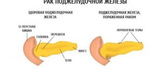
What pathologies does ultrasound of the pancreas show?
With such an examination, it is possible to identify some diseases in the early stages, as well as determine pathologies of internal organs. Most often, ultrasound confirms the following pathologies.
Decoding of identified diseases and pathologies:
- Lipomatosis. Replacement of healthy tissues with fat cells. It is determined by increased echogenicity and lighter areas of the organ.
- Chronic pancreatitis. The thickness of the walls of the duct is increased, it is often unevenly expanded. In addition, the gland is enlarged or its contour is changed.
- Tumors and cysts. An increase in the size of the organ, displacement of the head and disproportionate sizes. Normal tissue is replaced by fibrous or tumor.
- Inflammatory processes in the head. An increase in size, a narrowing of the duct and an additional change in the echogenicity of the organ.
Depending on the condition of the organ, additional examinations may be required, for example, taking a puncture and further laboratory examination.
The essence of the ultrasound diagnostic method
The high-frequency sound produced by the ultrasound machine's sensor is absorbed by some structures of the body and reflected from others. The reflected signal is captured by the sensor and displayed on the monitor in the form of a black and white picture. Hyperechoic tissues repel the ultrasound wave and are displayed in white, hypoechoic tissues transmit most of it and are indicated in black on the screen.
The gland is characterized by average echogenicity, comparable to the liver. On the monitor of the ultrasound machine it is visible in shades of gray. Its duct has lower echogenicity. When the function of an organ is impaired, its echogenicity and structure change. These changes are visible during ultrasound.
Visualization by ultrasound can be difficult in obese people, since a thick layer of subcutaneous fat makes it difficult to see the entire organ. Its head and body are best visible.
Decoding the results
Of course, only a specialist can make an accurate diagnosis, but approximate results can be determined independently. To do this, it is necessary to analyze three main indicators.
- The shape of the gland should be sausage-shaped or in the shape of the letter S.
- The size of the pancreas depends on individual indicators and varies from 14 to 22 centimeters. The width should not exceed three centimeters. The length of the head ranges from 2.5 to 3.5 centimeters.
- The contours should be smooth and clear, without blurry areas. Any imaging problems should be considered on a case-by-case basis.
- The normal structure of the organ is fine-grained (in some cases coarse-grained is acceptable) and homogeneous.
What can ultrasound show, and how reliable is its information content?
Ultrasound results according to commonly used terminology:
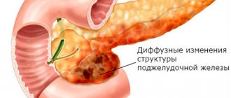
- Diffuse changes in the parenchyma are not a disease, but a symptom of a possible pathology of the digestive or endocrine system. The pancreas with diffuse changes may indicate pathologies of diabetes mellitus or the cardiovascular system, age-related sclerotic changes, obesity or obesity. Replacement of healthy parenchyma tissue with adipose or connective tissue in the case of obesity or pancreatitis entails diffuse changes, which is revealed by ultrasound
- Echogenicity is the ability of the tissue to which ultrasound is directed to reflect it. The difference between them gives the monitor a picture consisting of stripes colored gray in different shades. The echogenicity may be uniform or not. The density of organs depends directly on it. Ultrasound allows liquid substances to pass through; this condition is called echonegativity. Liquid formations are called anechoic. The pancreas has increased echogenicity when healthy glandular cells die. The size, contour, shape, homogeneity of the parenchyma against the background of increased echogenicity indicate various ailments. An increase in size is an indicator of swelling of the gland, a decrease in size is evidence of the replacement of healthy connective tissue. Loss of homogeneity may indicate cysts or calcifications.
- The pancreas has a change in structure, lack of clarity of the contour. This gives rise to the statement about pancreatitis. The acute phase can give complications of a necrotic nature, threatening to develop into pancreatic necrosis. Such lesions are visualized on ultrasound echo - dense zones with unclear contours.
- The resulting cavities with liquid substance form abscesses. Their multiplicity, merging, turns into foci of pancreatic necrosis.
- Excellent visualization of tumors. They have a heterogeneous oval-shaped structure. The advantage of ultrasound is to determine the exact localization of the formation.
The sensor operation is completed with a black and white graphic image. The signal intensity allows you to acquire different tones in the range from 64 to 256 shades of the black and white scale. The maximum intensity is shown in white, and the minimum echo signal in black. Echo-positive are white lesions on the screen, echo-negative are black. Among the shades:
- White accompanies a sharp disinfected suppuration in the form of acute pancreatitis, which is associated with interference with the release of enzymes, a decrease in blood circulation in the organ, and swelling of parenchymal tissue. This is accompanied by an increase in overall size and a decrease in tissue density. Since in this state there is no reflection of impulses from the structure, the device’s sensor sees it as transparent or white. The interpretation of the picture of the pancreas will appear in white colors on the monitor.
- The light-colored pancreas in the monitor image is inherent in lipomatosis, the process of replacing healthy cells with fat cells. It is observed when the patient's weight increases.
- Damage to the parenchyma by uneven foci leads to a mottled picture of the pancreas.
- Massive degradation throughout the entire volume of the organ in the form of scarring and fibrous changes creates a picture of a black pancreas.
Thus, an accessible and fairly simple examination provides extensive information. Therefore, when diagnosing, you need to start from simple to complex. If ultrasound is not enough to determine an accurate diagnosis, then other research methods can be added.
Why is ultrasound chosen to examine the pancreas?
Ultrasound of the pancreas is a type of instrumental diagnostics that, using ultrasound waves, allows you to visualize the gland and study it in detail. Ultrasound is successfully used not only in endocrinology, but also in many other areas, including urology, cardiology, traumatology, neurology, etc. But why is this research so widespread? To answer this question, it is not enough to name just one factor, since ultrasound has several very useful properties.
- The first is his safety. The complete safety of ultrasound examination of the pancreas makes it possible to diagnose patients of any age, regardless of age, gender or health status. Such diagnostics can be carried out even in pregnant women if damage to this organ is suspected. Also, diseases of the pancreas can take quite a long time to be treated, which will require regular examinations, and ultrasound is quite suitable here, since it can be safely performed as many times as needed.
- When examining the pancreas, ultrasound makes it possible to study the organ in different sections, which expands the scope of diagnosis compared to radiography. In addition, ultrasound diagnostics is carried out in real time, that is, it is not the image that is evaluated, but the image that the sonologist receives in real time.
- Ultrasound diagnostic devices and the examination itself are available to almost everyone due to affordable prices and the prevalence of these same devices. All this, together with the relative ease of diagnosis, makes ultrasound one of the best methods for the primary detection of pancreatic pathologies.
Examination of the pancreas using ultrasound
Ultrasound examination, or sonography, allows you to determine the presence of tumors of the gland, identify its structure and compare the size of the gland with the norm.
In order to correctly visualize this organ, the diagnostician is guided by the large arterial and venous trunks of the spinal column.
Abdominal organs in cross section
As a rule, an increase in size is observed with swelling of the pancreas in acute pancreatitis. Local enlargement of part of the gland can be observed with a tumor, cancer or cyst. In chronic pancreatitis, the dimensions may not change. A shrinkage of the gland occurs when the organ atrophies due to impaired blood supply or viral infection.
Standards for the study of adults
In health, this organ of the digestive system has the following characteristics:
- The largest part is the head. If its size exceeds 35 mm, then we can talk about pathology.
- The dimensions for the body of the pancreas should not exceed 25 mm.
- Normal dimensions for the tail are no more than 30 mm.
- The length of the gland can be from 16 to 23 cm.
Tip: The size of the pancreas is an important indicator in the diagnosis of acute pancreatitis and pancreatic necrosis.
In chronic pancreatitis, the indicator of homogeneity or heterogeneity of its structure is of greater importance. Proper preparation for the examination will make it easier for the diagnostician to visualize the organ. Come to the ultrasound on an empty stomach. In addition, the day before you should take a sorbent and a laxative, and also exclude legumes from your diet.
Standards for studying children
The size of this organ normally depends on the age and weight of the child. At birth, its length is about 5 cm, then gradually increases, reaching the adult norm by about 16-18 years. In order to draw correct conclusions about the presence or absence of pathology, the norm is most often calculated using special tables.
Tip: An important indicator of the organ’s functioning is the diameter of the Wirsung duct. In chronic pancreatitis, it increases and does not respond to the introduction of secretin. Normally, this figure averages 1.5-2 mm. Do not forget to check with the ultrasound doctor whether he has included data on the condition of the duct in the study protocol.