DESCRIPTION OF THE INVENTION TO THE PATENT
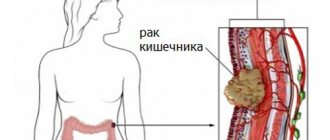
Fistulas are a cause of colon cancer.
The invention relates to medicine, in particular to abdominal surgery, and can be used to treat the inflammatory process in the anastomotic area after gastrectomy.
There is a known method for treating anastomositis after gastrectomy, which involves parenteral administration of a solution of nutrients and evacuation of stagnant gastric contents [1]. However, treatment of anastomositis using this method requires at least two weeks, and in severe forms of anastomositis it is difficult for patients to tolerate and sometimes requires repeated surgery.
There is also a known method for treating anastomositis after gastrectomy, which consists of introducing a depot form of an antimicrobial agent into the gastric stump and fixing it in the area of the anastomosis [2]. However, the use of this method is limited by the fact that many currently known antimicrobial agents affect the enzymatic functions of the tissues of the macroorganism and therefore can have a toxic effect on it.
The closest in technical essence to the proposed invention is a method for treating anastomositis after gastrectomy, which involves removing stagnant contents and introducing an anti-inflammatory solution into the gastric stump [3].
In this method, a 20% solution of sorbitol, which is a hexahydric alcohol, is used as an anti-inflammatory agent. When entering the body due to the absorption capacity of the gastric mucosa, sorbitol, due to its high biological activity, undergoes metabolic transformations, oxidizing to sorbose, and loses its anti-inflammatory properties.
Therefore, when treating an inflammatory process using the existing method, repeated (up to 8 doses) administration of sorbitol into the gastric stump is required, which lengthens the period of relief of the process.
The technical objective of the proposed invention is to reduce the treatment period for anastomositis.
This objective is achieved by the fact that in the known method of treating anastomositis after gastrectomy, which involves removing stagnant contents and introducing an anti-inflammatory solution into the gastric stump, a perfluorocarbon emulsion, for example, “Perftoran” is used as an anti-inflammatory agent [4].
These features of the proposed invention represent its difference from the prototype and determine the novelty of the proposal. These differences are significant, since they ensure the creation of the achieved technical result reflected in the technical problem, and are absent in known technical solutions with the same effect.
The occurrence of peptic ulcers after gastroenterostomy was first described by N. Braun in 1899, and after gastrectomy by N. Haberer in 1929. In the literature over the past 20 years, authors cite the frequency of post-gastroresection peptic ulcers from 1 to 4% [3]. 90 - 98% of peptic ulcers of the anastomosis develop after resection performed for duodenal ulcers. This is due to the fact that duodenal ulcers are usually accompanied by gastric hypersecretion and hyperchlorhydria, and the preservation of the secretory function of the gastric stump at levels comparable to preoperative levels is the main reason for the development of peptic ulcers.
There are four main reasons for the preservation of acid production in the gastric stump: 1) economical resection (less than 2/3) without vagotomy, with preservation of the acid-producing zone; 2) leaving part of the antral mucosa on the stump of the duodenum during resection according to the second Billroth method; 3) hypertonicity of the vagus nerves and incomplete vagotomy, if it was performed in combination with economical gastrectomy; 4) endocrine diseases - primary hyperparathyroidism, Zollinger-Ellison syndrome, Wermer syndrome, etc. Of these reasons, the first two are most common. They are technical errors of the operation. The most common mistake is economical gastrectomy for duodenal ulcers without vagotomy.
It not only does not reduce the increased acidity and peptic activity of gastric juice, but also creates conditions for the constant exposure of aggressive gastric contents to the mucous membrane of the jejunum or duodenum as a result of removal of the pylorus. Another typical mistake is leaving part of the antrum on the duodenal stump. This most often occurs during exclusion resection, when the antral mucosa in the remaining portion of the gastric wall is not excised. The antrum, cut off from the flow of acidic gastric contents, is a constant source of gastrinemia, and the levels of serum gastrin can be so high, and the course of the disease is so severe that it is possible to distinguish this pathology from Zollinger-Ellison syndrome before surgery only by determining gastrin levels using the secretin test. The causes of hypergastrinemia when the antrum is disconnected are in the absence of the inhibitory effect of hydrochloric acid on gastrin-producing cells of the antral mucosa and in vagal denervation [1].
Information about hypertonicity of the vagus nerves as a reason for the persistence of acid production in the gastric stump in the literature is scarce. It has been noted that even with significant resections and the absence of endocrine disorders in 5-10% of patients, acidity remains high. Attempts to identify a group of patients with hypertonicity of the vagus nerves using various secretory tests, including those with false feeding, are unlikely to be of significant help, if only because of the difficulties of interpreting the results in conditions of a resected stomach (on the one hand, rapid evacuation of acid, on the other - reflux of alkaline intestinal contents into the stump of the stomach). However, based on the favorable immediate and long-term results of isolated vagotomy for peptic ulcers, we can conclude that such patients have vagal hypertonicity [1].
The next group of causes of peptic ulcers is endocrine diseases. They are rare, but their significance is determined, for example, by the fact that patients with Zollinger-Ellison syndrome have a history of surgery and suffer from recurrent ulcers. According to some data, in 75% of patients with Zollinger-Ellison syndrome, ulcers have a typical localization, and there may be no clinical or laboratory specific signs. Attempts at radiological diagnosis of the syndrome are based on a number of signs: 1) a large atonic stomach with rough folds or hypersecretion in a small stump; 2) large atonic duodenum; 3) increased peristalsis, rapid passage through the small intestine, swelling of the mucous membrane; 4) unusual localization of the ulcer with signs of penetration. The only fairly accurate method for preoperative diagnosis of Zollinger-Ellison syndrome is a radioimmunological study of gastrin levels in peripheral blood.
Another important issue when identifying Zollinger-Ellison syndrome is to localize the source of hypergastrinemia before surgery, as this can radically change treatment tactics. The fact is that, firstly, there are two types of Zollinger-Ellison syndrome: type 1 – without a tumor at all, caused by hyperplasia of gastrin-producing cells in the antrum of the stomach, and type 2 – when the source of gastrin (tumor-gastrinoma) is localized outside the antrum. In the first type, a cure can be achieved using simple gastrectomy with vagotomy. The difficulties in localizing gastrinomas in the second type are that the tumor can be microscopic (in the wall of the duodenum), multiple, malignant and give metastases, which also produce gastrin.
With Wermer's syndrome, the parathyroid glands, pancreas, pituitary gland, adrenal glands, and less commonly the thyroid gland are most often affected. Sipple syndrome develops medullary thyroid carcinoma, medullary pheochromocytoma and hyperparathyroidism; with Schimke syndrome - the same thing, plus a protruding jaw, thick lips, a Marfan-like appearance, multiple neuromas on the mucous membranes, gastrointestinal neuromatosis, flat feet. Hyperparathyroidism, included in the concept of polyendocrine adenomatosis, can be primary and also lead to gastric and duodenal ulcers. Non-parathyroid tumors (including gastrinoma) can produce parathyroid hormone and thereby cause hypercalcemia and hypophosphatemia. In the differential diagnosis of primary hyperparathyroidism in this case, only selective catheterization of vessels with blood sampling directly from the parathyroid glands and determining the level of parathyroid hormone in it can help [1].
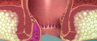
With a typical clinical picture, diagnosing peptic ulcers is not difficult. Severe hungry (including night) pain in the epigastric region, decreasing after eating or taking soda, atropine, nausea and vomiting (fecal when the ulcer is complicated by the development of a gastrocolic fistula), diarrhea, weight loss, recurrent gastrointestinal bleeding allow one to suspect the presence of an ulcer in the gastrointestinal anastomosis [1-3]. Often, peptic ulcers are much more malignant than the primary ulcer for which surgery was performed. Peptic ulcers are located, as a rule, on the intestinal side of the gastroenteroanastomosis, near the mesenteric edge of the intestine, which explains the tendency of such ulcers to bleed, which is often very severe. Often, two ulcers exist simultaneously in the area of the anastomosis (Fig.).
Rice. Two peptic ulcers identified during endoscopic examination of the gastrointestinal anastomosis area
These ulcers sometimes reach large sizes, they penetrate into neighboring organs and tissues (mesentery of the small and colon, pancreas, liver, diaphragm), which often explains the very pronounced pain syndrome.
However, the clinic may not be so bright. Pain may be moderate or even absent altogether. In such cases, a peptic ulcer usually manifests itself as gastrointestinal bleeding.
X-ray diagnosis of peptic ulcers is complex and reveals on average 50-65% of all anastomotic ulcers. This is due to the presence in the area of the anastomosis of various deformations and pockets in which the ulcer can be masked and which in turn can simulate it.
Peptic ulcers are best identified by endoscopic examination, but endoscopy can be of little help if the device cannot be passed into the afferent or efferent loop, i.e., below the anastomosis. During endoscopic examination, in the vast majority of cases, an ulcer is found on the afferent or efferent loop of the small intestine.
In some cases, with a clear clinical picture and when hyperacidity is detected in the gastric stump, the ulcer itself is not detected either radiographically or endoscopically, and sometimes even during surgery. A peptic ulcer can sometimes be superficial, heal easily with conservative treatment, but then quickly recur, sometimes causing complications, most often bleeding [1].
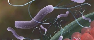
Conservative treatment of peptic ulcer of the anastomosis includes prescribing H2 receptor blockers and proton pump inhibitors to patients. If H. pylori is suspected, antibiotics are prescribed. Most patients respond to drug treatment. Surgical treatment is indicated for patients for whom conservative therapy is ineffective, with gastrinoma, with complications such as profuse bleeding, perforation, narrowing of the anastomosis, gastrocolic fistula [3].
Repeated operations on the stomach and intestines for peptic ulcer disease are undertaken infrequently and are complex interventions. Of the large number of operations proposed for the treatment of diseases of the operated stomach, two main groups can be distinguished: one is aimed at eliminating all kinds of functional (pathophysiological) or mechanical disorders - the so-called corrective operations; the second pursues the goal of removing the affected part or an entire organ. An important point in both operations is the creation of conditions for digestion that are as close as possible to physiological ones, for which, for example, they strive to restore the duodenal passage by reconstructing the Billroth I anastomosis or through the use of one of the options for gastrojejunoduodenoplasty [1].
The range of operations used for peptic ulcer of the gastrointestinal anastomosis is quite wide. This includes reresection using both Billroth methods in various modifications, truncal vagotomy (transthoracic and subphrenic) and selective gastric, as well as various combinations of vagotomy and reresection.
The choice of surgery for peptic ulcers depends primarily on its cause. In this sense, a full preoperative examination is of great importance, which must necessarily include, for example, determination of gastrin levels in the blood serum, so as not to miss Zollinger-Ellison syndrome. For example, the simplest operation for peptic ulcers is transthoracic vagotomy. It effectively reduces gastric secretion and promotes rapid healing of ulcers. But this operation does not allow performing the necessary corrective intervention on the abdominal organs, for example, with secondary afferent loop syndrome due to peptic ulcer or with concomitant dumping syndrome.
Resection of the gastric stump is not necessary if it is small - in such cases, vagotomy (and, if necessary, removal of the remaining antrum) has a quick and lasting therapeutic effect. Removal of the ulcer itself is not necessary in principle, but this issue is resolved individually in each specific case - with a large penetrating ulcer that sharply deforms and stenoses the anastomosis area, it is better to perform reresection, although mobilization of the gastric stump in such conditions is always quite difficult. In addition, the risk of bleeding from the left ulcer in the immediate postoperative period cannot be completely excluded.
In case of a large gastric stump (after economical resection of 1/2 of the stomach or anthrumectomy), reresection of the gastric stump is necessary. But if the previous operation was performed according to the first Billroth method, you can limit yourself to vagotomy. It should be noted that after resection according to Billroth I, peptic ulcers occur much less frequently than after resection according to Billroth II, but they are also severe, with penetration, repeated bleeding and severe pain. If resection is performed with them, the same difficulties with treating the duodenum may arise as with low penetrating duodenal ulcers. In such situations, selective proximal vagotomy (SPV) is the method of choice.
Gastrectomy remains the treatment of choice for Zollinger-Ellison syndrome. When identifying Zollinger-Ellison syndrome in a patient with a duodenal ulcer, most surgeons currently tend to take a more conservative approach. Two main surgical methods are proposed: 1-treatment with gastric secretion blockers, then gastric resection in combination with vagotomy, then again long-term, sometimes lifelong treatment with H2 receptor blockers; 2—first treatment with H2-receptor blockers, then PWS, then again with H2-receptor blockers. When a solitary adenoma is detected, everyone prefers to remove it.
Peptic ulcers in hyperparathyroidism and Wermer's syndrome should be treated by excision of hormonally active tissue (parathyroidectomy or subtotal resection of the parathyroid glands).
The most severe situations occur when a peptic ulcer is complicated by perforation or profuse bleeding. The natural desire to complete an emergency operation quickly can lead to technical errors. Of course, in conditions of pronounced adhesions, penetration of the ulcer and in severe general condition of the patient, reconstructive reresection is not worth doing. The method of choice in such cases is suturing the ulcer and truncal vagotomy. After removing the patient from a serious condition, it is necessary to fully examine him and decide on further treatment, possibly a new reconstructive operation [1].
We analyzed the case histories of 5 patients diagnosed with peptic ulcer of the gastrointestinal anastomosis, hospitalized in the department of surgical gastroenterology of Clinical Hospital No. 3 in Yerevan since April 1987. to August 2000 All patients are men aged 35 to 54 years. In 2 patients, the peptic ulcer of the anastomosis was complicated by bleeding, and one patient had afferent loop syndrome and cholelithiasis. Patients underwent gastric resection according to Billroth II for duodenal ulcer (3 patients) or stomach (2 patients). Patients presented the following complaints: pain in the epigastric region (5 patients), nausea (3), vomiting (3), discomfort in the abdomen (3), general weakness (2), belching (1), pain in the right hypochondrium (1 patient ). Patients with a bleeding peptic ulcer of the anastomosis had pain in the epigastric region, general weakness, nausea, vomiting of coffee grounds, tarry stools , and a feeling of discomfort in the abdomen. The above complaints in patients appeared from 4 months to 8 years after gastric resection according to Billroth II.
The peptic ulcer of the anastomosis in patients was identified endoscopically, within several hours from the moment of admission to the clinic. During esophagogastroduodenoscopy (EGDS) in patients with peptic ulcers, ulcers measuring 0.3-2.5 cm were found on the wall of the anastomosed loop of the small intestine. In one case, endoscopy revealed reflux of bile from the afferent loop of the small intestine into the gastric stump. All patients received conservative treatment, some refused the proposed operation and were discharged from the hospital with improvement within 2 to 24 days.
Thus, EGDS is the most reliable way to diagnose peptic ulcer of the anastomosis. Endoscopic examination also makes it possible to reliably judge the size of the gastric stump, i.e., in some cases it reveals the cause of peptic ulcers.
Literature
- Chernousov A.F., Bogopolsky P.M., Kurbanov F.S. Surgery for gastric and duodenal ulcers. M., 1996, p. 202-208.
- Christiansen S., Ram M., Sachatello C., Griffen W. Management of gastrocolic fistula. Am. Surg. 1981; 47. p. 63-66.
- Glasgow RE, Mulvihill SJ Postgastrectomy syndromes. 1997, p. 149-150.
Types of stomas
Stenosis is divided into 3 main types. The classification depends on the location of the narrowing.
Depending on the pathology, the doctor selects the most optimal method of treatment. Whenever possible, the surgeon always tries to preserve as much of the organ as possible.
gastrostomy; intestinal: ileostomy, colostomy; tracheostomy; epicystostomy.
The shape is convex and retracted. There are single-barreled and double-barreled. Depending on the duration of use: temporary and permanent.
Depending on the location, colostomies are classified into several types: transverse, ascending and descending.
Transverse colostomy.
A transversostomy is formed in the upper abdomen, in the transverse colon.
To avoid nerve damage, the transverse stoma is placed closer to the left splenic flexure.
A transverse colostomy is indicated for intestinal blockage or oncopathologies, traumatic injuries and diverticulitis, and congenital colon anomalies.
The location of the colostomy is determined by the doctor, taking into account the specific clinical picture of each patient.
The most common type of small bowel obstruction is obstruction due to adhesive disease. For the colon, this is the blocking of the intestinal lumen by a tumor.
Adhesive intestinal obstruction
Types, stages, tools
The term gastrectomy means complete removal of the stomach. But different stages of techniques have been developed to preserve organ fragments. Therefore, the types of operations are distinguished:
- Distal subtotal - the part attached to the intestine is removed;
- Proximal subtotal – removal of a fragment in the upper third with omentums and lymph nodes;
- Total – complete removal followed by anastomosis;
- Sleeve – to combat obesity, removal of the body and fundus of the stomach, suturing a narrow canal. After the operation, the patient eats less, as the feeling of fullness of the organ quickly comes.
Subtotal resection is performed with small bowel plastic surgery.
The operation takes place in 4 stages:
- An incision in the abdominal cavity;
- Examination of the condition of the stomach - the surgeon determines where the tumor is located;
- Mobilization of the stomach;
- Anastomosis - the anastomosis is fixed into a tight knot so that the connection does not separate.
Surgical surgery for gastric resection uses the following instruments:
- Laparotomy set – for dissection of the abdominal cavity;
- Presses, expanders;
- Dissector;
- Clamps.
The instruments are also used for peptic ulcers.
Symptoms
According to the currently most common classification, such disorders can be divided into
- organic,
- functional
- and combined complications after gastrectomy.
Functional disorders after gastrectomy include: early and late (hypo-hyperglycemic) dumping syndromes and conditionally - afferent loop syndrome, caused by a violation of its evacuation activity (it sometimes has an organic cause), post-gastro-resection asthenia (dystrophy) and anemia.
Symptoms of chronic afferent loop syndrome after gastric removal
Chronic adductor loop syndrome is divided into
Stenosis in adults manifests itself with almost the same symptoms as in children. But in adults, the symptoms are more protracted and more pronounced.
At the onset of the disease, patients begin to suffer mainly from severe pain in the upper abdomen.
Immediately after, nausea, gag reflexes and vomiting itself begin. The patient usually begins to vomit immediately after eating; the masses may contain an admixture of bile.
Stenosis very often has such a manifestation as an almost complete absence of stool. Since the patient’s body does not receive the required amount of fluid, the person suffers from dehydration. Gradually, the amount of urine decreases to such an extent that the body experiences anuria.
With congenital stenosis, the symptoms immediately manifest themselves clearly. Children primarily suffer from profuse vomiting. Vomiting may occur even before the baby starts feeding. The condition of children with stenosis is constantly deteriorating.
Over time, vomiting manifests itself even more aggressively, impurities of feces and blood appear in it, the smell becomes extremely unpleasant and sour.
The main symptom of the disease is the appearance of vomiting, which is observed immediately after the birth of the child (in the first days of birth). Vomiting may occur before breastfeeding. With stenosis, the vomit is mucous or watery.
The disease makes itself felt by the repetition of pain in the upper abdomen, followed by vomiting and nausea. Vomiting may begin immediately after eating or for no reason.
A lot of bile can be seen in the evaporated masses. There is no natural stool.
A person changes in appearance - the skin dries out and acquires a grayish tint. This is due to the fact that the body does not have enough fluid.
Over time, the symptoms worsen, the vomit acquires an unpleasant odor due to impurities of feces and blood. The stomach is constantly bloated, frequent fainting, attacks of suffocation, lack of air and dizziness.
These are all signs of stenosis.
In children with a congenital anomaly in the form of stenosis, vomiting begins after birth. The vomit turns green due to bile. These symptoms appear a short time after the baby is born. The disease can be identified by another characteristic sign - the absence of stool in a newborn baby. The reason is a narrowing of the lumen of the colon.
Clinical manifestations. The disease manifests itself with very characteristic symptoms.
These are abdominal pain of a cramping nature, bloating, nausea, vomiting, inability to pass gas, lack of stool, and a disturbance in the general condition. The clinical form of the disease can be acute, when all of the listed symptoms are sharply expressed, and chronic, in which they appear periodically and there are no sharp disturbances in the general condition.
Lifestyle after resection
Recovery and rehabilitation takes from six months to a year. After removal of the stomach for cancer, the patient requires special care in the first 2-3 days. Nutrition is provided through a tube and intravenously. Fluid is also restored through a vein.
Then the period of self-feeding begins. Diet plays a huge role. How much you can eat and what you can eat is determined by your doctor. However, some general dietary guidelines can be given.
- You need to eat minimal portions of food. This way the intestines will not be overloaded, and the patient’s well-being will not deteriorate.
- Meals should be fractional. You need to eat often and in small portions. You should eat 6-9 times during the day.
- You are allowed to eat fruits, vegetables, cereals, etc. All foods must be pureed and steamed.
With such nutrition, body weight will not fall to critical levels. There is no need to gain it, so the question of how to gain weight is unnecessary. It will be at a stable level, the main thing is to create a nutritious diet. This issue is resolved individually with a gastroenterologist and nutritionist.
Diagnostics
Diagnosis of stenosis occurs taking into account the collection of anamnesis, medical history, as well as on the basis of instrumental and laboratory studies.
To accurately diagnose the disease, an extensive examination of the abdominal cavity is used.
To make an accurate diagnosis, an extensive medical examination is performed. For diagnosis, new laboratory tests and instrumental procedures are used (x-rays using contrast agents, ultrasound of the abdominal cavity and colon).
A diagnostic scheme is selected to determine how much the disease has progressed and what methods are best to treat it so as not to harm the person.
How is a gastrectomy performed?
The course of the gastrectomy operation involves making an incision at the lower end of the duodenum, as well as lengthening it towards the esophagus. The end of the duodenum connects directly to the small intestine. The duration of the operation usually does not exceed 5 hours, and after gastrectomy, a hospital stay of at least 2 weeks is required.
Rehabilitation after stomach cancer is based on abstaining from eating food and drinks for 3-5 days. A revamped digestive system can be deadly if the connection between the rectum and esophagus leaks.
It is important to know! To check for leaks, a method such as X-ray irradiation is used. The consequences can be very varied, so you should refrain from consuming food and water.
Treatment
Newborns require immediate surgical intervention when stenosis is detected. The duration of the operation largely depends on the location of the pathological process. Before surgery, the patient must clear the digestive tract of excess liquid and gases. For this purpose, a thin tube is inserted through the mouth, allowing the liquid contents of the gastrointestinal tract to be evacuated.
Treatment of children
Both acute and chronic forms of the disease are in most cases treated surgically. Only at the very beginning of the disease, when the general condition of the patient is not yet disturbed, after examination, conservative measures are carefully applied - gastric lavage, cleansing enemas; in case of atony, peristalsis is stimulated with drugs (injections of proserin, neostigmine).
If the treatment is ineffective within a few hours or the cause is a tumor, adhesions, anomalies, or mesenteric thrombosis, surgical treatment is performed.
Lymph node dissection for stomach cancer
This procedure is considered mandatory for all radical effects on the main digestive organ. It is performed for significant reasons and has no relationship with the presence of metastases in the regional lymph nodes. Lymph dissection for stomach cancer is the complete removal of all lymphatic vessels and nodes, as well as the adjacent fatty tissue.
There are several options for this surgical procedure, which are directly related to the volume of tissue to be removed:
- D1. Regional lymphatic collectors located in the ligamentous apparatus of the stomach are cut out.
- D2. Both those in the immediate vicinity and perigastric collectors related to secondary metastasis and located in the vermis trunk, along the arterial branches, must be eliminated.
- D3. In addition to the above pathological phenomena, the lymph nodes of the third stage of metastasis located along the aorta and esophagus are also removed.
Not so long ago, the generally accepted scope of this procedure, carried out without fail, was considered option D1. In modern clinical practice, during radical interventions to remove late gastric cancer, lymph node dissection in the D2 volume is almost always performed, which helps to increase the five-year survival rate among patients.
Palliative surgery or surgery to alleviate the patient's condition
If a malignant neoplasm of the main digestive organ could not be detected in a timely manner, and its development has reached the final, inoperable stages, oncologists try to alleviate the patient’s condition and improve his quality of life. Palliative operations are used for this purpose. In case of stomach cancer, such interventions are called symptomatic and make it possible to effectively eliminate certain life-threatening and very severe symptoms, which temporarily alleviates the condition of the cancer patient.
The following are used as palliative surgery:
- Cytoreduction intervention. It is carried out to reduce the total number of abnormal cells, which prevents the spread of the destructive process provoked by a cancerous tumor in adjacent tissue structures and blood vessels. The operation involves excision of part of the stomach and removal of the primary source of pathology.
- “Cauterization” of the tumor. Using high-frequency or endoscopic laser ablation, mutated cells are destroyed and the risk of breakthrough gastric bleeding is reduced.
Performing such symptomatic operations allows the use of radiation and chemotherapy for stomach cancer that has reached an inoperable stage. Patients are also administered antitumor vaccines and monoclonal antibodies, which leads not only to stabilization of the disease, but also to an extension of the period of life, which will be of a higher quality.
Types of operations
The consequences of the operation may not always have a positive result, and complications after it are not uncommon:
- Special sterile operating room conditions, disinfected surfaces and instruments minimize the risk of infection. But if sterilization measures are not followed, the wound may become infected. In this case, redness, suppuration of the suture, fever, and weakness are observed.
- Internal bleeding is dangerous because, unlike external bleeding, it does not appear immediately.
Intussusception
Type of underlying disease that led to resection; Type of surgery and the course of the operation itself; The patient's condition in the postoperative period; Absence/presence of complications; Proper adherence to the diet and type of nutrition.
stage of the disease; complexity of the resection; compliance with the doctor’s recommendations during the recovery period.
Complications and pain after resection
After completion of the laparoscopic operation, the patient will remain in the hospital for approximately a week. You may experience pain for several days after the intervention.
If necessary, the doctor will prescribe painkillers. Immediately after the tumor removal procedure, the patient can eat, but the food should be soft.
You are allowed to return to your normal diet on the 5th day after the procedure. In order not to provoke the development of complications, you need to follow a gentle regime - lie down more and not make sudden movements.
You should abstain from sex for 6-8 weeks after surgery, and you should also avoid physical activity. It is important to ensure that the area of skin where laparoscopy was performed is dry and clean.
If the patient has had a colostomy or ileostomy, he or she will be instructed on how to care for the stoma without the assistance of a specialist. If an ileostomy was performed, then over time the patient is prescribed another surgical intervention, the purpose of which is to connect the ends of the intestine.
Sutures can only be removed by a doctor and no earlier than 14 days after laparoscopy. Afterwards, the patient is referred to oncology to undergo a course of maintenance therapy.
If after surgery your body temperature rises (more than 38 °C), you should immediately consult a doctor to find out the cause.
high pace of life; irregular meals; poor quality food.
Dissection of adhesions
Insertion of a central venous catheter to monitor central venous pressure and parenteral infusions. Bladder catheterization to control diuresis. Installation of a nasogastric tube.
Conservative therapy is also a method of preoperative preparation (if surgery is still required).
Removing an obstacle. If possible, eliminate the disease that led to this complication. Maximum possible actions to prevent postoperative complications and relapse.
The main stages of the operation and the surgeon’s tactics
1. Anesthesia. Usually this is endotracheal anesthesia with muscle relaxants.
2. Access – most often a wide median laparotomy.
The postoperative stage in such patients is a very important moment of treatment, no less significant than the operation itself.
If signs of intestinal obstruction appear, you should consult a surgeon. It is this specialist who determines the scope of necessary treatment measures.
Depending on the cause of development, timeliness of diagnosis, and general condition of the patient, surgical intervention can be urgent or planned. Before the intervention, the patient is prepared. In the case of a planned operation, it can be started at home and continued in the hospital; in case of an urgent operation, it can be done for several hours in a hospital.
Surgeries for intestinal obstruction are volumetric interventions with a long postoperative period. It is determined by the time of complete healing of the wound and the maximum possible restoration of the body.
How is the operation performed?
The second name for gastrectomy is extirpation. It takes place in stages under general anesthesia. The progress of the operation is determined by the degree of development of the cancer process. A surgical incision is made and the blood vessels are ligated. Then the organ is cut off from the esophagus and intestines in the area of attachment. In addition to the stomach, regional lymph nodes and omentums are removed. Then an anastomosis is formed by combining the esophagus and small intestine.
After the operation, a cytological examination of the swabs is performed. The absence of malignant cells is an indicator of a successful operation. Surgery is useless for metastases if the course of the cancer is complicated by ascites and endocrine disorders. Tuberculosis is also considered a contraindication.
In the preoperative period, the patient must adhere to a diet and daily routine in order to lose excess weight. You should stop taking acetylsalicylic acid and drugs based on it. The day before the operation you need to spend without food and cleanse the intestines with an enema.
Disability is given after complete removal of the stomach. Radical surgery is performed when conservative treatment does not help. As a result, an important link in digestion is lost, so following a diet is vital for the patient.
Indications for surgery
Intussusception
A tracheostomy is an artificially created hole in the neck with a tube removed, which is installed with the aim of recreating damaged human respiratory functions. If there are disturbances in the functioning of the respiratory system and the impossibility of performing an independent act of inhalation and exhalation, the patient often undergoes an emergency tracheal ostomy.
The epicystoma is removed from the bladder to the surface of the abdominal wall using a special catheter. The indications for such manipulation are the patient’s inability to urinate naturally for various reasons. There are temporary and permanent epicystostomies.
A colostomy can be temporary or permanent. Children most often undergo a temporary stoma.
Anorectal incontinence; Blockage of the intestinal lumen by tumor formation; Traumatic damage to the colonic walls such as gunshot or mechanical wounds; Severe cases of colonic pathologies such as diverticulitis or ischemic colitis, cancer or peritonitis, polyposis and ulcerative colitis, abscesses of the intestinal walls with perforation, etc.
(if it is not possible to carry out radical intervention); With rectosigmoid resection, if after the operation the sutures are ineffective.
How to avoid the consequences of surgery?
It should be noted that patients who have undergone gastrectomy experience a host of complications.
The consequences of such a rather serious surgical effect can be very dangerous:
- Anastomositis. An inflammatory process that occurs at the junction of the gastric stump with the duodenum.
- Anemia. Anemia is caused by postoperative internal bleeding.
- Peritonitis. Inflammation of the peritoneum.
- Death.
To ensure that these complications do not arise after surgery such as distal subtotal gastrectomy, after removal of stomach cancer, each individual patient is assigned an individual rehabilitation program. The most important stage of its implementation is the early postoperative period. The main methods to prevent the development of negative consequences, prevent the recurrence of stomach cancer after surgery and speed up a person’s recovery are a diet specially developed by the doctor, the exclusion of any physical activity and wearing a bandage.
Also, to improve the patient’s condition, further chemotherapy is mandatory. After surgery for stomach cancer, this treatment method eliminates any residual abnormal cells that migrate throughout the body through the bloodstream. This type of treatment, although it provokes a large number of side effects in a person, is recognized by doctors as necessary. It is thanks to him that, with the correct prescription and implementation of medicinal procedures, it is possible to avoid the risks of developing side effects.
Organ-preserving operations
Organ-preserving methods are gaining increasing recognition in the world practice of treating patients with cancer. The indication for such an operation is the detection of early forms of malignant neoplasm.
Early gastric cancer is considered a neoplasm that affects the submucosal layer of the stomach wall and its mucous membrane, regardless of the absence or presence of metastases in the lymph nodes.
If early gastric cancer is detected, it may be possible to perform an organ-preserving intervention - endoscopic resection of the affected area of the mucous membrane within healthy tissue.
The procedure for performing the operation is as follows. After the clear dimensions of the affected area are determined using special dyes, the planned boundaries of the intervention are marked - this is done through electrocoagulation. Next, the surgeon performs hydropreparation of the tissue (this is necessary to ensure better visual control of the layers and prevent possible perforation of the stomach wall). A special electric knife is passed through the instrumental channel of the endoscope, after which, under visual control, resection of the affected area of the mucous membrane and submucosal layer is carried out - down to the muscle layer.
During the rehabilitation period, the patient is under the dynamic supervision of a doctor, receiving the necessary drug therapy. Compared to standard surgical treatment, this method is more economical and less invasive, and also allows to shorten the patient’s hospital stay and speed up rehabilitation.