What is a liver fluke?
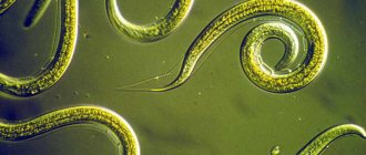
The liver fluke is a flatworm that belongs to the class of digenetic flukes. It parasitizes the bile ducts, as well as the liver of humans and other warm-blooded animals.
Liver flukes are various types of worms, including:
- Lanceolate fluke.
- Eastern fluke.
- Giant liver fluke.
- Hepatic fasciola.
- Cat fluke, etc.
The giant fluke and hepatic fasciola are dangerous to humans. These two types of flukes cause a disease called fascioliasis.
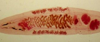
The giant fluke has quite impressive dimensions; its body can reach 76 mm in length and 12 mm in width.
The liver fluke has a flat, leaf-shaped body. There are two suckers on the head of the parasite. The liver fluke can reach 20-30 mm in length and 12 mm in width. Immediately after the parasite's short esophagus there are two loops of intestine that end blindly. Worms are hermaphrodites; inside their body there are unpaired ovaries, a small uterus and two branched testes. The parasites release large oval eggs covered with a yellow shell into the external environment.
Outbreaks of parasitic infestations can be both widespread and sporadic. The statistical coverage of the population affected by fascioliasis ranges from 2.5 to 17 million people. The main symptoms of human infection with the liver fluke are: fever, urticaria, nausea, pain in the right hypochondrium.
The permanent host of the liver fluke is most often domestic animals (cattle, small cattle, horses, rabbits, donkeys and sheep); humans are less often infected. The liver fluke can parasitize the body of wild animals, including kangaroos, beavers, deer, squirrels, etc.
The next liver fluke that parasitizes the human body is a trematode of the genus opisthorchis, which provokes a disease called opisthorchiasis.
An adult is a flat parasite whose body can reach 18 mm in length and 2 mm in width. The anterior end of the worm's body is pointed, the suckers are located on the peritoneum and on the upper part of the body (oral sucker). The digestive system of the parasite is not closed; there is an excretory canal at the rear end of the body.
Trematodes have both male and female reproductive organs. For reproduction, one individual parasitizing the body is enough.
This parasitic invasion is widespread throughout the world, but the palm belongs to Russia. In some regions, the number of infected people reaches 75%, which is explained by food traditions (consumption of dried, lightly salted or frozen fish). Cases of opisthorchiasis are registered in Belarus, Kazakhstan, Ukraine, Western European countries, etc.
Another worm is the liver fluke, which can parasitize the human body - the lanceolate fluke. This fluke causes a disease called dicroceliosis.
The parasite reaches 15 mm in length and 5 mm in width. Just like other liver flukes, lanceolate flukes are hermaphrodites. They lay eggs that have already developed miracidium. The larva hatches after it enters the body of the first intermediate host.
It should be noted that dicroceliosis is a rare disease among humans. Medicine knows only isolated cases of parasitic infestation by this liver fluke. The final host of the lanceolate fluke is most often small and large cattle.
Find out more: Liver fluke systems
Content:
The first signs and symptoms of liver fluke
The first signs of a liver fluke are associated with the incubation period of the development of the parasite.
There are three stages of parasitic infestation:
Symptoms of the liver fluke during the incubation period
The incubation period can last from 7 days to 2 months. It depends on how many parasites have entered the body. During this time, the infected person does not experience any signs of illness.
Symptoms of liver fluke in the acute stage
This stage is also called the migration stage.
It is characterized by general toxic and allergic symptoms, including:
- Increased body temperature. The mark on the thermometer may remain at the level of low-grade fever, or may rise to 39-40 °C. The temperature can rise in waves, or it can be remitting, when daily fluctuations exceed 1-2 degrees.
- The patient experiences weakness, headaches, and general malaise.
- The most common allergic reaction is urticaria, which is accompanied by severe itching. The development of Quincke's edema is possible.
- Dyspeptic disorders are characteristic, including: pain in the right hypochondrium, nausea and vomiting.
- The liver increases in size and jaundice develops. When palpated, the organ responds with pain.
- Chest pain is a sign of developing myocarditis of an allergic nature. Possible increase in blood pressure and increased heart rate.
- The number of eosinophils and leukocytes in the blood increases.
The acute phase can last several weeks; upon completion, the symptoms of the disease disappear.
Symptoms of liver fluke in the chronic stage
Chronitization of the parasitic disease occurs 3-6 months after the invasion.
In this case, symptoms indicating damage to the liver and biliary tract come to the fore:
- The patient experiences frequent paroxysmal pain in the right side.
- During an exacerbation of the disease, jaundice develops.
- The liver remains constantly enlarged in size.
- The longer the untreated infestation persists, the worse the prognosis. The formation of liver cirrhosis, hepatitis, and anemia is possible.
- Secondary infection is dangerous, which can cause liver abscess, purulent cholangitis and cholecystitis.
Medicine knows of cases where liver flukes developed in the mammary glands, in the brain, in lung tissue, in the larynx and under the skin. Prolonged stress of the immune system makes a person susceptible to diseases of various etiologies.
Find out more: Developmental stages of the liver fluke
Diagnostic methods
Due to the fact that the liver fluke has an unusual development cycle, it is extremely difficult to diagnose.
Fascioliasis is characterized by a dangerous feature: making a diagnosis of the acute phase is extremely problematic, since the life cycle of the liver fluke has not yet completed and the eggs, which are released with feces only after 4 months, are not excreted from the body. Then doctors focus on the medical history; when making a diagnosis, they have to separate fascioliasis from diseases caused by other types of helminths.
To confirm the diagnosis, laboratory tests of the patient's stool are performed twice. If yellow eggs with a cap on one side are found inside, this is the main factor for the final conclusion. In case of doubt, the bile of the infected person is examined.
It is very effective to conduct a blood test, the analysis of which, if infection has occurred, shows a noticeable excess of leukocytes and a decrease in the total amount of protein. But at the stage of a chronic disease, this procedure is practically useless, since all blood cells return to normal.
To identify the parasite in the body, a comprehensive diagnosis is required. The main method is a blood test for biochemical composition. It is studied for the level of eosinophils, alkaline phosphatase and bilirubin levels.
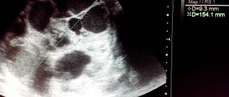
An ultrasound of the liver reveals the exact presence of the parasite in the organ. Nearby organs are also examined. Stool analysis is only effective in the 3rd month, when the helminth can lay eggs. After the same period, a study of duodenal juice secreted by the liver can be carried out.
Additionally, the patient may be prescribed an x-ray of the pancreas. If necessary, computed tomography and MRI are performed.
Routes of infection with liver fluke
Infection with liver fluke and giant fluke occurs when eating plants (garden and wild) that were watered with water from fresh water bodies, provided that there were infective worm larvae in it. Infection is possible by drinking unboiled water or by swallowing water while bathing.
Infection with the liver fluke, which causes opisthorchiasis, occurs when a person eats infected fish that has not undergone high-quality heat treatment.
Infection with the liver fluke of the genus Fluke lanceolata occurs through accidental ingestion of infested ants, for example, during the consumption of berries, meadow grasses, vegetables from the garden, etc.
Thus, the route of infection with the liver fluke is food. That is, in order for any liver fluke worm to begin parasitizing in the human body, it will need to get into the gastrointestinal tract.
Find out more: Developmental cycle of the liver fluke
Prevention measures
To prevent parasites from entering the body, it is recommended to thoroughly wash plants used for food. It is important not to drink raw water from various bodies of water and not to eat unheated fish and meat. It is required to maintain personal hygiene and regularly examine the animals living at home.
In order to prevent and avoid infection of the body with liver fluke, the following measures should be observed:
- thoroughly rinse vegetables and fruits under running water; after this procedure, be sure to pour boiling water over them;
- Before use, you need to boil raw water;
- It is important to buy meat and fish products only from trusted suppliers;
- Before cooking meat and fish, they must be thoroughly thermally processed;
- avoid eating raw fish, especially for oriental dishes, the same popular sushi;
- eat smoked, dried and dried fish products with caution;
- carry out annual deworming of pets;
- maintain personal hygiene.
These recommendations apply to all citizens and can be easily followed individually. A set of antiparasitic measures is also being carried out by the state. Authorities carry out measures to prevent contamination of water bodies with fecal matter, preventive work on deworming livestock, and also carry out sanitary education work among the population.
If you suspect a liver fluke disease, you should undergo a series of tests and undergo a full diagnostic examination. Only after this the doctor will be able to prescribe adequate treatment for the patient. In order to protect yourself from such an unpleasant parasitic infection, you should follow basic preventive rules.
- The development cycle of the liver fluke in brief
- Flukes class, preparation for the Unified State Exam in biology
- Portal vein of the liver - the norm according to ultrasound and other diagnostic methods
- ICD 10 – Liver failure, not elsewhere classified (K72)
Treatment of liver fluke
Treatment of liver fluke consists of two stages. The first stage is preparatory, and the second is aimed at directly eliminating the parasite from the human body.
The first stage of treatment for liver fluke
Treatment of this parasitic infestation is carried out in a hospital setting, although outpatient management of the patient is possible if the patient is in satisfactory health. If the disease is diagnosed in the acute phase, the patient is transferred to a gentle diet and prescribed desensitizing drugs. To eliminate allergic manifestations, medications such as Suprastin, Tavegil, Zyrtec, Zodak, etc. can be prescribed.
As for diet, when getting rid of liver fluke, table number five according to Pevzner is considered optimal. Spicy, smoked and fried dishes are being removed from the menu. It involves steaming, boiling and baking foods. After completing the second stage of treatment, the patient is recommended to consume more foods rich in fiber, which will enhance intestinal motility and improve bile discharge.
To remove toxins from the body, the patient is prescribed Smecta, Polyphepan and other sorbents. To reduce body temperature, you can take Ibuprofen or Butadione. In addition, these drugs act as anti-inflammatory drugs. Glucocorticosteroids are prescribed against the background of myocarditis.
The second stage - antiparasitic treatment of liver fluke
Antiparasitic therapy begins after the symptoms of the acute phase of the disease subside.
Drugs prescribed for the treatment of liver fluke:
- Chloxyl (taken for 3-5 days).
- Praziquantel (Biltricide). The drug is taken once.
- Triclabendazole. Depending on the severity of the infection, a single or double dose of the drug is possible.
Choleretic drugs are prescribed to quickly remove dead parasites from the human body, in addition, they prevent stagnation of bile. These may be drugs such as Holiver and Holosas.
Control diagnostics with stool analysis and duodenal intubation are carried out 3 months later and another six months after treatment.
Taking antibiotics is necessary in case of purulent complications. Surgical intervention with drainage of the liver, bile ducts, etc. is not excluded.
The third stage is restoration of the body after treatment of liver fluke
Since the liver fluke disrupts the functioning of the digestive organs and the functioning of the body as a whole, patients will need to undergo a rehabilitation period.
It may include the following activities:
- Normalization of the functioning of the gallbladder and liver with the help of a course of choleretic drugs (Holiver, Holosas, Allohol, Holagol, etc.).
- Improving liver function, protecting and restoring it with the help of hepatoprotector drugs. These can be products such as Ursosan, Galstena, Silymarin, etc.
- Normalization of digestive processes with the help of enzyme preparations (Pancreatin, Creon, Panzinorm, etc.).
- Improving metabolism with multivitamins.
The earlier the disease is detected, the faster and easier it is to achieve a full recovery. If the parasite infestation is highly intense, or if a secondary bacterial infection is added, then the prognosis worsens significantly. Even the death of the patient is not excluded.