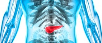
MRI of the pancreas allows you to obtain a layer-by-layer image of the organ and surrounding tissues, and is used both to make and to clarify the diagnosis, when it is impossible to make a final conclusion based on the results of computed tomography (CT) or ultrasound examination. This is one of the relatively safe diagnostic techniques. It is suitable for examining the pancreas in women in late pregnancy and children, when examination methods using x-rays are contraindicated. The magnetic fields generated during MRI are considered harmless.
Principles of MRI of the pancreas
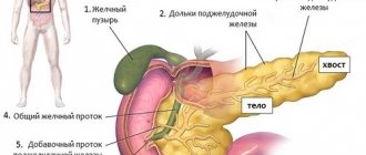
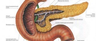
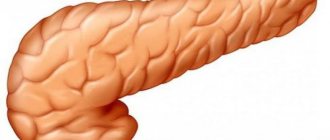
The pancreas is a parenchymal type of organ. Therefore, it is most often examined using ultrasound diagnostics. If difficulties arise in making a diagnosis, an MRI of the pancreas is prescribed. The examination involves exposing the body to a magnetic field. It is evenly distributed in all structures and tissues. But the image quality depends on the power of the device.
There are two types of magnetic resonance devices.
- Closed form. The power of this device is 1 Tesla. Therefore, such a model shows informative results.
- Open form. The power does not exceed 0.5 Tesla. Therefore, the image quality is slightly reduced.
Using tomography you can see:
- shape and size of the organ;
- structure and density;
- the presence of pathological formations;
- peculiarity of vascularization of the pancreas;
- condition of the ducts.
Tomography of the pancreas without the use of a contrast agent makes it possible to detect changes in structural tissues whose size exceeds two millimeters. If it is necessary to strengthen the magnetic signal and detect small formations, the patient should take a barium solution.
Preparation
Magnetic resonance imaging of the pancreas belongs to the category of planned medical procedures that require certain preparations that do not make any special adjustments to the patient’s usual lifestyle.
- Three days before the scheduled examination, the patient should begin following a special gentle diet that prevents increased gas formation in the intestines. The following should be absolutely excluded from the daily diet: dishes from legumes (peas, lentils, beans, beans);
- bread made from rye flour;
- baked goods;
- cabbage dishes;
- sweet vegetable and fruit juices;
- soft drinks saturated with carbon dioxide;
- salted, fried, smoked and too fatty foods;
- alcoholic drinks;
- strong brewed tea and coffee.
During this period, the patient should stop using medications (mainly tinctures and balms) containing ethyl alcohol.
- Magnetic resonance imaging should be performed on an empty stomach. If the procedure is scheduled for early morning, the patient should have dinner before 19:00; if at a later time, the fasting period should be at least five hours. On the day of the test, you should also completely stop smoking and drinking any liquids.
- During the 24-48 hours preceding the MRI, the patient's body should not be subjected to studies that require the injection of contrast agents into the main pancreatic duct.
- If the study is prescribed for a patient in a medical hospital, preparation for it, depending on the specific internal regulations and the power of the equipment used, will be somewhat different. Some patients may be given a cleansing enema, while others may have stomach contents drained through a tube. This forced measure is used in cases of congestion in the upper parts of the digestive tract.
- Before conducting the study, the patient is obliged to inform the attending physician about the presence of any metal prostheses or electronic devices implanted in his body, since this circumstance is included in the list of absolute contraindications for its implementation.
- The doctor must also be informed about the presence of any allergic reactions (especially to contrast agents ever introduced into the patient’s body).
- Before lying on the tomograph couch, the patient must remove all jewelry (including piercings) made of metal, and also remove removable metal dentures. Mobile phones and credit cards must be left in the locker room.
- The hearing aid (if the patient uses one) must also be removed before starting the procedure.
- When going for an MRI procedure, the patient should wear underwear made from natural materials.
- The patient must come for the examination somewhat earlier than the appointed time, since half an hour before the start of the examination, the specialist is obliged to carry out an allergy test for the effect of a contrast agent administered intravenously. Thanks to this substance passing through the blood vessels, all pathologies occurring in the pancreas and its excretory ducts are visualized.
Proper preparation contributes to obtaining the most reliable research results.
The attending specialist can refer the patient for an MRI of the pancreas based on undoubted indications for its implementation. The referral must be certified by the signatures of members of the medical commission of the given medical institution.
If the examination falls into the category of budget procedures paid for from medical insurance funds, the patient is required to have a standard package of documents with him, consisting of a SNILS certificate, a passport and a new medical policy.
Main indications for diagnostic examination
Magnetic and computed tomography of the pancreas is prescribed for those patients who complain of some symptoms such as:
- sharp and aching painful feeling in the stomach;
- intestinal colic, bloating and flatulence;
- chronic diarrhea or constipation;
- weight loss and lack of appetite.
Doctors also identify a number of main indications in the form of:
- detection of pathological areas during ultrasound diagnostics;
- pancreatitis of acute, chronic or calculous type;
- suspected tumor in the pancreas;
- development of intraductal hypertension and blocking the outflow of pancreatic juice;
- cysts in the organ;
- purulent lesion of the pancreas.
This examination method is considered safe. It has a minimum of contraindications, so it can be prescribed to children and women during pregnancy.
Preparation before the examination
MRI is usually performed routinely.
In rare cases, diagnosis is carried out urgently. Most often, the procedure is performed in the morning. To make the results more accurate, several rules must be followed. Preparation for MRI of the pancreas consists of the following recommendations.
- Avoid alcohol-containing drinks. The last drink of alcohol should be at least one day before the appointed date.
- 3-4 days before diagnostic procedures, it is necessary to adjust your diet. Foods that increase the formation of gases in the intestinal tract are excluded from the diet. Therefore, it is worth completely abandoning carbonated drinks, cabbage, store-bought juices, bakery and confectionery products, and legumes.
- The last meal should be 8 hours before the test.
- Before diagnosis, all metal objects are removed: earrings, bracelets, chains. If the patient has metal staples inside, cardiac stimulators or wires in the spine, then the doctor must be warned about this in advance, otherwise adverse consequences cannot be avoided.
If an MRI of the pancreas is performed with contrast, then it is necessary to do a test for allergic reactions. When the patient is in the hospital, preparation may vary. Before diagnosis, the doctor may give an enema or give a laxative.
MRI technique and preparation for the procedure
MRI of the pancreas is performed in 2 ways - with and without the introduction of a contrast agent. If an MRI with contrast is prescribed for the gland, the patient is given an iodine-containing substance into a vein before the study, which allows a more complete picture to be obtained. Contrast is used to detect malignant and benign formations.
The day before the procedure, patients must adhere to a diet. Avoid alcohol, fried, spicy, fatty and salty foods. Before the tomography, you should not eat or drink tea or coffee (8 hours before the procedure).
Before starting the examination, you must remove all metal objects and jewelry from yourself.
The patient lies down on a mobile table, which moves inside the tomograph ring. There are MRI machines that do not examine the entire human body, but only a separate part of the body. You cannot move during the examination, otherwise the readings will not be entirely accurate. The procedure takes from 10 to 30 minutes. The tomograph makes noise, which is repeated several times during the examination. To prevent patients from experiencing discomfort, they are offered special headphones.
Important information: Symptoms and treatment of pancreatic inflammation
Carrying out the procedure
Many people are interested in what an MRI of the pancreas shows. This type of diagnostic examination is simple to use and can identify any organ pathology.
To carry out the diagnosis, the patient needs to lie down on a special couch. After this, the device will automatically push it into the large capsule. This happens if a closed procedure is carried out. In the open form of diagnostics, a spherical tube is placed above the person, inside which a tomograph is located.
When an MRI with contrast is performed, a catheter is inserted into the person. The solution will be supplied through it. If the patient has fears of closed spaces, then open equipment is more suitable.
Being in a closed capsule, the patient and the doctor will always have a connection. The person will not have to do anything. The main thing is to lie still and not move too much. The device is noisy. To prevent the patient from experiencing discomfort, he is asked to wear special headphones.
The study lasts from 30 to 60 minutes. Some clinics allow the presence of loved ones.
Contraindications
MRI has a very limited list of contraindications, but they still exist.
- Magnetic resonance imaging cannot be performed for those whose body contains metal structures - pins, implants or crowns, IUDs, pacemakers, etc.
- Tattoos made in artisanal conditions can also cause discomfort - the ink may contain heavy metals.
- If you are overweight (above 250 kg), it may be impossible to conduct an examination - in this case, the doctor prescribes another diagnostic method.
- Epilepsy and claustrophobia may prevent the patient from remaining still for the examination. For these diseases, you should consult your doctor.
- Also, in consultation with the doctor, an MRI is performed during pregnancy.
If magnetic resonance imaging with contrast is required, it is important to ensure that there is no individual intolerance to contrast agents.
Restrictions to the procedure
CT scan of the pancreas with contrast is practically no different from conventional diagnostics.
A barium solution makes it possible to more clearly localize the affected areas. Doctors identify several contraindications such as:
- the presence of metal objects on the body that are difficult to remove. This list includes fragments, tooth crowns, fastening staples, fixed-type dentures;
- the presence of a pacemaker, heart valves, artificial joints;
- gestation period in the first and third trimester. At this time, the fetus and the woman’s body are vulnerable. Therefore, the procedure may adversely affect the development of the child. The second trimester is considered the safest of all;
- mental disorders;
- allergic reactions to contrast agent;
- the patient weighs more than 150 kg. This is explained by the fact that the couch is not designed for such a heavy load. And the magnetic field may not pass through the entire body.
The procedure is also prohibited by hemolytic anemia, chronic renal failure, cardiac dysfunction, and the presence of metal tattoos. The reason for refusal to perform the procedure may be the patient’s poor health.
Methodology
The MRI procedure is performed in a special room equipped with a tomograph. Some clinics ask patients to completely remove all clothing and put on a hospital gown to prevent unwanted images from appearing on the images.
The patient lies down on the movable platform of the tomograph, after which the nurse uses soft straps to secure his arms and legs. Small pads are used to support the head. This is necessary in order to eliminate the possibility of accidental movements that could cause blurred and inaccurate images during scanning.
If the test is performed with the introduction of contrast, the nurse will insert an intravenous catheter connected to a special device that delivers the drug at a certain pressure. To perform the MRI procedure, contrast agents based on salts of the rare earth metal gadolinium are used.
Survey results
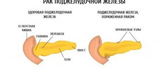
What can an MRI and CT scan of the pancreas show? Using this type of diagnostic examination, you can identify:
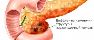
- inflammatory processes of acute and chronic type;
- neoplasms of malignant and benign types;
- anomalies of congenital and acquired type.
Using tomography, you can obtain data that will influence the course of further actions. In some cases, pathologies require surgical intervention.
After the examination, you can obtain information about:
- increase in organ size;
- inflammatory process. There are three types of them;
- form of neoplasm. If they have a round shape, then they are benign;
- organ contour. It must be clear. If the contour is uneven or cut, it indicates the presence of a malignant formation;
- metastasis to neighboring tissues and organs.
The patient does not receive the results of the study immediately, but a day after the examination.
Difference between computed tomography and magnetic tomography
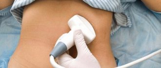
Most often, patients first undergo ultrasound diagnostics. This method is considered the most harmless, but not always reliable. There, all organs of the digestive system are examined at once: stomach, liver, gallbladder, intestines. If complex forms of the disease are suspected, the patient is sent for magnetic resonance or computed tomography.
These two types of research are not particularly different from each other. They are both reliable and also have the same readings. Only when contrast is used, computed tomography requires the injection of a solution through a vein. Without its use, it will not be possible to make an accurate diagnosis. Another difference is that a CT scan of the pancreas is not prohibited if there are metal objects in the body.
Magnetic tomography can be done with or without contrast.
Poor preparation may affect image quality. Before any type of diagnosis, you need to follow a strict diet, avoid eating gas-forming foods and pre-cleanse the intestines.