Main functions
The pancreas is an organ located in the abdominal cavity. It is part of the digestive system and produces important substances that help break down food. These are hormones and enzymes. The pancreas is one of the main organs of the endocrine system, because its hormones, which immediately enter the bloodstream, play a large role in carbohydrate, fat and protein metabolism.
Functions and hormones
The structural features of the pancreas make this organ so important in the human body. It is directly involved in the digestion process, as it performs external and internal secretory functions.
This organ produces pancreatic juice, which contains a lot of enzymes that help neutralize acidity in the stomach. And the hormones produced (insulin and glucagon) control blood sugar levels.
It also produces lipocaine, which also plays an important role in the functionality of the body. It synthesizes phospholipids, which leads to blocking fat deposits in the liver and stimulating oxidative processes.
Location
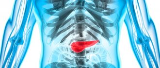
Where is the pancreas located in humans? Why are all diseases of this organ, especially tumors and cancerous processes, diagnosed at a late stage? Why can’t the size of the pancreas be determined during examination? This is because it is located deep in the abdominal cavity, and therefore various lesions of the pancreas are rarely palpated. This explains why most symptoms of cancer of this organ do not appear until the tumor grows large enough to affect the function of the gland itself or other nearby organs, such as the stomach, upper small intestine, and liver.
The pancreas, which measures about 25 in length, is located behind the stomach.
How does she look?
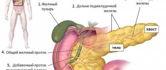
The pancreas consists of a head, body and tail. The dimensions of the pancreas are as follows: length - 18-25 cm, diameter - from 3 cm in the head area and 1.5 cm in the tail area. Where is the pancreas located in a person, how does it relate to other organs in terms of location and functions - a surgeon or gastroenterologist can give you a clear answer to this question. These specialists deal with diseases of this gland, which is important for the body.
The head of the pancreas is located on the right side of the abdominal cavity and is adjacent to the duodenum. A duct from the pancreas connects the organ to this intestine. The narrow end of the pancreas, called the tail, extends toward the left side of the body.
The internal structure of the pancreas is spongy; its shape vaguely resembles a fish, which is located horizontally across the abdomen. The head is the most voluminous part; it lies on the right side of the abdomen, near the place where the stomach passes into the initial part of the small intestine - the duodenum. It is here that chyme, the partially digested food that passes from the stomach into this intestine, mixes with the juice from the pancreas.
The body is located behind the stomach, and the tail deviates posteriorly and comes into contact with the spleen, left kidney and adrenal gland.
There is a pancreatic duct that runs through the pancreas from the tail to the head. It collects ducts from all groups of glandular tissue cells. Its end is connected to the bile duct, which comes from the liver and delivers bile to the duodenum.
Localization of the pancreas in the human body
Compared to other organs, the pancreas is located in the most rational way and is located in the abdominal cavity.
Anatomically, the spine runs behind the gland, in front is the stomach, to the right of it, below and above is the duodenum, and to the left is the spleen. The abdominal aorta, lymph nodes and celiac plexus are located in the posterior part of the body of the pancreas. The tail is located to the right of the spleen, near the left kidney and left adrenal gland. The sebaceous bursa separates the gland from the stomach.
The location of the pancreas relative to the stomach and spine explains the fact that in the acute phase, the pain syndrome can be reduced by the patient sitting and leaning slightly forward. The figure clearly shows that with this position of the body, the load on the pancreas is minimal, since the stomach, displaced under the influence of gravity, no longer affects the gland with its mass.
Internal structure of the pancreas
There are two main types of tissue that are found in the pancreas: exocrine and endocrine. About 95% of the gland's tissue is exocrine tissue, which produces enzymes to aid digestion. Normal food processing is impossible without the pancreas working productively. The norm for juice production is about 1 liter every day.
5% of the pancreas is made up of hundreds of thousands of endocrine cells called islets of Langerhans. These grape-shaped cells produce important hormones that not only regulate pancreatic secretions but also control blood sugar levels.
Pancreas. Sources of development, structure and functions. Pancreas
Produces pancreatic juice. This juice contains enzymes: trypsin, chymotrypsin, amylase (breaks down carbohydrates), lipase (breaks down fats). The exocrine part is 97% of the mass of the pancreas. Endocrine function is associated with the production of the main hormone: insulin, as well as glucagon,
somatostatin, VIP hormone and pancreatic polypeptide.
These hormones are of great importance in the regulation of carbohydrate, fat and protein metabolism in tissues. Lack of insulin leads to diabetes.
Endocrine part – 3% of the mass of the pancreas. Develop
During embryogenesis, the pancreas is formed from the epithelium of the midgut, which grows into the mesenchyme. The secretory section is formed from the epithelium, and the vessels and connective tissue layers are formed from the mesenchyme.
. The exocrine part is already detected at the end of the 3rd week, and the endocrine part
– by the end of 3 months of embryonic development.
STRUCTURE
The pancreas is a complex, branched gland, with pronounced lobulation. The outside is covered with a thin connective tissue capsule, from which septa extend inward, which are less pronounced. Excretory ducts and blood vessels are located in the interlobular connective tissue septa - these are interlobular formations. In the lobules there are exocrine secretory sections, endocrine (in the form of islands) and intralobular excretory ducts (intercalary and common intralobular ducts).
Exocrine part . It is represented by the secretory department - the acinus. This is a formation in the form of a sac consisting of 10-12 cells. The cells are cone-shaped. The nucleus is in the basal part. Here
as well as the synthetic apparatus (granular EPS, mitochondria). Therefore, the basal part is colored basophilic and is homogeneous. Secretion granules accumulate in the apical part; they are more oxyphilic in color. Therefore, the apical part is oxyphilic - zymogenic (zymogen = proenzyme). The isolated zymogen is converted into an active enzyme in the cavity of the duodenum.
The secretion comes from the secretory department into the intercalary duct. They are short and can directly exit the secretory department. They can be located on the side of the secretory department. (They can be inserted into the secretory section. In this case, centro-acinous cells - intercalary duct cells - appear in the center of the secretory section). Intercalary ducts can be material for the formation of new secretory sections. This is especially true in the first years after birth or when the pancreas is damaged.
Larger excretory ducts are lined with prismatic epithelium. The excretory ducts contain thin layers in the lamina propria. The interlobular excretory ducts are larger in the area of the head of the pancreas, smaller in the body area, and may not be detected in the tail area. These excretory ducts are lined with prismatic epithelium. The lamina propria, goblet cells are prominent, and there are bundles of muscle cells that act as a specific sphincter, especially at the site of entry into the duodenum.
Regeneration of the exocrine region in adults is almost not expressed. Due to the small amount of connective tissue, foci of necrosis quickly generalize, and inflammation spreads throughout the organ.
The endocrine part is no less important, because Every 20th person suffers from diabetes. The endocrine part is presented in the form of islets of Langerhans-Sobolev
.
The number of islets is up to 1.5 million, each islet has 20-40 cells .
There are 5 types of cells in endocrine islets. 70-75% - B cells are cells that produce insulin
- the main hormone of these islets. They are basophilic stained and occupy the central part of these islands. The grain size is coarse. Insulin secreted in the islets acts on receptors in liver cells and muscle structures. In liver cells, each cell contains up to 150 thousand insulin receptors. When exposed to these receptors, the permeability of the cytomembrane to glucose changes, and sugar enters the cell, from which glycogen is formed. In this way, insulin lowers blood sugar. Its deficiency leads to increased sugar (diabetes mellitus).
A cells are acidophilic stained. Located in islands along the periphery. There are 20-25% of them. Contains large acidophilic granules. These granules contain the hormone glucagon
. There are receptors for it (up to
200 thousand receptors per cell). Glucagon, acting on the receptor, triggers the mechanisms of intracellular breakdown of glycogen, and glucose is released into the blood. Glucose is an energy material.
D cells produce somatostatin
, there are 5% of them. They block the secretion process: both the exocrine and endocrine parts of the pancreas.
D' cells . They produce a vasointestinal peptide, which lowers blood pressure and dilates blood vessels, which indirectly increases blood circulation and secretion.
PP cells . Produce pancreatic polypeptide. Strengthens the secretion of the glands of the stomach and pancreas.
The blood supply to the pancreas is represented by arteries that branch into the capillary network. The outflow goes through the veins, the lymphatic vessels are well defined. Innervation is carried out by the autonomic and nervous systems.
What does it produce?
What does the pancreas do? Enzymes, or digestive juices, produced by this organ are needed in the small intestine to further break down food after it has left the stomach. The gland also produces hormones such as insulin and glucagon and releases them into the blood in order to regulate glucose, or sugar, levels in the body.
The pancreas is capable of producing the right substances at the right time and in the right quantity in order to properly digest the food we eat.
After food enters the duodenum, the exocrine tissue secretes pancreatic alkaline juice containing a number of enzymes. They break down food into small molecules that can be absorbed in the intestines:
• trypsin and chymotrypsin - for the digestion of proteins;
• amylase, capable of breaking down carbohydrates;
• lipase - to break down fats into fatty acids and cholesterol.
The endocrine tissue of the pancreas, or islets of Langerhans, consists of several cells that secrete hormones directly into the bloodstream. Insulin is a hormone secreted by the beta cells of the gland in response to increased blood sugar levels. The hormone also helps in moving glucose from the blood into muscles and other tissues so they can use it as a source of energy. In addition, insulin helps the liver absorb glucose and store it as glycogen when the body needs energy during stress or exercise.
Glucagon is a hormone secreted by the alpha cells of the gland when there is a decrease in sugar in the bloodstream. Its main task is the breakdown of glycogen into glucose in the liver. This glucose then enters the bloodstream to restore sugar levels to normal.
Anatomy of the human pancreas.
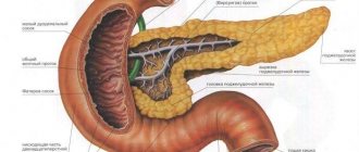
The structure of the human pancreas is represented by a lobed, comma-shaped organ of gray-pink color. It is located behind and slightly to the left of the stomach. If a person is placed on his back, this organ will be under the stomach, based on this, the name “pancreas” appeared. The body, head and tail of the pancreas are distinguished.
The head of the pancreas is the part of the organ that directly adjoins the duodenum. At the border of the body and head there is a recess in which the portal vein runs. The body of the pancreas has the shape of a triangular prism. The front part is directed towards the back wall of the stomach and slightly upward. Posterior - to the spine, it is in contact with the inferior vena cava, abdominal aorta, and celiac plexus. The lower surface is directed downward and slightly forward, located slightly below the mesentery of the colon.
The tail of the gland is pear-shaped and runs towards the hilum of the spleen.
The duct of Wirsung runs throughout the gland, flowing into the duodenum.
Major diseases
There are few diseases of the pancreas: pancreatitis, benign tumors and cancer.
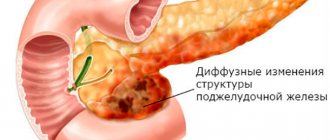
Intense pain in the pancreas is often associated with acute pancreatitis. In any case, it is difficult to identify and assess the condition of this organ if you know where the pancreas is located in a person. Other signs that it is pancreatitis: jaundice, itchy skin and unexplained weight loss, enlarged pancreas on further examination. If you experience pain in the pancreas area, consult your doctor. The very definition of the term “pancreatitis” is inflammation of the organ when enzymes begin to digest the pancreas itself. It can be acute or chronic, but both forms must be diagnosed promptly as it can lead to additional health problems.
Chronic pancreatitis
This disease is a long-term inflammation (more than three weeks) of the pancreas, which leads to permanent damage. One common condition is chronic heavy drinking or drug use. There are other reasons that cause attacks of acute pancreatitis. These may include cystic fibrosis, high levels of calcium or fat in the blood, blockage of the bile duct by stones or tumors, and autoimmune disorders.
Symptoms include upper abdominal pain, nausea, vomiting, weight loss and oily stools. Such stool, or steatorrhea, does not appear until more than 90 percent of the pancreatic tissue is damaged.
Chronic pancreatitis requires a low-fat diet and cessation of alcohol and smoking. If chronic pancreatitis is left untreated, it tends to worsen over time and medications will be needed only for pain relief. Treatment of such pancreatitis is possible only surgically: this is stenting or removal of the head of the pancreas due to the fact that tumors most often arise in it.
There is a connection between pancreatitis, most often chronic, and pancreatic cancer. Recent studies have shown that the incidence of pancreatic cancer increases 2-5 times in patients with chronic pancreatitis when various unfavorable factors are associated.
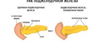
Cancer
It is difficult to diagnose this disease in its early stages. Unfortunately, cancer symptoms can be vague: abdominal pain, jaundice, severe itching, weight loss, nausea, vomiting and other digestive problems. An enlarged pancreas is detected only with ultrasound and MRI.
It is impossible to determine changes in the pancreas due to the fact that this organ is inaccessible for palpation. Even tumors, as a rule, cannot be felt by touch. Due to the difficulty of early diagnosis and the speed at which cancer spreads, the prognosis is often poor.
Risk factors for the development of cancer are: smoking, long-term diabetes and chronic pancreatitis. The cancer process usually begins in the cells that produce digestive juices or in the cells that line the ducts. In rare cases, pancreatic cancer begins in the cells that produce hormones. To diagnose cancer, doctors usually do physical exams, blood tests, CT scans, endoscopy, ultrasound, and biopsies. Treatment options include surgery, radiation and chemotherapy to specifically attack cancer cells without harming normal tissue.