Performing an ultrasound and reading the results
As you know, the ultrasound diagnostic method consists of monitoring the interaction of ultrasonic waves with body tissues.
Increased echogenicity (ability to reflect) indicates a higher density, and vice versa. The results are visualized on the screen, thus making it possible to “examine” internal organs without the slightest harm to human health. Carrying out an examination, the diagnostician evaluates the shape and size of the organ, its density as a whole and the presence of individual dense areas (that is, structure), the condition of the intra- and extrahepatic ducts, the vascular pattern and the walls of the liver veins.
https://www.youtube.com/watch?v=LJWkV7rjyB4
Subsequently, based on the symptomatic picture shown by the ultrasound examination, a specialized specialist determines what pathology such a compaction in the liver may indicate. Based on the results, it is also determined which doctor will treat the patient: a gastroenterologist, an infectious disease surgeon, an oncologist, or (in case of minor deviations), a therapist.
To be able to read the results of an ultrasound scan yourself, you should have an idea of the norm. If the conclusion says that the parenchyma is finely grained and has a normal level of echogenicity, this means (if we talk only about this parameter) that the liver is healthy.
Seals can be diffuse in nature (that is, evenly distributed throughout the entire volume of the liver parenchyma), or localized in one/several specific areas.
For example, a local compaction detected on ultrasound may indicate the presence of a neoplasm, stone or calcification in the liver.
The presence of small nodes of irregular (eg, triangular) shape makes one suspect metastases of malignant tumors.
A rounded seal enclosed in a capsule is most likely a cyst or abscess.
A uniform, “same type” increase in echogenicity is observed, in particular, with fatty hepatosis and hepatitis.
In some cases, the degree of progress of the disease should be taken into account; if at one stage the consistency is determined to be dense, then at another the liver may turn out to be hard (as they say, “stony density”), or, conversely, the echogenicity will decrease.
Signs of the most common diseases.
For cirrhosis
- tissue homogeneity is impaired,
- echogenicity is increased, with a tendency to attenuate in the deep sections,
- the observed nodes, or lesions, cover the entire organ,
- “mosaic” structure (due to regeneration foci),
- corners are widened (rounded),
- surface roughness,
- symptom of hepatomegaly (enlargement) in the initial stage,
- reduction, up to wrinkling, in the final stages,
- blood flow abnormalities (portal hypertension, thickened wall of the portal vein of the liver).
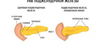
With a tumor
A compaction with a clear localization often indicates a neoplasm. Informative in terms of diagnosis is the determination of the type and structure of the lesion, as well as a number of other signs, for example, changes in the veins or vessels of the liver.
Diagnosis of neoplasms, in particular, determination of their nature (benign or malignant), can only be done by an experienced specialist, and sometimes by a group of specialists.
Benign tumors do not metastasize, but they can degenerate, which means examination should be carried out regularly.
A hypoechoic rim around the circumference of the formation is a sign of metastasis.
Also, metastases of tumors located in other organs can grow in the liver.
When neoplasms are detected, the doctor, as a rule, refers to a CT or MRI; these instrumental methods allow the primary diagnosis to be detailed. You may need to conduct a biochemical blood and urine test. In difficult cases, fine-needle puncture or laparoscopy is performed.
For the diagnosis of viral hepatitis, the results are not a reliable criterion, especially for determining the type (A, B, C...), but they complement the clinical picture.
In the acute phase of hepatitis C, general compaction, diffuse inflammatory foci, disruption and heterogeneity of the structure are observed. The ducts are dilated, hepatomegaly is detected.
In chronic hepatitis, in the progress stage, there is an increase in echogenicity, the size is increased, the tissue has a heterogeneous structure, the contour is unclear.
With severe fatty hepatosis, pathological changes are also diffuse in nature: the scanner displays uniform compaction and increase in size. The edges are rounded, the vein pattern is clearly visible. When pressing with the sensor, virtually no deformation occurs.
You should pay attention to the condition of the pancreas and spleen, and also conduct a blood test. To clarify the diagnosis and prescribe a course of treatment, you must consult a gastroenterologist.
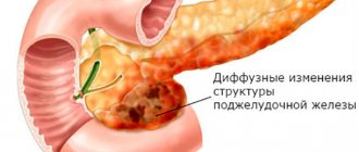
Features of diffuse changes in the liver and pancreas in a child
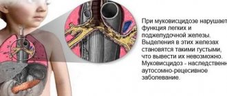
As you know, the pancreas is affected by a fairly large number of external factors - metabolic disorders, genetically inherited characteristics and diseases, intestinal diseases, as well as hormonal changes. An enlarged pancreas during a child's examination should be a signal for parents to conduct a full examination of the child's abdominal cavity - the liver, its ducts, echo - examination of other internal organs. Of course, you also need a certain diet for the child and an examination that will reveal possible focal lesions of the internal organs. Under no circumstances should you panic, but you cannot ignore these changes either - this means exposing your child to enormous risk.
A competent doctor will immediately prescribe blood tests - both serological and biochemical. In addition, the child will need to undergo a computed tomography scan of the abdominal organs and, in some cases, even a puncture, which will allow material to be collected for histology. The correctness of the chosen treatment and diet will depend on the completeness of the examination.
Possible causes of compaction
A thickening of the liver on ultrasound is a change in its normal structure. This phenomenon accompanies inflammatory reactions caused by various processes. The parenchyma can be compacted uniformly or single pathological foci appear in it. When receiving such results, differential diagnosis is carried out between several diseases:
- hepatitis - inflammation of the liver caused by mechanical injuries, viral agents, alcohol or drug intoxication, as well as any other types of poisoning;
- hepatosis (fatty liver) is a pathological condition in which inclusions of adipose tissue are located between normal hepatocytes;
- cirrhosis is the terminal stage of liver disease, which occurs with damage to cells and their replacement by connective tissue scar;
- tumors - can be benign or malignant, grow from different types of tissue, require differentiation from cysts and abscesses.
The primary cause of compaction may be associated with an unhealthy lifestyle, congenital or acquired diseases of other organ systems. The condition of the liver is also affected by metabolic disorders (diabetes mellitus, thyroid dysfunction), and infectious processes in the body. Often such symptoms are accompanied by excess weight and hormonal imbalances.
REFERENCE! An ultrasound of the liver is performed in conjunction with an examination of the abdominal organs. Diagnostics includes analysis of the condition of the stomach and intestines, pancreas, gallbladder and other organs as indicated.
What are diffuse changes and why do they occur?
What is diffuse change in the liver and pancreas? This is a disorder that develops in the structure of tissue. The organs lose their massiveness, appearance, and size. It is unacceptable to attempt to treat the disease without permission, because the symptoms of the disorder are similar to hepatomegania.
This means a simultaneous increase in the volume of the spleen, liver, and various methods of therapy will be required.
The causes of diffuse changes in the liver and pancreas are considered to be:
- altered metabolic process;
- infectious pathologies;
- inflammatory phenomena;
- gallstone disease.
Similar manifestations such as pancreatitis, alcohol addiction, stress also cause changes in the parenchyma of the pancreas and liver. Disorders can be provoked by painful development in the gallbladder, this is chronic cholecystitis.
When the body is affected by negative phenomena, the victim develops steatosis, which involves the presence of fatty inclusions in the tissue.
Causes of seals
Very often, when examining the abdominal organs and gastrointestinal tract, diffuse thickening of the liver is diagnosed. The pathology is common among people aged 45-50 years. Doctors call this slight thickening or enlargement of the liver hepatosis. The disease is also called fatty degeneration.
In severe cases, liver hardening may indicate hepatitis, liver failure, or cirrhosis. A variety of factors can lead to diffuse changes. So, the cause may be alcohol abuse. Large amounts of alcoholic beverages kill liver cells. A person is in constant toxic poisoning. Smoking also has an extremely negative effect on the condition of the entire body.
Liver hardening often develops due to poor nutrition. A large amount of fatty foods, a minimum of vitamins and minerals lead to pathologies of the liver and the entire intestinal tract. Other reasons include:
- Diabetes;
- Hormonal disbalance;
- Dysfunction of the endocrine system;
- acute infectious diseases;
- Viral hepatitis.
It is also worth noting the long-term use of certain medications. Antibiotics and hormonal drugs pose a danger. Synthetic estrogen-based hormones provoke not only compaction, but also polycystic liver disease. Therefore, these medications are prescribed in courses with breaks.
Characteristic symptoms of disorders
There is a whole range of signs that suggest the presence of a pathology leading to tissue degeneration.
The development of disorders is accompanied by the appearance of pain and discomfort in the right side. The occurrence of discomfort is accompanied by increased physical exertion on the patient. In addition, this condition can be the result of eating large amounts of food rich in fat.
Painful sensations become constant, their intensity can increase under the influence of irritating factors.
Signs of degenerative processes can manifest themselves in any age group; sometimes such manifestations are detected in pediatric patients.
The main symptoms of the development of the pathological condition are:
- Feeling nauseous for no reason.
- Decreased appetite.
- The occurrence of belching with bitterness after eating.
- Frequent heartburn.
- Periodic occurrence of vomiting.
- Significant weakening.
- Rapid fatigue when exerting physical activity on a person.
- The appearance of frequent headaches and migraines.
In the male population, pathological changes can provoke problems with sexual function. When such a disorder develops, women experience hormonal imbalance. In the presence of diabetes, hormonal imbalance leads to exacerbation of the disease, which significantly affects dystrophic changes in the liver and pancreas parenchyma.
In patients suffering from this disorder, a change in the color of the skin and whites of the eyes is detected. Sometimes, with prolonged development of the disease, darkening of urine and discoloration of feces are detected.
Forms of pathological changes in the parenchyma
There are several types of the disease. All types of lesions can be divided into three groups.
The first group is minor lesions. This form of pathology is common among the middle-aged population. Most often, such forms of disturbance occur at different stages of hepatitis; unfavorable factors provoke such disruptions.
The second group – lesions of the parenchyma of moderate severity, occur due to poisoning by frequent consumption of alcoholic beverages and consumption of unhealthy foods. Taking vitamin complexes, as well as switching to eating natural foods and avoiding processed foods, helps correct the situation.
The third group - severe lesions, are observed in patients suffering from diabetes. Quite often they occur in the presence of obesity and tumors in humans.
There are several types of parenchyma changes. Doctors distinguish the following types:
- hypertrophic;
- sclerotic;
- swelling;
- dystrophic.
Each type has its own characteristic reasons for the formation of changes.
New information: Skin rashes due to diabetes
Diffuse forms of pancreatic pathology manifest themselves:
- Moderate increase in parenchyma density. This pathology is caused by the occurrence of acute pancreatitis in the patient. This form is accompanied by tissue lysis produced by digestive enzymes. When performing an ultrasound, an increase in size is observed, the pathology is accompanied by the appearance of swelling of the walls.
- By reducing the thickness of the functional tissue, the disorder develops when the patient has chronic pancreatitis. The causes of such phenomena are considered to be disturbances in the functioning of the gastrointestinal tract.
In addition, lipomatosis may occur. This form of pathology is caused by the appearance of a fatty layer.
Regardless of the form and type of illness, when the first suspicion arises, you must contact your doctor. Only a full medical examination can clarify the cause of the pathological processes leading to tissue deformation.
Liver ultrasound data
Ultrasound data can suggest the causes of liver hardening. The basic operating principle of the device is the effect of ultrasonic waves on body tissues. They all have different densities and structures, so they have different abilities to repel these waves. This feature is visualized on the monitor in the form of lighter or darker areas. Thus, human bones are the densest, so they look completely white.
Compaction in the liver is pathological areas of lightening of the parenchyma. They may have their own characteristics:
- diffuse induration or localized focus;
- formation with smooth edges or has an irregular shape;
- density may vary (liver stones will be the lightest on the monitor);
- presence or absence of a capsule.
REFERENCE! Ultrasound is not the most accurate method for diagnosing liver disease, since most of it is hidden behind the costal arch. If the presence of neoplasms or other pathologies is suspected, an MRI or CT scan is prescribed. Such diagnostic methods allow you to obtain a detailed three-dimensional image of the organ in several projections.
Prevention
Instead of wasting time and money on treating the cause of liver lumps, it is better to take care of preventing their occurrence. Preventive measures are:
- normalization of body weight;
- exclusion of alcoholic beverages from the diet;
- correction of the diet: limiting the consumption of fatty, fried, salty, smoked foods;
- maintaining an active lifestyle;
- improving the immune system by taking vitamins and hardening the body;
- timely detection and treatment of pathologies of viral and infectious origin, including chronic ones;
- normalization of metabolic processes (protein, fat).
If it was not possible to avoid the development of a disease of the gland, which occurs with compaction of the parenchyma, it is recommended to follow all the prescriptions of the attending physician. This is the only way to achieve a speedy recovery and prevent negative consequences.
To avoid recurrence of liver compactions, it is necessary to follow preventive measures. If a person notices even slight discomfort from the digestive system, it is necessary to consult a gastroenterologist. You should definitely monitor the quality of food. After all, liver health depends on what we consume.
A healthy lifestyle is a must even after the liver has completely recovered. A person needs to give up bad habits such as alcohol and smoking. Daily activity is also important. A sedentary lifestyle contributes to the development of bile stagnation in the bile ducts. Therefore, regular exercise and moderate physical activity will protect the liver and strengthen overall immunity.
If diffuse liver compactions have been previously noticed, you should avoid contact with chemicals, toxins, and household chemicals. Inhaling the vapors of these substances is strictly prohibited. We get a lot of chemicals through food. In this regard, it is worth carefully studying the composition of food, thoroughly washing vegetables and fruits before eating. Regular examination by a doctor will allow a person to monitor the condition of his body and avoid possible complications.
If the liver is compacted, what this means and what are the causes of this condition can be determined using ultrasound. In most cases, this indicates a serious illness. Diffuse changes in the gland, if it is healthy, should not be present. Otherwise, a comprehensive diagnosis is required, the results of which determine the source of the compaction.
Useful video about the pancreas
Please, help! I did a blood test and revealed an increased level of ASAT 57, Alat 43. Ultrasound showed diffuse changes in the liver parenchyma, deformation of the gallbladder, the lower edge is sharp (hepatic veins are not dilated, a slight tendency to rounding), pronounced diffuse changes in the pancreas (chronic pancreatitis cannot be excluded) The attending doctor prescribed, no joke, corn silk (!!), and said to come back for an examination in a year. By the way, this time I didn’t test for AntiHVC, but in general the result was positive before, hepatitis C. Where to go? How to proceed? Or is the doctor right and I’m just panicking??
Diffuse changes in the liver, or more precisely, in its parenchyma (the main component of its tissue) occur quite often, even in young children. However, they are not the norm and, as a rule, indicate dysfunction of this organ.
Diffuse changes in the liver parenchyma are not even an ultrasound diagnosis, but rather a symptom; during an ultrasound examination, liver tissue unevenly reflects the ultrasound signal due to the presence of multiple areas of compaction in them, scattered literally everywhere. In most cases, diffuse changes in the liver and pancreas are detected simultaneously, because the function of both organs is closely related.
What does diffuse liver changes mean?
The liver is formed by specialized cells - hepatocytes. They detoxify poisons entering the body, they secrete bile containing enzymes for the breakdown of fats in the digestive tract; numerous reactions occur in the liver cells, converting substances that the body does not need into those that are safe for health and removing them with the help of bile. So, here hemoglobin is broken down from worn-out blood cells, iron is returned to the blood, and pigments are formed from the protein part of hemoglobin molecules, which then color the stool brown.
Liver cells are not always able to cope with the poison entering the body. Sometimes, despite all their efforts, they end up poisoned by it and die, and then in their place appears something that always replaces damaged cells in the human body - connective (fibrous) tissue. It cannot perform the functions of liver cells, and has a different echogenicity compared to them. Ultrasound signs of diffuse changes in the liver are due to the presence of these transformed tissues. Naturally, the more liver cells die, the more whitish “dust” and mesh on the ultrasound the doctor sees against the background of the ultrasound image of this organ.
Diffuse changes in the echostructure of the liver can also be caused by hepatitis viruses. If hepatitis A most often passes without a trace, hepatitis B, and especially hepatitis C, can become chronic and cause permanent death of liver cells.
Moderate diffuse changes in the liver gradually increase when the problem is ignored, ongoing intoxication (for example, alcohol), and the absence of treatment for existing hepatitis. More and more liver cells die, connective tissue grows more and more, entire layers of fibrous tissue are formed, some liver cells simply turn into fat cells, and are unable to process the poison, accumulating it in themselves. Fat deposits in the liver are called fatty hepatosis, and uneven diffuse focal changes in the liver parenchyma are called cirrhosis.
In such advanced cases, the edge of the liver is rounded, it increases in size, and hepatomegaly develops. Diffuse changes in the liver of this nature, fatty hepatosis, are almost impossible to cure.
Diffuse changes in the structure of the liver are accompanied by impaired blood flow in it, and portal hypertension develops. It causes bleeding from dilated veins of the esophagus, hemorrhoids, ascites, and all manifestations of cirrhosis of this organ. It often ends in the death of the patient from these complications. And of course, such a liver is not able to cope with its work. This means that now the toxins will remain and poison the body, resulting in constant intoxication, weakness and poor health.
Diffuse focal changes in the liver, dystrophic, heterogeneous changes in its structure are often accompanied by yellowing of the skin and mucous membranes. Impaired excretion of hemoglobin breakdown products manifests itself as jaundice, itchy skin and abnormal, light-colored stools. Digestion is also disrupted - a feeling of heaviness after eating, upset stool, nausea become the usual companions of a sick person.
Minor diffuse changes in the liver parenchyma in a child are often associated with biliary dyskinesia, pancreatopathy due to poor nutrition. The consumption of sweet soda, chocolate, lack of a routine, and “adult” dishes on the children’s table lead to the child’s liver being unable to cope with digestion. This threatens in the future, already in adulthood, with diseases of the liver and gall bladder, and often then chronic cholecystitis develops. But diffuse changes in the liver in a newborn are usually associated with an infection in utero or intoxication of the fetus.
We have figured out what diffuse changes in the liver mean, now let’s summarize when this happens.
Signs of diffuse changes in the liver parenchyma - causes
Echo signs of diffuse changes in the liver can be detected in the following diseases:
1. Chronic hepatitis (viral hepatitis B, C, D, toxic alcoholic, drug-induced hepatitis, etc.) 2. Chronic pancreatitis 3. Chronic cholecystitis, both calculous and non-calculous 4. Anomalies in the structure of the gallbladder
In addition, on ultrasound, diffuse changes in the liver are often detected due to poor nutrition (abuse of fried, fatty foods, alcohol consumption, when working with potentially hazardous substances, when taking medications for a long time, including oral contraceptives. Severe diffuse changes in the liver most often indicate about the presence of a real disease, and moderate diffuse changes in the liver parenchyma are more characteristic of the effects of malnutrition and external intoxications.
Diffuse changes in the liver and pancreas
Sonographic signs of diffuse changes in the liver are very often combined with pathology of the pancreas. This is due to the fact that the excretory ducts of the pancreas and liver are connected, and both organs take an active part in the digestion of food.
Hepatomegaly and changes in the edge of the liver
Signs of hepatomegaly and diffuse changes in the liver indicate that either the process of fibrosis (death of hepatocytes and their replacement with connective tissue) has gone very far, or that there is swelling of the liver due to its inflammation. Be that as it may, in any case it is very serious.
Diffuse liver changes: treatment
If your ultrasound reveals changes in the liver tissue, further examination will follow to clarify the reasons, and after the examination, appropriate treatment will be prescribed.
In any case, you will need a diet. You'll have to forget about alcohol, fried and fatty foods. Diet No. 5 according to Pevzner is usually prescribed.
In case of liver pathology, many medications are contraindicated; intoxication should not be allowed, as this will worsen your condition and can lead to cirrhosis. The development of cirrhosis threatens many complications, some of which often cause the death of the patient.
Comments
The normal structure of the liver is homogeneous and weakly echogenic, through which bile ducts and large vessels can be seen, the echo signs of which are clearly visible on ultrasound. If the structure of the liver tissue is changed, this means that we are talking about diffuse changes in the liver. Such organ damage can indicate both very minor changes in the body and quite serious diseases, focal lesions that threaten human life.
Additional examinations can help determine the level of diffuse changes that have become noticeable on ultrasound - blood biochemistry, liver biopsy, clinical studies of the pancreas, as well as diagnostics of other organs located in the neighborhood.
Diffuse changes in the liver parenchyma is a rather superficial term that hides a dozen diseases. But let's still figure out what this definition means. The main types of diffuse changes that the liver structure undergoes can be diagnosed by ultrasound. These include hepatitis, lipid infiltration, chronic hepatitis. The severity of the inflammatory process directly correlates with the size of liver edema. In this case, echogenicity is reduced, and the level of sound conductivity increases. A special place is occupied by focal liver lesions - tumors and cysts.
If we classify diffuse changes in the liver parenchyma, its ducts and vessel walls depending on the nature of the changes, then we can talk about fibrous, sclerotic, dystrophic, edematous, hypertrophic liver damage.
Diffuse changes in the liver parenchyma can also be divided into mild (moderate) and severe:
- The first include diffuse liver changes such as fatty hepatosis, stagnant cirrhosis, and metastases.
- Pronounced diffuse changes in the liver imply that the patient has sarcoidosis, liver cirrhosis (not congestive), an abscess, or toxic fatty hepatosis.
Understanding Normal Liver Values
A healthy liver on ultrasound has a homogeneous structure. It is fine-grained, echogenicity is within normal limits. During the analysis, the necessary measurements can also be taken. There are certain standards for what the size of the liver should be in an adult:
- height of the right lobe - up to 12.5 cm, left - up to 10 cm;
- liver length - 14-19 cm;
- the liver protrudes beyond the edge of the costal margin by a maximum of 1 or 2 cm.
A lobule is a structural unit of the liver. The entire parenchyma of the organ consists of such formations. they connect and form the lobes of the liver - this structure can already be distinguished on ultrasound. The specialist also measures the diameter of large vessels and bile ducts. It should be understood that the norms for an adult and for a child will be different, but compaction is not an indicator of the norm at any age.
So what is it - lumps in the liver? What does such a change in structure mean and why does it occur?
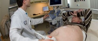
Liver ultrasound procedure
Ultrasound is performed using ultrasound. These waves pass through the tissue, creating visualization on the machine's monitor. Depending on how dense the liver is, the intensity of the shadows is displayed in the image. This procedure allows you to track changes in structural abnormalities and blood movement.
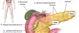
The bile ducts are a collection of tubular channels that are involved in the movement of bile produced by the lobes of the liver. The secretion enters the duodenum through pressure created by the gland. The structure of the biliary system is simple, but multicomponent. It begins with small canals in the liver, which gradually connect into the common hepatic duct. After connecting with the bladder, both of them flow into the gallbladder, which has valves for the portioned supply of bile.
The standard acoustic size of an organ is determined by individual lobar and segmental parameters in total. Each section of the gland has its own specific indicators, which together make it possible to identify and trace any pathological processes.
The liver is the largest gland in the digestive tract. Therefore, a doctor cannot examine each of its segments at the same time. Diagnostics is carried out in stages for the lobar parts (quadrate, tail, right and left).
| Structural part | Size |
| right lobe – thickness | 11.2-12.6 cm |
| right lobe – vertical oblique dimension | up to 15 cm |
| right lobe – length | 11-15 cm |
| left lobe – thickness | up to 7 cm |
| left lobe – height | up to 10 cm |
| gland - length | 14-18 cm |
| iron - width | 20.1-22.5 cm |
| gland – sagittal size | 9-12 cm |
| Structural part | Size |
| common duct | 6-8 mm |
| portal vein | up to 13 mm |
| vena cava – diameter | up to 15 mm |
| distance from the orifices to the hepatic veins | 6-10 mm |
| hepatic artery - located at the hilum of the gland | 4-7 mm |
The diagnosis is determined taking into account the comparison of the obtained numbers with acceptable indicators. The structural appearance of the parenchyma, contour, and echogenicity of the tissue are taken into account.
The standard acoustic size of an organ is determined by individual lobar and segmental parameters in total. Each section of the gland has its own specific indicators, which together make it possible to identify and trace any pathological processes.
| Structural part | Size |
| common duct | 6-8 mm |
| portal vein | up to 13 mm |
| vena cava – diameter | up to 15 mm |
| distance from the orifices to the hepatic veins | 6-10 mm |
| hepatic artery – located at the hilum of the gland | 4-7 mm |
Diagnosis and treatment of liver tissue heterogeneity
After detecting heterogeneity in the structure of the liver and pancreas, the patient needs to undergo additional studies to make an accurate diagnosis.
He may be prescribed a complete blood count, an MRI of the abdominal organs, a CTM, a blood test for tumor markers, an angiographic study, or a liver puncture.
Only a doctor can decide how to treat diffuse changes. Moreover, treatment in all cases involves eliminating the causes that caused this pathological condition. If inhomogeneities arose due to the patient’s lifestyle, then he needs to adjust his diet and give up bad habits. In addition, to restore liver tissue, the doctor will prescribe hypoprotectors.
For hepatitis and other infectious diseases, treatment involves taking antiviral drugs. Also, in all cases, maintenance therapy is prescribed, which includes taking immunomodulators, vitamins and liver preparations. Diet No. 5 complements the treatment.
In its normal form, liver tissue is a weakly echogenic homogeneous structure. When examined in the liver tissue, you can see blood vessels with bile ducts, which have increased echogenicity. Signs of diffuse changes in the liver parenchyma indicate that the liver tissue has been completely changed. Such signs are typical for both minor changes and very severe lesions. Therefore, it is necessary to conduct a laboratory examination to determine how affected the diseased organ is.
Ultrasound of the liver
The more pronounced the inflammation process, the greater the swelling of the parenchyma. Echo signs of diffuse changes in the liver are reduced, and sound conductivity increases. Ultrasound verdict “Diffuse changes in the liver” can occur in the following diseases:
— obesity, diabetes mellitus and chronic alcoholism
. In this case, fatty degeneration of the liver occurs, it becomes larger in size, the echogenicity of its structure is increased when compared with the norm;
— chronic hepatitis
. In this case, the liver is greatly enlarged, but the structure of the parenchyma may be homogeneous;
— cirrhosis of the liver
. The structure of the parenchyma in this disease becomes diffusely heterogeneous and there are many areas of increased, decreased or average echogenicity;
— cysts and tumors
. The density of liver tissue always has local changes.
- headache;
- nausea;
- general weakness;
- there is a bitter taste in the mouth;
- fatigue and irritability;
- constant change of mood.
Treatment
1. Examination and identification of the primary disease
. Such a concept as “diffuse changes in the liver parenchyma” is not a disease in itself and this particular condition cannot be treated. After any illness, moderate diffuse changes in the liver parenchyma occur; treatment of this condition consists of identifying the reason why all this happened. And this is the underlying disease that needs to be treated. First of all, the doctor orders blood tests: serological, biochemical, tumor markers. Next, depending on the preliminary diagnosis, the necessary instrumental examination methods are prescribed: MRI and CT, liver puncture to obtain a histological sample, that is, a piece of liver tissue, radioisotope scanning.
2. Right way of life
. If the disease is caused by bad habits, then you need to quit smoking and indulge in alcohol. If this is due to poor nutrition, then the necessary diet is prescribed. Proper nutrition is considered very important in liver treatment and dietary table No. 5 is prescribed.
3. Medications
. If liver changes occur due to viral infections, then the necessary antiviral drugs are prescribed. Hypoprotectors may be prescribed; they promote the regeneration of liver cells.
4. Traditional methods
. In the restoration of the affected organ, folk recipes also play an important role. Pumpkin with honey, plum juice, chicory decoction, mixtures of various herbs and much more are used for treatment.
When undergoing an ultrasound examination of the abdominal organs, patients often hear the conclusion “diffuse changes in the pancreas,” which determines the morphological state of the organ. The same term can also be heard in relation to the liver, which has a parenchymal structure. What is it and what can it lead to?
When examining with ultrasound, doctors evaluate the density of the organ tissue, its size and shape, and the presence of possible formations. The homogeneity of the liver or pancreas parenchyma indicates the absence of pathological processes.
If there is a violation of the normal dimensions of the organ or the echogenicity is uniformly reduced (increased), then they speak of the development of diffuse changes.
Defects of this nature indicate the presence of diseases associated with the gastrointestinal tract or aging of the body.
Hepatitis, fatty degeneration
Hepatitis and hepatosis of the liver have similar echographic signs. On examination, the liver has a homogeneous structure, the vessels and bile ducts are clearly visible. When pressing on an organ using a sensor, the patient experiences an exacerbation of pain, but no significant deformation is observed.
Examination of the liver is complicated by the peculiarities of its location - most of it is hidden behind the ribs
There are several factors that may indicate inflammation or fatty infiltration:
- the liver is compacted and enlarged in size, its capsule is tense;
- with chronic hepatitis, uniform inflammation of the parenchyma and its compaction occurs;
- in the acute stage of inflammation, pathological foci with a higher density may appear;
- with hepatosis, the organ becomes uniformly enlarged and thickened, its edges smooth out.
To clarify the diagnosis, a study of the pancreas, stomach and intestines is prescribed. You should also focus on blood test results. If hepatitis is suspected, additional serological tests are performed to exclude a viral origin.
Reasons for the development of the pathological process
The liver can be enlarged for various reasons, these include:
- development of viral hepatitis;
- regular alcohol intoxication;
- introduction of metastases to other organs;
- diseases of the cardiovascular system;
- stagnation of blood in the veins.
- long-term use of medications;
- self-medication.
In addition, liver enlargement can be a consequence of chronic conditions; as a rule, heredity contributes to this.
Changes in the structure of pancreatic tissue most often occur due to the development of such reasons as:
- severe swelling;
- progressive pancreatitis;
- replacement of organ tissue with adipose tissue;
- tissue proliferation or scarring as a result of the developed inflammatory process.
Clinical signs
Compaction of the liver parenchyma is often observed in combination with other clinical manifestations. These are often pronounced, suggesting a developing disease. The first symptom is pain, which occurs against the background of increased temperature, yellowness of the skin and mucous membranes, discoloration of feces and darkening of urine.
If a chronic pathology occurs in the organ, general intoxication symptoms are more pronounced: malaise, increased fatigue even with minor physical activity, disruption of work and rest, and changes in mood. In this case, liver compaction is a characteristic sign.
Significant clinical manifestations are yellowing of the skin and mucous membranes, splenomegaly (increase in the size of the spleen), and itching syndrome. The first symptom occurs against the background of the development of hepatitis in the acute stage, cirrhosis of the gland. The chronic form of hepatitis is not characterized by this symptom.
Nonspecific symptoms of liver damage include:
- the appearance of spider veins in the upper body (on the arms, abdomen);
- redness of the skin on the palms;
- absence of papillae on the tongue, its atypical smoothness;
- moderate tremor of the hands and tongue in a protruding state.
On ultrasound, a healthy liver should have a uniform, fine-grained structure, without compaction or deformation. The area of the bile ducts and vessels is characterized by hyperechogenicity.
With diffuse compaction, the patient may exhibit clinical signs of pathology and organ dysfunction.
Candidate of Medical Sciences, doctor V. M. Savkin:
Cleansing the liver will help rejuvenate the body in a few days and give you an additional 15 years of life...
With liver pathologies, the following symptoms appear:
- nausea and vomiting;
- disruption of the gastrointestinal tract;
- skin rashes;
- pain in the right hypochondrium;
- strong weight loss in a short time;
- yellowness of the skin and eye sclera;
- general malaise;
- internal bleeding;
- organ hypertrophy;
- change in the color of urine and feces;
- dropsy (accumulation of a large amount of fluid in the abdominal cavity).
Secondary diffuse compaction
The liver is an organ that is closely connected with the entire body. Therefore, diseases of other systems cause changes in its tissues. With heart pathology, the outflow through the hepatic veins slows down, and congestion develops. It leads to the development of fibrosclerotic processes, which manifest themselves as follows:
- against the background of a normal shape and clear edges, the size of the liver increases;
- the capsule is not clearly differentiated;
- the lower edge of the liver is rounded;
- the structure of the parenchyma is heterogeneous;
- the inferior vena cava and hepatic veins are dilated;
- reduced ability to conduct sound;
- increased echogenicity;
- Additionally, fluid is determined in the abdominal (ascites) and pleural (pleurisy) cavities.
In some cases, ultrasound identifies a “honeycomb” pattern - the liver parenchyma is pierced by strands of connective tissue intertwined like a network. Echocardiography is likely to show dilation of the heart chambers.
Cirrhosis of the liver
Changes in the structure of the liver during cirrhosis are determined by its stage. So, at the first stage, the organ will increase in size due to inflammatory processes and the presence of regeneration foci. A sign of the terminal stage of the disease and complete impairment of liver function is its reduction and wrinkling. At the same time, a significant enlargement of the spleen can be observed.
Characteristic signs of liver cirrhosis on ultrasound:
- unevenness of the structure, the presence of both denser foci (areas of proliferation of fibrous tissue) and sections where the density is reduced (remnants of normal functional tissue);
- smoothing edges;
- the appearance of tuberosity on the capsule;
- disturbance of the blood supply to the organ: thickening of the wall of the portal vein, increased blood supply to the vessels of the portal tract.
REFERENCE! In case of liver cirrhosis, additional tests are performed to determine its cause. This pathology develops gradually, going through several stages. Often it is a consequence of untimely treatment of hepatitis.
Symptoms of diffusion in the liver, pancreas
Moderate diffuse changes in parenchymal organs may not be clinically manifested. The following signs of liver damage include:
- pain in the right hypochondrium;
- hepatomegaly - enlargement of the liver, which can be detected by palpation;
- poor appetite;
- bitterness in the mouth;
- general weakness, decreased performance, emotional instability;
- headache;
- nausea;
- skin itching;
- rash on the body;
- fluctuations in blood pressure;
- increase in abdominal size;
- flatulence;
- darkening of urine, discoloration of stools;
- jaundice;
- tendency to edema;
- in women - menstrual irregularities, in men - decreased potency;
- slight increase in temperature;
- weakened immunity, which is manifested by frequent colds.
Symptoms of acute hepatosis develop rapidly. Characterized by general intoxication, dyspepsia, yellowing of the skin. Liver failure develops. Without immediate hospitalization, hepatosis can be fatal.
With chronic hepatosis, fat accumulates in the liver structure, hepatocytes die, and fibrous tissue grows in the organ. In the future, cirrhosis with characteristic symptoms may develop.
Pancreatic diffusion is manifested by symptoms:
- decreased appetite;
- bowel dysfunction (constipation, diarrhea);
- heaviness, pain in the stomach;
- nausea, vomiting;
- pale skin.
Depending on the diseases of the organs that provoke structural changes in the pancreas, the following is observed:
- burping;
- heartburn;
- heaviness in the left hypochondrium;
- dry mouth.
Diffuse damage to the pancreas is often recorded with pancreatitis.
The acute form of the disease requires immediate treatment. It is accompanied by severe pain radiating to the left scapula, nausea, vomiting, increased body temperature, and moderate jaundice of the sclera. Dyspeptic symptoms occur (diarrhea, bloating, heartburn), hemorrhages near the navel, and bluish spots on the body.
Reasons for changing the structure
A hardening of the liver may be a sign of organ damage. Pathology can be located both inside the organ and outside its borders.
The most common reasons include:
- alcoholism;
- long-term therapy with antibiotics and other strong medications;
- poor nutrition;
- cirrhosis;
- neoplasms;
- sclerosing type cholangitis;
- hepatitis of various etiologies;
- dystrophy that developed against the background of fatty infiltration;
- dysfunction of the digestive system, which developed against the background of problems with the pancreas;
- chronic form of infectious lesions of other body systems;
- congenital and acquired metabolic abnormalities;
- extrahepatic diseases (diabetes mellitus, etc.).
Lumps clearly visible on an ultrasound image may indicate oncological formations. Diffuse changes in the structure can also be caused by prolonged abuse of fried and fatty foods, as well as working in harmful conditions.
With cirrhosis, different areas of the liver exhibit different echogenicity. Also, this disease is characterized by a heterogeneous structure, changes in the normal parameters of the organ and portal vein.
Hepatitis
With hepatitis, there is an increase in echogenicity, an increase in liver volume, growth of fibrous tissue and compaction of the periportal tracts. This pathology affects the entire organ, so the ultrasound image reveals diffuse tissue damage.
During the acute stage the following are observed:
- seals;
- increase in organ volume;
- the vascular pattern is enhanced;
- heterogeneous structure;
- dilated bile ducts;
- foci of inflammation.
A natural remedy has been found that causes aversion to alcohol!
Elena Malysheva: “Surely you know that until recently the only effective remedy for combating alcohol addiction was...”
Neoplasms
A common cause of dense parenchyma on an ultrasound image is benign or malignant neoplasms.
On ultrasound, a benign tumor appears as a dense area with clear edges. Sometimes lesions with blood or fluid are also detected.
Malignant neoplasms in the liver can be metastases that appear as a result of oncology of other internal organs. On ultrasound they appear as numerous areas of compaction. When they are identified, additional examinations and tests are prescribed to help establish an accurate diagnosis.
Round capsules with liquid contents detected on ultrasound may indicate the presence of a cyst, abscess or parasitic liver disease. To clarify, additional diagnostic measures are carried out.
Diffuse compaction of the liver parenchyma is diagnosed using ultrasound. Before such an event, a physical examination is carried out using palpation. The doctor assesses the size of the organ by feeling the abdomen and its density.
Additional studies include:
- Blood analysis. Bilirubin and liver enzymes are determined for quantitative indicators. Toxic components and antibodies to viral and bacterial infections are detected.
- Computer, magnetic resonance imaging. Such procedures, like ultrasound, allow you to examine the condition of the organ in detail and find pathological compactions and neoplasms.
- Gland biopsy. Bio-material taken from the organ through puncture is subjected to histological diagnosis.
Causes of changes in the pancreas
A change in the structure of the pancreas may have natural causes - the organ changes echogenicity as the body ages. Disturbances in the structure of the gland can also indicate a variety of phenomena:
- circulatory disorders;
- infectious diseases;
- metabolic problems;
- intoxication;
- chronic diseases.
Echogenicity
allows you to evaluate the density of the organ - normally it is homogeneous. Thus, an increase in the indicator is characteristic of an inflammatory process with edema. Pancreatitis in any form causes an increase in echogenicity, so ultrasound alone can suggest a diagnosis. Below are the main problems of the pancreas, which are expressed by diffuse changes in the parenchyma.
| Disease | Echo signs by ultrasound |
| Diabetes | Lipomatosis (replacement of normal tissue with fat cells), diffuse compaction, reduction in the size of the gland, sclerosis, dystrophy. |
| Chronic pancreatitis | Dilation of the Wirsung duct, tuberosity of the contours of the organ, decreased echogenicity, increased size of the gland. |
| Acute pancreatitis | Increased size, increased echogenicity, unclear contours, the presence of cysts, and sometimes an abscess. |
| Cancer | Identification of a cavity or lesion with heterogeneous density. |
Small changes in the gland can appear due to poor nutrition, alcohol abuse, obesity, smoking, after infections and poisoning, and with cardiovascular diseases.
Neoplasms
Signs of liver disease in women
Diagnosis of liver tumors using ultrasound is an insufficiently informative method. However, there are characteristic signs that indicate the need for further examinations (MRI, CT). Unlike other diseases, neoplasms are visualized as limited areas of compaction. They may differ in shape and size, depending on their good quality. Ultrasound also determines the degree of damage to neighboring tissues - benign tumors do not grow through large vessels.
Neoplasms can be suspected based on several echographic signs:
- a compaction with clear edges of regular shape is a benign tumor (it is then differentiated from a cyst and abscess);
- the presence of a rim around the tumor, in which the density is reduced, is a symptom that the tumor is a metastasis and not the primary focus;
- possible deformations of blood vessels;
- small foci of increased density may be metastases from distant organs.
Malignant tumors do not have a clear structure. The monitor visualizes an area with impaired echogenicity: there may be hyperechoic and hypoechoic foci. Liver cancer does not have clear outlines, so it is difficult to determine the shape of the tumor.
A healthy lifestyle is one of the best ways to prevent liver disease
Condition correction methods
Treatment of diffuse changes, including increased density of the gland, is prescribed taking into account the type of disease that caused the symptom. In any case, a special diet aimed at decompressing the liver is required.
Nutrition therapy for a child and an adult involves eliminating fried, smoked, fatty, and too salty foods from the diet, which burden the organ and interfere with its normal functioning. It is recommended to limit coffee, chocolate and pastries, sweets. Prohibited foods also include radishes, radishes, legumes, sorrel, sausage, butter and margarine, white bread, sauces, and alcohol.
Allowed food for the period of treatment of diffuse changes:
- crackers;
- vegetable broth;
- dietary meat;
- viscous porridge.
With the help of medications, you can get rid of the inflammatory process and accelerate the regeneration of organ cells. For this purpose, hepatoprotectors are used, for example, “Gepabene” based on plant components. Active substances enhance the production of proteins and phospholipids and prevent the appearance of fibrosis.
You can cleanse the body with the help of enterosorbent drugs. These activate the activity of the organ and contribute to the removal of toxic compounds from this environment. Choleretic medicines such as Milk Thistle, Heptral, and Allohol are used.
Diffuse changes in parenchymal tissues
Signs of diffuse changes in the liver parenchyma practically do not appear; pronounced symptoms are observed in patients when the disease is at an advanced stage and is actively progressing.
The liver is a large gland in the human body. The organ includes many liver cells - hepatocytes. Anatomically it consists of 2 lobes, which are separated by channels that remove bile secretions and blood vessels.
Causes
DIP is not an independent (separate) disease, but the result of abnormal transformations in the organ associated with various pathologies.
The etiology of development is determined by factors, diseases and conditions:
- Consumption of alcoholic beverages frequently and in large quantities.
- Nicotine addiction.
- Poor nutrition (consumption of fatty foods, fasting).
- Genetic pathologies.
- Long-term use of powerful medications (antibiotics and medications with hepatotoxic effects).
- Autoimmune disorders, viral forms of hepatitis.
- Cirrhotic processes.
Diffuse changes in the liver parenchyma appear in people of any age group. Against the background of DIP, parenchymal tissues modify their structure. If they are detected on ultrasound, a number of additional studies are carried out to accurately establish the cause and disease.
Clinical manifestations
In most clinical pictures, mixed transformations do not manifest themselves in any way; negative symptoms are observed somewhat less frequently.
Clinical manifestations:
- Discomfort and heaviness in the area of the liver projection, pain is rare.
- Heaviness and discomfort in the right shoulder.
- Yellowness of the skin, mucous membranes (not very pronounced).
Ultrasound is performed for diagnosis. Diffuse transformations are detected not only in primary liver diseases, but also due to systemic failures - amyloidosis. This is a systemic disease, as a result of which a specific glycoprotein is deposited in soft tissues and organs, which leads to impaired functionality.
ethnoscience
https://www.youtube.com/watch?v=4-JDeRWFWrM
You can supplement the basic treatment prescribed by your doctor with folk remedies. They actively use herbal infusions, which are available in any pharmacy kiosk. Some recipes:
- Oats (500 g), birch buds (50 g), lingonberry leaves (40 g). The ingredients are mixed, poured with boiling water in a volume of 3 liters, and left to infuse for 24 hours. After this time, the infusion is filtered and taken orally, 100 ml three times a day.
- Rosehip (4 tbsp). The fruits are poured with hot water (500 ml) and left to infuse for 12 hours. The finished product is taken orally, half a glass four times a day.
- Turmeric (0.5 tsp), honey (1 tbsp). Mix the ingredients until a homogeneous mass is obtained. Consume at a time. Number of receptions per day – 3.
- Garlic (2 shares). Cut into thin slices, add warm water and leave to infuse for 12 hours. The resulting remedy is drunk on an empty stomach.
- Rosehip for the liver and gallbladder
- Liver cleansing preparations: inexpensive and effective, list of the best medicines for removing waste and toxins, for the gallbladder, review of reviews
- Symptoms and treatment of gallbladder and liver disease
- Diet for liver disease: nutrition during treatment, what not to eat, recipes for recovery