Statistics
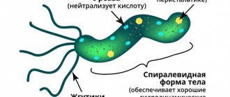
The frequency of diverticula of the thoracic esophagus is 20 times higher than similar lesions in the cervical and abdominal parts. The overall prevalence is 40% of all diverticula of the digestive system. During X-ray examination, pathology is found in 2% of individuals.
Most often, esophageal diverticulum appears as a single formation (90% of cases), but in 1/10 of the patients multiple protrusions are found. It has been established that men over fifty years of age who suffer from chronic diseases of the digestive system (gastritis, peptic ulcer, pathology of the biliary tract) are more likely to get sick.
There are 3 possible areas of localization of diverticula (4 - subdiaphragmatic space)
ethnoscience
If a patient is diagnosed with esophageal diverticulum, treatment with folk remedies is one way to get rid of the problem. However, in this case you need to be extremely careful, because self-medication can lead to undesirable consequences.
- Coarsely ground wheat bran. These are the main enemies of diverticula. One and a half tablespoons of the ingredient need to be brewed in 100 ml of boiling water. Then everything is infused a little and filtered. You need to start taking the medicine with 6 teaspoons per day. Gradually the dose should be increased to 6 tablespoons per day. The course of treatment is 4 months.
- Vegetables. To prepare the medicine, you need to take white cabbage and beets. You need to pass raw vegetables through a vegetable cutter, and squeeze out the juice from the chopped mass. You need to roll the cake into small balls (about the size of a hazelnut). These balls must then be dried in the oven. You need to take 6 of them per day, half an hour before meals. The balls should be washed down with water. The course of treatment is 3-4 months.
- Linseed oil. You need to take 100 grams of flaxseed, grind it in a coffee grinder to a powder. Next, the resulting mass should be filled with half a liter of unrefined vegetable oil. After this, the mixture is infused for 21 days in a dark place. And only after this the medicine is ready for use. You need to take not only the liquid, but also the thickener - 1 teaspoon per day. The course of treatment lasts approximately six months.
Features of the esophagus affecting the course of diverticulitis
The upper boundary of the thoracic section of the esophageal tube is the conventional line of the second thoracic vertebra from the posterior mediastinum (the space surrounding the heart). The lower one coincides with the esophageal opening of the diaphragm. The entire segment is 16–18 cm long in an adult. It is separated from the spine by a thin layer of fatty tissue.
It is in close contact with the inner layer of the pleura (mediastinal region). Passing from top to bottom, the esophagus is first located to the left of the trachea, then passes the area of the aorta and azygos vein, at the level of the fourth thoracic vertebra, located next to the left main bronchus and the bifurcation of the trachea.
Here, the left atrium of the heart and the pericardial wall, the aortic arch, and the subclavian artery are adjacent to the esophagus in front. Along its entire course, the esophagus is accompanied by the recurrent nerve and multiple groups of lymph nodes. The close proximity to important organs of the chest leads to their damage due to diverticula of the esophagus.
Symptoms
Esophageal diverticula have several classifications, based on which we try to identify the degree of complexity and neglect of the disease. So, diverticula are divided into:
- True - pathology formed from the entire layers of the esophageal wall.
- False - a diverticulum that appears due to protrusion of the lining of the esophagus through the muscle layer.
The mechanism of development of diverticula can be:
- Pulsion;
- Traditional;
- Pulse-traditional.
The location of diverticula can also be quite varied and unpredictable. Thus, in medicine, the following types of pathology are distinguished:
- Tsenkerovsky;
- Bifurcation, midesophageal;
- Supradiaphragmatic;
- Subdiaphragmatic.
Important: The symptoms of the pathology are fully based on all of the above characteristics. Quite often, esophageal diverticulum is confused with other gastrointestinal diseases, which leads to inevitable complications. Therefore, if suspicious symptoms are detected, a doctor’s consultation is necessary.
Most often, esophageal diverticulum begins to actively manifest itself in an already quite advanced state. Pathological manifestations look like this:
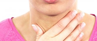
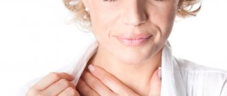
- Dysphagia – difficulty swallowing;
- Sore throat;
- Sensation of a foreign object in the esophagus;
- Regurgitation of food;
- Cough (mostly dry);
- Changes in tone and timbre of voice;
- The presence of increased salivation;
- Nausea and vomiting;
- Weakness and malaise.
In the presence of medium-sized diverticula, the symptoms are not as pronounced, but they may also be present:
- Attacks of night cough;
- Nausea;
- Feeling unwell;
- Weakness.
If there is a protrusion in the lower esophagus, the following points may be added to the symptoms:
- Dyspnea;
- Palpitations;
- Bronchospasm;
- Heart pain.
It must be remembered that depending on the type of protrusion, symptoms can vary significantly. For example, pulsion and pharyngoesophageal diverticula of the esophagus, with small sizes, also do not tend to manifest themselves, but as they develop, they significantly disrupt the functioning of not only the main organ, but also the entire organism as a whole.
Causes
Based on their origin, esophageal diverticula are divided into congenital and acquired. Congenital formation is formed due to a violation of the formation of layers of the wall of the esophageal tube. In a certain area, insufficiently dense muscle tissue appears, which cannot withstand the load and leads to protrusion.
The process of formation of diverticula is also called diverticulosis. Acquired diverticula develop due to inflammatory processes in neighboring organs (lungs, pleura, pericardium) and in the esophagus itself, injuries. Diverticula appear over a long period of time:
- esophagitis;
- mediastinitis;
- gastroesophageal reflux disease;
- fungal infection;
- tuberculosis of regional lymph nodes;
- esophagospasm;
- achalasia;
- cicatricial narrowing of the esophagus.
The mechanism of protrusion formation can be pulsation, traction or mixed.
- Pulsion diverticulum is always associated with dysfunction of the motility of the esophageal wall, spastic contractions of the muscle layer, and a subsequent increase in intraesophageal pressure. This leads to stretching and bulging at the weakest point.
- Traction mechanism - caused by fusion and abnormal fixation of the esophageal wall to the inflamed lymph nodes of the mediastinum. As a result, the muscle layer stretches and then bulges.
With a mixed type, both mechanisms of action take place.
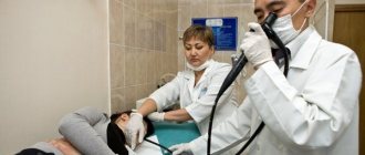
The surgeon diagnoses and treats pathology
Causes of diverticulum formation in the esophagus
- In 1% of cases, esophageal diverticula (Zenker) are detected during contrast examination.
- In 70% of cases, diverticula are located within Killian's triangle (diverticula of the cervical esophagus or Zenker's diverticula);
- In 22% of cases, they are located at the level of the tracheal bifurcation (traction diverticula);
- In 8% of cases, esophageal diverticula are located directly above the diaphragm (supradiaphragmatic, or epiphrenic, diverticula);
- Zenker's diverticula form at a weak point between the oblique and transverse muscle bundles of the inferior pharyngeal constrictor;
- The likely cause is incomplete relaxation of the upper esophageal sphincter;
- Traction diverticula usually develop due to inflammation of the lymph nodes in tuberculosis and histoplasmosis;
- Supradiaphragmatic esophageal diverticula may be associated with peristalsis disorders;
- A rare form - intramural pseudodiverticula - arise due to the expansion of the axillary lymph nodes and represent multiple fistula-like protrusions.
Classification of diverticula
Depending on the morphological structure of the wall, diverticula are divided:
- for false ones (pseudodiverticula) - there is no muscular membrane in the area of prolapse, in fact they do not differ from hernias, they are always acquired in nature, usually shapeless, the traction mechanism is involved in their origin, they occur against the background of adhesive-cicatricial deformities in the area of inflammation or neoplasm;
- true - correspond to the structure of the wall of the esophagus, most often congenital, caused by the formation of a cyst-like structure in the embryonic period.
Pseudodiverticula include any cavities (abscesses, cysts, neoplasms) that have broken through from adjacent tissues into the esophagus. Typical cases (Bischoff pseudodiverticulum) caused by damage to the wall of the esophagus due to a retropharyngeal abscess or suppuration of a branchioma tumor are described.
Diverticula are classified according to location:
- pharyngoesophageal (pharyngoesophageal, borderline, Zenker's diverticula, transitional, cervical);
- bifurcation (epibronchial, parabronchial, upper thoracic);
- supradiaphragmatic (epiphrenal, lower thoracic).
Since topographic location causes peculiarities of manifestation and differences in treatment approaches, we will provide a separate description of each form.
Features of pharyngoesophageal diverticula
The species is found most rarely (3–5% of all cases). According to the mechanism of formation, they are classified as pulsational. These include 75% of all pulsion diverticula in the esophagus. They occur 3 times more often in men than in women. Surgeons believe that the large size of the larynx and pharynx contributes to this.
Localization is typical on the posterior wall of the pharynx and esophagus. Here an anatomical weak point is formed between the muscle bundles (Killian's triangle). Uncoordinated contractions with simultaneous mechanical pressure from a bolus of food form an outward protrusion in the area in the form of a bag.
This species is characterized by very slow growth
Sizes range from cherry size to giant. Inside the diverticulum, a body and a neck are distinguished; they are lined with a mucous membrane and can accumulate up to a liter of fluid, depending on their size. Symptoms of Zenker's esophageal diverticulum depend on the size. They are usually divided into 3 stages. In the first stage, all symptoms are unstable, the disorders are only functional, nonspecific.
The patient complains of periodic symptoms:
- sore throat;
- changes in salivation, causing dry mouth or, conversely, accumulation of saliva;
- coughing;
- discomfort when swallowing;
- “lump in the throat” due to excitement or food.
Patients undergo long-term treatment for pharyngitis. Upon examination, the doctor notes pain and tension in the masticatory muscle (Pottenger's symptom). In the second stage, clinical manifestations are caused by stagnation of mucus, food particles, and air in the diverticulum cavity. The patient's general condition is not impaired and is considered satisfactory.
A careful examination reveals an asymmetrical thickening on the neck; a soft protrusion disappears upon palpation, but grows against the background of food intake. If a person has eaten a liquid dish, then tapping is accompanied by the sound of splashing; upon palpation, a rumbling sensation is felt. Auscultation reveals a characteristic bubbling sound.
The patient notes difficulty swallowing both liquid and solid food, moderate belching at night when lying down, and putrid odor from the mouth.
Pressure on neighboring organs provokes additional symptoms (compression syndrome):
- cough at night;
- change in voice timbre;
- shortness of breath;
- In the morning, patients find mucus on the pillow.
It is considered typical after eating to develop an attack: the face turns red, suffocation occurs, dizziness to the point of fainting. Improvement occurs after vomiting. Stage of decompensation - the patient’s condition suffers, the person loses weight, all symptoms are significantly pronounced, complications appear.
A person has difficulty swallowing liquid and solid food
What are the differences between bifurcation diverticula?
Bifurcation localization in the structure of esophageal diverticula occupies from 70 to 80%. They most often occur in women aged 40–60 years. According to the mechanism, they are traction or mixed, purely pulsion are very rare. If the size does not reach 2 cm in diameter, then patients do not feel discomfort.
With a large size, narrowed neck, inflammation often occurs, so symptoms appear: pain behind the sternum or in the epigastrium, radiation to the back, difficulty swallowing, regurgitation, bad breath, fever. Pain is considered an obligatory (absolute) symptom of diverticulitis.
Features of epiphrenic diverticula
They are pulsion in origin and are most often located in the supradiaphragmatic segment of the abdominal esophagus. The frequency of detection of diverticula in the structure is from 10 to 15%. The largest number of cases were found among women in the age group 50–60 years, in 2/3 it is combined with cardiospasm.
It is believed that weaknesses in the lower segments of the esophageal tube, uncoordinated contraction of the muscles of the cardia of the stomach and esophagus, increased pressure inside the esophagus, and hiatal hernia are involved in the formation. The morphological structure of the wall contains all layers of the esophagus. More often the anterior or left lateral part protrudes.
They do not reach large sizes (usually no more than 2–3 cm). Thus, ½ of cases are detected during examination and are asymptomatic. Signs of large diverticula are caused by irritation of the vagus nerve, pressure on the anterior wall of the esophagus.
Among the symptoms:
- difficulty swallowing;
- heaviness at the bottom of the sternum;
- feeling of a “stuck lump”;
- nausea;
- increased regurgitation;
- putrid odor from the mouth.
With significant sizes it is possible:
- pain in the heart area;
- cardiopalmus;
- dyspnea;
- manifestation of “gurgling” in the epigastrium.
Treatment
If the diverticula are small and do not manifest themselves symptomatically, then a conservative treatment method is provided.
- First of all, a diet that is gentle on the esophagus is prescribed. Food that can mechanically damage the walls of the organ is excluded. Dishes with a puree-like consistency are provided.
- Before eating, a person must take a comfortable position, the food is completely crushed and washed down with small sips of liquid.
- In order to prevent inflammatory processes, the esophagus should be washed and drained from time to time. This is done using warm boiled or mineral water.
- Drug therapy.
If the diverticulum is large and causes significant discomfort to a person, it is removed surgically.
Minimally invasive operations have become increasingly popular, after which the patient recovers quickly. In addition, the digestive system returns to normal in a short time. Such operations include:
- Endoscopic clipping of diverticulum.
- Laser stitching.
The operations are considered difficult and require special preparation, so you should contact experienced surgeons.
Diverticulum can occur not only in humans, but also in animals, such as dogs.
Traditional medicine in the fight against esophageal diverticulum involves the use of an appropriate diet. First of all, treatment should begin with coarsely ground wheat bran. Brew bran in boiling water in a ratio of 1-2 tablespoons per 100 grams of water. You need to start taking the medicine with 6 teaspoons, gradually increasing the dose, and by the end of 4 weeks it should be 6-8 tablespoons of the drug per day.
Vegetable pulp has a good effect. Especially beetroot or white cabbage. To do this, grind the vegetables and squeeze out all the juice from them, roll the cake into balls the size of a walnut. Such balls are dried in the oven, and should be taken daily before meals in the amount of 5 to 7 pieces, washed down with a small amount of water. This treatment lasts on average six months or a little less.
Use homemade flaxseed oil to treat esophageal diverticulum. To do this, grind 100 grams of the seed in a coffee grinder and pour 500 ml into it. vegetable oil. The product is infused for 3 weeks in a dark place. Take 1 teaspoon of the product together with the seed. The course lasts up to six months.
These prescriptions are used only to eliminate diverticulum, but do not treat concomitant diseases. Therefore, do not self-medicate without prior diagnosis and consultation with a doctor.
We next consider esophageal diverticulum. Treatment is also something that needs to be discussed in detail. So, if this is a true diverticulum, getting rid of the problem can take place in three main directions:
- Symptomatic.
- Operational.
- Non-operative.
What will the doctor advise you to do in this case? Since diverticula can be detected in later stages of the disease, in this case the doctor will immediately recommend rinsing the esophagus. This is necessary in order to get rid of the stagnant masses. However, this procedure will not cure the disease. Non-operative treatment in this case will be ineffective, because it will not help cope with food stagnation and the growth of diverticulum.
What to do if the patient has a false diverticulum of the esophagus? Treatment in this case will be aimed specifically at eliminating the inflammatory process (which is the cause of this problem). In this case, surgery will not be required. And all efforts of doctors should be aimed at ensuring that the diverticulum does not turn from a traction form into a pulsation form. If food masses get stuck in the esophagus, they need to be removed.
Diverticula with mild symptoms are treated with conservative methods under medical supervision. The patient is recommended to strictly adhere to dietary table number eight. After consuming food, it is necessary to carry out the following procedure: drink a small amount of liquid, strain, take a draining position, rinse the cavities with a non-concentrated antiseptic solution, these actions help to empty the esophageal diverticulum.
Surgical intervention is used only in cases with serious complications and defects, which are accompanied by dysphagia, complicated course (penetration, perforation, bleeding, etc.) or acute pain. In these cases, excision of the esophageal diverticulum is performed, and in case of a small diverticulum, it is removed by intussusception - directing the diverticulum into the lumen of the esophagus and securing it to the esophageal wall.
Diagnostics
The most accessible method is targeted tomographic examination of the esophagus using an X-ray machine. It allows you to take pictures at different depths, identify signs of a diverticulum, determine the shape, size, angle of deviation of the neck, and the presence of inflammation.
Contrast allows you to identify protrusion of the esophageal wall
With large diverticula, the survey image shows cavities with a level of fluid, air, and a connection with the esophagus. Bifurcation diverticula are easily detected when they are small in size, since they usually have a wide entrance and are clearly contrasted.
For traction views, a contrast study may be required with the patient in a horizontal position. A sign of diverticulitis is considered to be a time delay of contrast for 2 minutes or more, revealing the layering of the contents of the body of the protrusion. Computed tomography - gives more accurate results, performed with or without contrast.
Esophagoscopy allows you to examine all parts of the esophagus, identify changes in the walls, and signs of inflammation. With fibrogastroscopy, you can notice gastroesophageal reflux (return of food from the stomach), identify diseases that caused the diverticulum, and take material for a biopsy. The procedure is carried out with caution, since there remains a danger of perforation of the wall.
To study the contractile function of the esophageal muscles, esophageal manometry is performed. In differential diagnosis it is necessary to distinguish it from heart pathology, therefore electrocardiography and Holter monitoring are needed.
Is it worth treating?
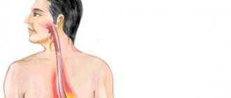
Esophagus affected by diverticulosis
Esophageal diverticula vary widely, so there is no single answer to this question. When their sizes are small and they do not cause any trouble, conservative treatment is carried out.
Large sacs must be removed surgically because there is a risk of complete blockage. In truth, such formations are not common.
- Cardiospasm of the esophagus symptoms treatment
- Esophageal problems symptoms and treatment
- Torn navel symptoms and treatment
- Sigmoid colon adhesions symptoms and treatment
What complications does diverticulitis cause?
Inflammation of the diverticulum over a long period of time can lead to:
Hiatus hernia
- to attacks of suffocation;
- phlegmon of the neck;
- chronic bronchitis;
- aspiration pneumonia, lung abscess;
- bleeding;
- abscess formation of a diverticulum;
- mucosal erosion;
- perforation into surrounding tissues;
- mediastinitis with esophageal-mediastinal fistula;
- esophageal polyps;
- cancerous degeneration.
Bifurcation diverticulitis has the rarest tendency to complications.
Complications
The danger of the defect lies in the progression of the disease under the influence of concomitant abnormalities. In patients with diverticula, the formation of polyps and multiple adhesions may begin. When the esophagus becomes inflamed, there is a risk of ulcers and perforation, which then leads to hemorrhage into the lumen of the organ. There is a possibility of transformation of the diverticulum into a malignant neoplasm. The defect can lead to an abscess and pneumonia.
Self-medication attempts at home often lead to such complications. This disease can be identified in a timely manner and high-quality drug treatment can be carried out with a favorable prognosis, but not everyone seeks help.
Treatment of esophageal diverticulum
The choice of treatment depends on the size of the diverticulum and the risk of complications. In case of minor forms and satisfactory health of the patient, observation and conservative therapy by a gastroenterologist is recommended. What matters is a diet based on maximum sparing of the esophageal mucosa.
It is recommended to give up dense foods, fried foods, pickles, alcohol, hot seasonings, very hot and cold drinks, prepare fairly crushed dishes, semi-liquid, boiled, stewed, steamed, pureed, take food in small portions, slowly, chew well.
After each meal, it is recommended to take measures to completely empty the diverticular sac. For this:
- drink water, compote, jelly, milk;
- strain several times;
- take a position favorable for drainage.
To prevent leakage at night, it is better to sleep on a high pillow. Surgical treatment consists of removing large diverticula. It is indicated in the presence or high risk of complications, severe symptoms. Under convenient conditions and small sizes, the diverticulum is immersed into the lumen of the esophagus (similar to the reduction of a hernia), the wall is sutured without plastic surgery.
The operation includes excision of the sac (diverticulectomy) followed by plastic surgery of the wall defect with a flap from the diaphragm or pleural layer
Complications
Without timely seeking help from a specialist who will diagnose and prescribe treatment, the progression of the disease will lead to the development of severe consequences requiring immediate surgery:
- ulceration of the mucous membrane;
- hemorrhages into the lumen of the esophagus - can be manifested by symptoms such as regurgitation or vomiting with blood;
- perforation;
- transformation of a diverticulum into an oncological neoplasm;
- multiple adhesions;
- aspiration pneumonia;
- lung abscess;
- formation of polyps.
In addition, independent treatment with traditional medicine can lead to the development of the above complications.