Etiology
A large number of predisposing factors can cause the occurrence of one or another lesion of the duodenum. These include:
- poor nutrition, addiction to junk food or consumption of low-quality products that cause poisoning, which is also the source of some ailments;
- maintaining an unhealthy lifestyle, namely abuse of alcoholic beverages and smoking;
- the course of the infectious process in the body;
- damage to the shell by a foreign object;
- infection with the bacterium Helicobacter pylori or other microorganisms that pathologically affect the gastrointestinal tract;
- the influence of stressful situations in which the body directs all its forces to preserve the emotional state;
- long-term use of certain medications that negatively affect the mucous membrane of the duodenum, thereby destroying it.
In addition, there are congenital forms of diseases of this organ, which makes it possible to assume the genetic nature of their development.
Functions
- Motor-evacuation.
It is the process of pushing food through the alimentary canal. The organ also serves as a reservoir; it releases bile acids and various pancreatic enzymes. - Digestive.
The initial stage of digestion occurs in the intestine, due to the action of bile acids and pancreatic enzymes. - Regulatory.
Caused by the regulation of bile acids and pancreatic enzymes. - Acid-base.
In the duodenum, the pH of the bolus of food is brought to optimal levels for its further transformation in other parts of the digestive tract.
The most common diseases of the duodenum.
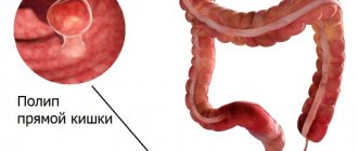
The first representative of this group of disorders is hookworm disease, a parasitic disease caused by several pathological microorganisms. Infection occurs from a sick person or pets, contaminated soil or hands.
This disorder has the following symptoms:
- itching and burning of the skin;
- the appearance of a characteristic rash on the skin;
- diarrhea;
- heartburn and soreness.
Congenital structural pathologies are diagnosed extremely rarely, but have various manifestations, for example:
- atresia;
- congenital stenosis;
- adhesions and diverticula;
- arteriomesenteric obstruction.
The symptoms of such disorders are practically no different from the manifestations of other duodenal diseases.
The duodenum plays an important role in the entire digestive process. Since it is the initial link of the intestine, the processes of absorption of nutrients from incoming food and liquid actively occur here. It brings the acid-base indicator of food to a level that will be optimal for subsequent stages of digestion in the intestines. It is in this organ that the stage of intestinal digestion begins.
Another integral phase of the work of this part of the intestine is the regulation of pancreatic enzymes secreted by the pancreas, as well as bile, depending on the acidity of the food bolus and its chemical composition.
The duodenum affects the proper functioning of the secretory function of the stomach, since the opposite interaction occurs. It involves the opening and closing of the pylorus of the stomach and humoral secretion.
The 12 duodenum carries the function of further promoting food gruel, treated with enzymes, into the next section of the small intestine. This occurs due to the massive muscle layer of the duodenal wall.
- Duodenitis is the most common ailment of the duodenum of acute or chronic type, manifested in the form of inflammation of the mucous membrane.
- Ulcer – develops as a result of chronic duodenitis. Chronic damage to the duodenum, in which ulcers form in the mucous layer.
- A cancerous tumor is a malignant neoplasm localized in different layers of the duodenal wall.
Duodenitis
More than 90% of patients develop chronic duodenitis. It can develop due to many factors, including:
- consumption of low-quality products;
- alcohol abuse;
- smoking;
- ingress of foreign bodies and toxic substances;
- other chronic intestinal diseases.
This disease manifests itself in the form of pain in the epigastrium of moderate intensity, weakness, belching, heartburn, nausea, turning into vomiting. Symptoms are often accompanied by fever.
If the disease is at an acute stage, the person feels pain and nausea and suffers from repeated vomiting. Acute bulbitis develops against the background of long-term use of a large group of drugs, or poisoning. In the chronic form, there is also an aching pain syndrome, sometimes it can be accompanied by nausea.
Patients also experience chronic duodenal obstruction, which occurs against the background of tumor processes, developmental anomalies and other disorders in the duodenum. It is expressed in a violation of motor and evacuation functions in this part of the intestine and is characterized by the following symptoms:
- heartburn;
- decreased appetite;
- feeling of heaviness and discomfort in the epigastric region;
- constipation;
- gurgling and bubbling.
The manifestation of this disease is influenced by the causes that caused duodenal obstruction, the stage of progression and how long ago the disease arose.
Peptic ulcer
The main cause of this dangerous disease is the reflux of acid from the gastric contents and its destructive effect on the mucous membrane of this part of the intestine. But this pathological process develops only when the surface layers of the intestine fail to cope with their protective functions. The ulcer is localized in the initial part of the duodenum and in the bulb, that is, in that zone of the intestine that is located at a minimum distance from the stomach.
Many gastroenterologists unanimously speak about the negative impact of frequent use of anti-inflammatory drugs, which lower the protective barrier of the mucous layer of the duodenum. These drugs are aspirin and dosage forms based on it, ibuprofen, diclofenac, etc. Therefore, if possible, you should limit the use of drugs in this group as much as possible.
Poorly treated or neglected duodenitis, alcohol abuse and consumption of foods harmful to the body can also cause the development of duodenal ulcers.
The Helicobacter bacterium also tends to infect not only the stomach, but also the mucous membrane of the duodenum. It is a fairly common cause of ulcerative pathology, opening the way for acid into the mucous layers of the intestine. In 19 out of 20 cases of the development of ulcers of this organ, it is the Helicobacter bacterium that is to blame.
The main dangerous complications of this disease of the duodenum are bleeding or perforation, which require emergency surgical assistance. Bleeding is fraught with dangerous loss of blood and filling of the abdominal cavity with it. Perforation is when food with all the enzymes and acids enters the abdominal cavity through an ulcerative hole formed in the intestine.
An ulcer, like other lesions of the duodenum, is diagnosed by an endoscopy procedure. Using this procedure, a gastroenterologist can visually assess the condition of all organs of the digestive system. A blood test may also be necessary, especially if we are talking about duodenal ulcer caused by the Helicobacter bacterium.
Unfortunately, at this point in time, in medical practice there is no accurate data on the causes of cancer in the body. But there is a certain category of risk factors that can provoke a malignant process in the body - and the duodenum is no exception. This disease can be caused by:
- genetic predisposition to cancer;
- addictions: smoking, drug use, alcoholism;
- diabetes;
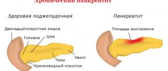
- chronic pancreatitis;
- kidney stones, bladder stones;
- eating large amounts of animal food.
According to scientists, components of coffee in combination with nicotine can also affect the development of duodenal cancer. Therefore, doctors do not recommend getting carried away with coffee: you should limit yourself to 2–3 cups per day as a maximum. Constant ingestion of carcinogens and chemicals that have a detrimental effect on the entire gastrointestinal tract can also cause duodenal cancer.
This disease is considered insidious because it is difficult to diagnose in the initial stages of development. The first signs of the disease can be easily confused with ordinary gastrointestinal disorders. Later, pain is added to these sensations during the development of oncology, especially when a person feels a feeling of hunger and heaviness. The patient feels weak, loses his appetite and experiences depression. These symptoms are associated with the process of intoxication.
A person with duodenal cancer has a much greater chance of a normal outcome if the tumor is detected in the primary stages of development. To make an accurate diagnosis, an endoscopy and a biopsy of the affected area of the intestine are performed, and a complex of laboratory tests is also connected to them (CBC, tumor marker CA 125, etc.). After this, an urgent operation must be performed to remove the tumor and the lymph nodes closest to it.
From all of the above, a simple and logical conclusion can be drawn. The duodenum is, like all organs, a very important part of our body. It performs complex and important functions in the digestive system, so every person should be attentive to their food preferences - if possible, exclude unhealthy foods from their diet and give up bad habits. After all, it is much easier to prevent diseases than to then go to the doctors and stay in the hospital in the hope of overcoming them.
All kinds of pathologies of the duodenum
Diseases of the duodenum mean inflammation in the mucous membrane, affecting its activity and the digestive chain as a whole. The occurrence of the inflammatory process is facilitated by pathologies that affect the performance of the entire organism.
Helicobacter can cause duodenal disease
Often, disease of the initial part of the small intestine is caused by Helicobacter pylori infection. This microorganism lives in the stomach without manifesting itself in any way. Its presence increases the secretion of stomach acid, which irritates the duodenum.
Diseases of a characteristic segment of the digestive system also develop against the background of stress or surgery. In some cases, provoking factors are addictions or the use of non-steroidal anti-inflammatory drugs.
Possible diseases of the duodenum are described below; you should remember their symptoms in order to start treatment on time and avoid serious consequences.
Neoplasms
Cancer of the segment of the digestive tract in question is diagnosed in rare cases, most often in elderly patients. Malignant tumors are formed from intestinal glands and epithelial cells.
The early stage of the disease is asymptomatic. The first signs of cancer appear after the tumor narrows the intestinal lumen or develops tumor intoxication. The patient has:
- pain of varying intensity;
- lack of appetite;
- increased fatigue;
- sudden weight loss;
- yellowing of the skin due to impaired bile excretion.
If a blood test shows a higher than normal concentration of cancer tumor markers, then there is a high probability of developing a malignant tumor. The effectiveness of treatment at an early stage is higher, so it is necessary to undergo an annual examination for cancer cells.
Erosion
Duodenal erosion is understood as the occurrence of an inflammatory process on the surface of the mucous membrane of a characteristic organ that does not penetrate into the muscle tissue. Accompanied by the formation of erosions. Thickening of the intestinal wall is noted. The disease is provoked by stress, addictions, poor nutrition, and certain medications.
Symptoms of the pathological condition include:
- problems with stool;
- burping;
- heartburn in the stomach;
- the occurrence of pain.
Duodenostasis
Duodenostasis is a disease that affects the motor function of the intestine. Its contents stagnate, a mushy mass accumulates, which consists of gastric secretions, enzymes and incompletely digested food.
The pathology is accompanied by the following symptoms:
- lack of appetite;
- pain in the stomach and right hypochondrium;
- nausea, sometimes accompanied by vomiting;
- formation of constipation.
Duodenitis of the duodenum
A fairly common pathology of the duodenum is duodenitis. The disease affects the walls and mucous membrane of the intestinal segment. The long course of the disease leads to its thinning. Often develops against the background of secretory insufficiency.
The disease manifests itself with several signs:
- distension of the stomach after eating;
- dull and persistent pain;
- nausea accompanied by vomiting;
- lack of appetite.
When palpating the epigastric region, painful sensations occur.
Anomaly of intestinal development
Congenital stenosis is an anomaly of intestinal development.
Developmental defects are quite rare in medical practice. One of these anomalies is congenital stenosis, diagnosed in the first hours of a baby’s life. This category also includes a diverticulum, which is a protrusion of the wall, lymphangiectasia.
The reason for the formation of the latter is unilateral lymphostasis. Often occurs against the background of malformations of other digestive organs, such as ulcerative colitis, Crohn's disease.
Peptic ulcer
Duodenal ulcer is a chronic inflammatory pathology. Crater-shaped wounds form on the mucous membrane of the organ. The size of the formations ranges from 5 to 10 mm in diameter.
Various factors contribute to the occurrence of peptic ulcers:
- the presence of Helicobacter pylori in the stomach and intestines, which destroys epithelial cells;
- aggressive action of hydrochloric acid, which violates the integrity of the mucous membrane;
- constant stress that provokes spasms of intestinal vessels, as a result of which the nutrition of cells deteriorates, and then their death occurs.
The clinical picture is expressed during exacerbations. The main symptom of an ulcer is night hunger pain that disappears after eating. Intestinal ulcers are characterized by heartburn, nausea, loss of appetite, frequent belching, and constipation.
Parasite infestation
The introduction of parasites into the body occurs along with food if basic hygiene rules are violated. For a long time, their damage to organs remains asymptomatic. Pests living in the duodenum are nematodes. Their larvae enter the body through the oral-fecal route or through pores in the skin. In addition to the organ in question, parasites also infect other parts of the body.
Symptoms of intestinal pest infestation are:
- skin rashes;
- diarrhea;
- itching;
- pain in the abdominal area;
- heartburn in the stomach.
Intestinal obstruction
There are many reasons for obstruction of the initial section of the small intestine:
- congenital anomalies;
- violation of intestinal rotation;
- vascular compression.
Gallstones can enter the stomach through a fistula between the organ and a characteristic segment of the digestive tract. The calculus migrates through the alimentary canal and often gets stuck in the small intestine. Before the formation of an anomaly, a person is bothered by a feeling of pain in the right hypochondrium.
Classification
Diseases of the duodenum are divided into inflammatory and non-inflammatory; bacterial and fungal lesions are diagnosed much less frequently.
The most common inflammatory disorder is duodenitis, the formation of which is caused by infection from other internal organs. In second place in frequency are erosive or ulcerative disorders. Often such ailments are combined with diseases of the liver and intestines, as well as some systems. The duodenal bulb most often suffers from the consequences of inflammatory processes in nearby organs.
The non-inflammatory group of disorders of this organ is several times less common than the previous one and represents the formation of oncological, cystic or benign neoplasms.
The risk of developing just such ailments increases with the patient’s age group. Among malignant tumors, sarcoma takes the lead, which often affects the lower pancreas.
Complications
They occur less frequently, but are painful and can be serious:
- bleeding from an ulcer varies from a trickle to life-threatening bleeding;
- perforation, or perforation of the wall of the duodenum, with this complication, food and acids enter the abdominal cavity, which causes severe pain and the need for emergency medical care.
Therefore, you need to know: if the duodenum is bothering you, the symptoms of the disease, even if they are very minor, should under no circumstances be ignored!
- The structure of the human intestine photo with description
- What pinworms look like in humans photo
- Erosion of the duodenal bulb treatment
- Where is a hernia located in humans?
Features of the structure of the organ (shape, location, fastening)
The shape of most people is varied, and even in one person, both the shape and the location of the duodenum can change over the course of a person’s life. It can be V-shaped, and resemble a horseshoe, loop and other shapes. In old age, or after weight loss, it is lowered compared to where the duodenum is located in young and middle-aged people and overweight people.
But most often it originates at the level of the seventh thoracic or first lumbar vertebra, located from left to right. Then there is a bend with a descent to the third lumbar vertebra, another bend with an ascent parallel to the upper part and the intestine ends in the area of the second lumbar vertebra.
It is attached to the abdominal organs by connective fibers located on the walls. The upper part of the duodenum has the fewest such attachments, so it is mobile - it can move from side to side.
The structure of the wall of the duodenum:
- The serous outer layer performs mechanical protective functions.
- The muscle layer is responsible for the peristalsis of the organ during the digestion of food.
- The submucosal layer contains nerve and vascular nodes.
- the inner layer is the mucous membrane, strewn with a large number of villi, folds and depressions.
This anatomical location of the organ has a huge impact on the characteristics and course of diseases that arise in it.
Why is it needed, how and where does the duodenum hurt:
- You ate at lunch, it doesn’t matter what, just a big one. The food you eat will stay in your stomach for about 6 to 8 hours.
- In portions it begins to accumulate in the upper part of the stomach. Then it is mixed and placed in layers.
- We must not forget about moderation in eating. You can overeat if you eat food hastily.
- Then it passes in small portions into the small intestine, connected to the stomach. The small intestine begins with the duodenum.
- But in it, with the help of juices produced by the pancreas, its enzymes, bile coming from the liver, the breakdown of food begins.
- Carbohydrates, proteins, and fats are actively processed.
- All walls of the duodenum are covered with a large number of villi. They all have their own blood vessels, capillaries.
- Well-broken substances are absorbed in them: such as glucose, amino acids, glycerol.
- Digestion occurs throughout the entire length of the small intestine.
- Undigested food remains move into the large intestine within 12 hours. This is where most of the water absorption occurs into the blood.
- It is twelve fingers long. If something doesn’t work in this area, all digestion is disrupted.
- Upper part (level of the first lumbar vertebra). It is also called an onion because of its round shape. Length five, six centimeters.
- Descending part (descends to the third lumbar vertebra).
- Horizontal part (level of the third lumbar vertebra).
- Ascending part (ascends to the second lumbar vertebra).
- Top bend.
- Bottom bend.
- The junction of the duodenum and the jejunum.
Gastroduodenitis
Inflammation of the lining of the duodenum or duodenitis is the most common pathology of this organ and is quite rarely independent.
Symptoms of this disease include:
- heaviness in the stomach;
- rapid satiety, despite the fact that a small amount of food was eaten;
- decreased or complete lack of appetite;
- attacks of nausea that end in vomiting only during exacerbations of the disease;
- pain in the abdominal area, often patients experience night hunger pains;
- the appearance of blood impurities in feces and vomit;
- severe weakness and dizziness are explained by the presence of anemia.
By the way your stomach hurts, you can recognize the location of the pathological process. For example, when the upper part of this organ is affected, the pain syndrome appears much faster after eating food than when the lower part is involved in inflammation.
This disorder exists in several forms - acute and chronic; depending on the nature of the course, the intensity of the manifestation of symptoms will differ.
The disease consists of the combined occurrence of gastritis and duodenitis. Inflammation begins in the stomach, spreads to its entire surface and spreads to the duodenum.
Similar to duodenitis, it can occur in acute and chronic forms. In an acute course, the main symptom is pain in the right hypochondrium, while in a chronic course, this symptom becomes more intense, atrophy of the membrane occurs, and the motor and secretory function of the affected organs is impaired.
How does the duodenum hurt?
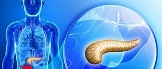
Considering the fact that the duodenum starts from the stomach, and the ducts of the gallbladder and pancreas also open into it, many of its diseases are associated with the malfunction of these organs:
- increased acidity of the stomach leads to the fact that hydrochloric acid begins to corrode the mucous membrane of the duodenum;
- Low stomach acidity means that rough food that has not been processed well gets into the intestine. This causes mechanical damage;
- with pancreatitis and cholecystitis, the production of digestive enzymes is disrupted, because of this, food is poorly crushed in the duodenum;
- With hepatitis and cirrhosis, blood circulation is impaired and, as a result, nutritional deficiency occurs.
But sometimes the occurrence of duodenal diseases is influenced not by existing pathologies of other organs, but by a person’s lifestyle. Snacking on the go and in a hurry, insufficient chewing of food, overeating, too long breaks between meals - all this negatively affects the functioning of the gastrointestinal tract (GIT).
You can identify the reason why an organ is suffering by how it hurts:
- duodenitis caused by Helicobacter pylori. The pain occurs at night and on an empty stomach. It disappears after taking antisecretory and antacid drugs, as well as after eating. Unpleasant sensations may be accompanied by heartburn, belching and constipation;
- duodenitis caused by diseases of the gallbladder and pancreas. Painful sensations occur in the right or left hypochondrium and intensify after eating fatty foods. Patients complain of bitterness in the mouth, nausea, and constipation, which is replaced by diarrhea;
- inflammation associated with stomach cancer or atrophic gastritis. Pain and heaviness in the stomach;
- peptic ulcer. Pain in the form of colic, which is a consequence of spasm of the smooth muscles of the muscles.
Duodenitis is an inflammation of the mucous membrane of the duodenum. The disease can be acute or chronic, which occurs with relapses. In almost all recorded cases of duodenitis, a chronic process is observed.
Poor nutrition, bad habits, chronic gastrointestinal diseases - all this can serve as an impetus for the activation of the inflammatory response. Patients are concerned about pain in the upper abdomen, nausea, belching, heartburn, and weakness. Inflammation of the duodenum can lead to peptic ulcers and even cancer.
Peptic ulcer disease is also accompanied by inflammation of the organ, only the appearance of ulcers on the surface of the mucous membrane is added to everything else. This is a chronic pathology with frequent relapses. If the disease is left to chance, it can lead to atrophic changes, as well as fistulas and bleeding.
A duodenal ulcer can even cause death. Poor nutrition, taking potent drugs, chronic duodenitis - all this can lead to ulcers. But the most common cause is still the bacterium Helicobacter pylori.
The infectious agent seriously damages the mucous membrane of the organ with its metabolic products. A characteristic symptom is hunger or night pain, which disappears half an hour after eating. The danger of a peptic ulcer is that it can develop into cancer.
Ulcerative lesion
Duodenal ulcers very often occur against the background of the pathological influence of the bacterium Helicobacter pylori, but this is not the only factor in the formation of the disease. Sources include increased secretion of hydrochloric acid, a weak immune system, stress and genetic predisposition.
Regardless of the reasons for the appearance of such a pathology, its symptoms will not differ. External manifestations include:
- severe pain is the very first sign of the disease, which has its own characteristics, namely, it often bothers patients at night or on an empty stomach;
- sudden onset of heartburn that does not go away on its own;
- burping – often causes severe discomfort to people;
- increase in the size of the peritoneum;
- attacks of nausea that occur regardless of the time of day;
- vomiting - characterized by the fact that it brings relief to the patient and allows him to slightly improve his well-being;
- bowel dysfunction, which results in constipation;
- blood impurities in stool;
- weight loss is caused by a reluctance to consume food, because it is after a meal that some symptoms are expressed.
Duodenal ulcer
Ulcerative lesion of the duodenum without timely surgical treatment can cause the development of severe complications that pose a threat to the patient’s life.
Causes of duodenal pathology
Causes of pathologies of the duodenum can be:
- gastric and intestinal diseases and pathological processes - inflammation of the gastric mucosa, viral infections, diarrhea, etc. Due to the increased secretory activity of the stomach, a lot of hydrochloric acid enters the duodenum; due to the reduced secretory activity, coarse unprocessed food enters the duodenum;
- Helicobacter pylori, which leads to increased production of gastric secretions, irritating the intestinal mucosa;
- pancreatitis and cholecystitis;
- liver diseases - hepatitis, cirrhosis;
- prolonged stress;
- previous operations;
- smoking and alcohol abuse, fast food;
- taking non-steroidal anti-inflammatory drugs;
- food poisoning;
- abuse of fatty and spicy foods;
- helminthic infestations;
- hereditary predisposition.
Dyskinesia
Duodenal dyskinesia or duodenostasis is a motor dysfunction of the duodenum, which is manifested by symptoms such as:
- pain that occurs after eating;
- constipation, which can cause intoxication of the body, and if treated incorrectly or untimely, lead to death;
- weight loss and food aversion;
- feeling of heaviness in the peritoneum;
- nausea, rarely ending in vomiting.
Such symptoms of duodenal disease are quite common, which is why only a gastroenterologist can make a final diagnosis.
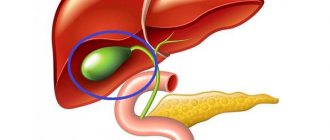
What systems surround the duodenum?
In front and above the upper part of the intestine are the following organs: the quadrate lobe of the liver, the neck and the body of the gallbladder. The hepatoduodenal ligament is located between the upper part of the intestine and the left lobe of the liver (lower surface). At the base of this ligament lies the common bile duct. The upper part of the duodenum, not covered by peritoneum, is in contact with the portal vein, common bile duct, and arteries (gastroduodenal and pancreatic-duodenal).
The posterior surface of the descending part of the intestine is in contact with the pelvis of the right kidney, ureter, and renal vessels. The hilum of the kidney is slightly covered by the duodenum. The descending part of the duodenum is in contact with: the right curvature of the colon, the ascending colon, and the head of the pancreas.
The common bile duct is often located in the parenchyma of the head of the pancreas, then it connects with the pancreatic duct and flows into the duodenum. On the surface of the intestinal mucosa there is a small longitudinal fold (up to 1 cm long), on the surface of which there is a large duodenal papilla, on which the pancreatic duct opens. Sometimes there is an accessory pancreatic duct that opens higher than the main one.
Between the descending part of the duodenum and the head of the pancreas, a groove is formed in which the common bile duct lies. The superior mesenteric vein and artery are adjacent to the lower part of the duodenum. These vessels are located at the root of the mesentery. Further along this section the transverse colon and loops of the small intestine are adjacent.
Sometimes the duodenum can be compressed by the superior mesenteric arteries, leading to obstruction. In the clinic, such cases are called arterio-mesenteric obstruction, which often occurs with prolapse of the small intestine and some other diseases of the abdominal organs. Retroperitoneal tissue and loops of the small intestine surround the ascending part of the duodenum. The lower part of the intestine crosses the aorta at different levels: above or below the bifurcation.
The intestinal walls are constructed as follows:
- serous membrane;
- muscle layer;
- mucous membrane.
Between the mucosa and the muscle layer is the submucosa. The mucous membrane is covered with many villi. The villi are lined with prismatic bordered epithelium, which significantly increases the absorption capacity of this part of the gastrointestinal tract. Between these epithelial cells are goblet enterocytes that produce glycoproteins and glycosaminoglycans. The hormones enteroglucagon, gastrin and secretin are produced by other cells - Paneth or intestinal enterocytes. The duodenal bulb has longitudinal folds, the rest of the small intestine has circular folds. Due to the common bile and pancreatic ducts, a longitudinal fold of the mucous membrane is formed.
The mucosa contains lymphatic follicles, lymphocytes and plasma cells. The submucosa contains duodenal or Brunner's glands, the ducts of which open on the surface of the intestinal crypts.
The muscular layer of the intestine consists of non-striated muscle fibers arranged in two layers. The outer layer is longitudinal, the inner layer is circular. Serosa covers the duodenum partially, for a short distance. The remaining parts of the intestine are covered with adventitia, which is loose connective tissue. Numerous vessels and nerves pass through it.
Which environment predominates in the lumen of the duodenum depends on the stage of digestion. Before digestion begins, the environment is slightly alkaline (pH from 7.2 to 8.0). When the acidic contents of the stomach enter the intestine, the reaction changes towards acidic, but is then quickly neutralized and restored to its previous levels. The acidic environment is neutralized by pancreatic juice and other digestive juices.
Oncology
Duodenal cancer has recently become more common and is characterized by the fact that it develops quite quickly. This means that it is very rare to find it in the initial stages of development.
Very often, malignant neoplasms are a consequence of cancer metastasis from other internal organs. This suggests that primary cancer is diagnosed quite rarely. Signs of such a disease are:
- pain, the intensity of which will increase depending on the increase in the size of the malignant neoplasm;
- acquisition of a yellowish tint by the skin and mucous membranes;
- change in the color of urine and feces - the former become darker, the latter become discolored;
- constant and causeless fatigue;
- sudden weight loss;
- skin itching;
- increase in temperature, up to a feverish state.
Features of duodenal ulcers
Ulcers are lesions of the duodenum. They can be compared to erosions. However, there are significant differences between these two types of damage. Erosion affects only the mucous membrane that lines the duodenum. The ulcer penetrates into the submucosal and muscular layers.
Research shows that ulcers are found in the upper part in most cases. They are localized near the pylorus of the stomach. The diameter of the damage varies. Most often there are ulcers in which this parameter does not exceed 1 cm. In some cases, large ulcers are found. Doctors in their practice encountered injuries to the duodenum that reached 3–6 cm in diameter.
Duodenal reflux
Duodenogastric reflux is accompanied by the backflow of the contents of the duodenum into the stomach cavity. This condition most often becomes a sign of many other diseases, for example, chronic gastritis or gastric ulcer. As an independent disease, it occurs in approximately 30% of all recorded cases.
Duodenal reflux is accompanied by:
- pain in the upper abdomen;
- heartburn and belching;
- nausea and vomiting;
- bitter taste and unpleasant odor from the mouth;
- the appearance of a yellowish coating on the tongue.
Common diseases
Pathologies of the duodenum are common. Depending on the type of disease, certain symptoms appear.
Duodenitis
This is an inflammation of the duodenal mucosa. According to ICD-10, the disease is classified as K29.8. Primary duodenitis occurs due to damage to the walls. Increased acidity and decreased protective properties cause irritation and inflammation of the mucous membrane. The provoking factor may be stress, unhealthy foods, food poisoning, or the effects of certain medications.
The main signs of the disease are:
- gagging, nausea;
- dull pain on the right under the ribs;
- feeling of fullness after eating;
- increased gas formation;
- loss of appetite;
- weight loss.
The disease can affect both the bulb and the postbulbar region, where the duodenum passes into the small intestine. Local duodenitis includes papillitis and diverticulitis. Secondary duodenitis develops against the background of gastritis, ulcers, infectious and inflammatory processes.
Duodenal ulcer
Peptic ulcer disease has a chronic course with periodic relapses, code – K26. The cause may be Helicobacter Pilory, a chronic form of erosion, low immunity, or genetic predisposition.
The following symptoms are observed with an ulcer:
- pain occurs on an empty stomach, or 2 hours after eating;
- severe heartburn;
- constant belching;
- relief after vomiting;
- night hunger pains.
An admixture of blood is detected in the stool, and anemia increases. A person constantly has weakness. You can see what a duodenal ulcer looks like in the photo.
Duodenal ulcer in humans
The ulcer can be traced during FGDS
Important!
Ulcer symptoms worsen several times a year. During this period, the process may spread to other parts of the digestive system.
Duodenal neoplasms
Duodenal cancer (C17.0) is rare. Develops from epithelial cells of the duodenal glands and intestinal sinuses. The area of the duodenal papilla is mainly affected. More often it is detected as tumor growth from neighboring organs.
People over 50 suffer more, especially men. There is a danger of the ulcer degenerating into a malignant tumor. The process involves the nearest lymph nodes, pancreas, and liver.
The neoplasm (cancer) is accompanied by very severe pain
The following symptoms appear:
- severe pain;
- mechanical obstruction;
- vomiting, dehydration;
- decreased appetite, gradual exhaustion;
- heavy bleeding;
- yellowness of the skin and sclera.
A characteristic symptom is itchy skin, which causes irritation and insomnia. This is due to high levels of bilirubin in the blood and stimulation of skin receptors.
Diagnostics
To establish the correct diagnosis, the patient must undergo a comprehensive diagnosis, consisting of:
- examination by a specialist of medical history and life history;
- performing a thorough physical examination and questioning of the patient;
- laboratory studies of blood, urine and stool tests;
- performing breath tests for the presence of the bacterium Helicobacter pylori;
- FGDS is an endoscopic examination for examining the internal surface of the gastrointestinal tract. During this procedure, a biopsy is indicated, which is necessary if the doctor suspects cancer;
- radiography using a contrast agent.
Diagnosis of inflammation
Before figuring out what to do and what drug therapy to prescribe for duodenal disease, the doctor refers the patient to undergo diagnostic procedures.
- An ultrasound is prescribed to identify pathologies of organs, their changes and the presence of formations.
- EGD is the main procedure that allows you to examine the gastrointestinal tract for the presence of inflammatory processes and pathological changes in the duodenum.
- X-rays using a contrast agent can reveal defects in the digestive system.
- The patient may be prescribed an endoscopy with the collection of biomaterial to identify the tumor.
- Taking gastric juice will allow you to study the acidity and composition.
- If a duodenal infection is suspected, a Helicobacter pylori test is performed.
- Using a coprogram, stool is examined.
- The patient undergoes a general blood test and an antibody test.
Treatment
Treatment of the duodenum in most cases consists of conservative methods, including:
- taking medications aimed at eliminating symptoms and pathogens;
- maintaining a gentle diet - patients are often prescribed diet tables No. 1 and 5;
- use of traditional medicine.
Surgical intervention is resorted to in cases of ineffectiveness of other treatment methods, as well as in serious condition of the patient.
The treatment regimen is prescribed individually for each patient.
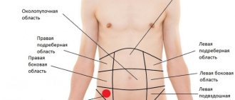
Where is the duodenum located and how does it hurt:
The duodenum is located in the epigastric region, above the navel. Presses on the anterior abdominal wall.
Two ducts enter the intestine from the gallbladder ducts, as well as from the pancreas. This place is considered the main one; all digestive enzymes go here. Amylase, lipase, protease begin the breakdown of food.
Based on this, there are five forms of duodenum:
- Up to 60% - horseshoe shape.
- Up to 20% - folded form.
- Up to 11% - V - shaped.
- Up to 3% - C - shaped.
- Up to 6% - ring-shaped.
- The mucous membrane itself (absorption of fats, amino acids, glucose).
- Submucosa.
- Muscular layer (motor-evacuation function).