This site was made by experts: toxicologists, narcologists, hepatologists. Strictly scientific. Tested experimentally.
Author of this article, expert: Gastroenterologist-hepatologist Ekaterina Kashukh
Briefly: Obstructive jaundice occurs due to obstruction of the flow of bile from the gallbladder. Bile contains a substance called bilirubin, which, when accumulated, turns a person yellow. Treatment and symptoms depend on the disease that caused the jaundice.
- Is obstructive jaundice dangerous?
- What happens to the liver
- What symptoms should you be wary of?
- Diagnosis of obstructive jaundice
- How to be treated
- Prevention of obstructive jaundice
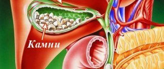
Obstructive jaundice (also called subhepatic or mechanical) is a disease that is directly related to disruption of the normal outflow of bile due to an obstruction outside the liver. There can be a large number of reasons why the disease occurs, but the most common according to statistics are cholelithiasis and space-occupying formations of the hepatobiliary system. Because of this, the level of bilirubin in the blood increases, which leads to yellowing of the skin and whites of the eyes, and itchy skin.
Is obstructive jaundice dangerous?
Obstructive jaundice is a dangerous condition, since a mechanical obstruction to the normal outflow of bile leads to disruption of the functioning of the liver and the body as a whole. The amount of bilirubin in the blood increases, which is toxic to the nervous system.
Reasons for the development of obstructive jaundice:
- malformations of the biliary tract and duodenum (cysts, diverticula, atresia);
- benign diseases of the biliary tract - cholelithiasis, narrowing (stricture) of the ducts;
- inflammatory diseases - acute cholangitis, acute pancreatitis, cholecystitis;
- tumors of the gallbladder and ducts, pancreas, duodenum;
- parasitic diseases of the liver and bile ducts - echinococcosis, alveococcosis, opisthorchiasis.
Gallstone disease often causes obstructive jaundice.
Complications and prevention
The described disease is often provoked by complications. This form of jaundice is considered the most dangerous.
Negative consequences:
- Cancer development
- Cirrhosis
- Severe forms of anemia
- Sepsis
- Inflammatory processes
- Chronic intoxication
To reduce the risk of pathology, as well as eliminate the likelihood of complications, it is recommended to follow prevention. It is aimed at preventing pathologies in which the outflow of bile is disrupted.
Prevention measures:
- Rejection of bad habits
- Competent treatment of infectious diseases
- Proper nutrition
- Removing worms
- Antiviral vaccination
- Compliance with hygiene standards
What happens to the liver
Obstructive jaundice develops due to a violation of the release of conjugated bilirubin through the bile ducts. Stagnation in the ducts leads to increased pressure in them, dysfunction and death of working liver cells (hepatocytes).
As hepatocyte function deteriorates, the uptake of bilirubin from the blood also suffers, causing the amount of unconjugated bilirubin to increase. Due to a decrease in the secretion of bile into the intestines, digestion is disrupted, and the number of “bad” microbes in the small intestine increases.
Features of the disease
The condition in question, also called resorption (acholytic, obstructive, subhepatic) jaundice, or extrahepatic cholestasis, develops due to the appearance of a mechanical obstacle that prevents the free flow of bile into the lumen of the duodenum. At the same time, the content of bilirubin in the plasma increases - a reddish-yellow pigment compound formed during the breakdown of red blood cells and excreted with bile secretion through the intestines. Hyperbilirubinemia develops, causing external icteric manifestations.
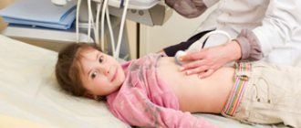
Symptoms of jaundice in adults indicate the severity of the disease. And for most newborns this is a common physiological phenomenon that develops in 60% of cases. It is safe for the body and does not require medical intervention. Obstructive jaundice in newborns is rare and is caused by congenital anomalies. This requires targeted treatment, and surgery may be required.
What symptoms should you be wary of?
Depending on the reasons that caused stagnation of bile or blockage of its outflow, obstructive jaundice can develop abruptly or gradually. In the first case, the cause may be an exacerbation of cholelithiasis, in the second - a tumor.
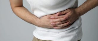
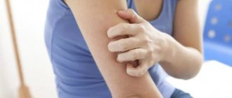
Often the development of yellowing of the skin and whites of the eyes is preceded by itchy skin, which intensifies with the appearance of jaundice. This means that the disease is progressing.
Symptoms such as:
- decreased appetite;
- pain in the upper abdomen;
- nausea;
- abnormal stool, discoloration;
- darkening of urine.
In general, jaundice is not a separate disease, but a syndrome within a particular disease. Therefore, the symptoms are determined by the underlying disease.
Nutrition
Proper therapeutic nutrition is very important for obstructive jaundice (ICD 10 K83.1). Before surgery, the diet is aimed at reducing the load on liver cells. In the postoperative period, the goal of a therapeutic diet is to accelerate the recovery process of the body as a whole.
It is necessary to maintain a drinking regime and drink at least two liters of liquid. Such a measure will speed up the process of removing bilirubin and reduce the load on the central nervous system, lungs and kidneys.
Diagnosis of obstructive jaundice
If obstructive jaundice is suspected, the patient is usually hospitalized. Next, a survey is carried out, including the following activities:
- general blood analysis;
- general urine analysis;
- blood chemistry;
- tests for viral hepatitis A, B, C, E;
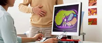
- Ultrasound of the abdominal cavity;
- or abdominal MRI if necessary;
- MR cholangiopancreatography if necessary;
- endo-ultrasound if necessary.
The severity of jaundice is determined by indicators such as bilirubin (its direct and indirect fractions), aminotransferases (ALT, AST), and alkaline phosphatase. The higher their level, the stronger the jaundice and the worse the consequences.
Diet
It is very important to follow a diet for this disease . It is important to eliminate unnecessary mechanical and chemical effects, therefore:
- cooking takes place without salt ;
- meals should be 5-6 times a day ;
- mucous soups into ;
- liquid porridges can be either milk-based or water-based;
- Vegetable juices, compotes, jellies are allowed ;
- consumption of wheat bread is limited ;
- meat can only be pureed ;
- sauces, mayonnaise, pastry creams are prohibited ;
- flour foods are limited ;
- The diet should contain fiber of plant origin (fresh fruits, vegetables).
Reference! It is important to ensure that the food is low-fat and steamed.
Obstructive jaundice is a manifestation of many diseases . It is important to diagnose this pathological process in a timely manner and carry out appropriate treatment. This will help avoid adverse effects that may arise from the said condition.
How to be treated
Treatment methods for obstructive jaundice are dictated by the disease that led to it:
- In case of acute inflammation (acute cholecystitis, acute pancreatitis), emergency surgical intervention is usually required.
- For tumors of the liver, bile ducts, and pancreas, treatment depends on the stage of the disease, the possibility of removing the tumor or only palliative intervention.
- Along with the main methods of treatment, in some cases, blood filtration procedures from toxic bilirubin and plasmapheresis are performed.
The prognosis for obstructive jaundice is also determined by the underlying disease.
Treatment
The main goals of treatment are:
- elimination of bile stagnation;
- prevention and treatment of liver damage.
Both conservative and surgical treatment methods are used.
Conservative treatment is based on:
- carrying out infusion therapy (Reopoliglyukin, Insulin with glucose, Hemodez);
- vitamin therapy;
- hepatoprotectors (Karsil, Essentiale, Gepral);
- amino acid preparations (Methionine, Glutamic acid);
- means that improve blood circulation (Trental, Methyluracil);
- inhibitors of gastric secretion (Omez, Ranitidine, Razo, Famotidine);
- antibacterial agents (Ceftriaxone, Imipinem).
Antibiotics are prescribed to prevent acute inflammation of the bile ducts . Otherwise, complications may occur in the form of liver abscesses (limited foci of inflammation), impaired functioning of the kidneys and brain. Surgical treatment is used to restore the patency of the bile ducts. This operation is performed by decompressing the bile ducts . This surgical intervention can be either minimally invasive and performed with minimal trauma (ERCP, bougienage, stenting), or direct, which requires opening the anterior abdominal wall. If after treatment an increased secretion of fat in feces is observed, then fat-soluble vitamin substances must be administered.
Prevention of obstructive jaundice
In some pathologies (for example, cholelithiasis), obstructive jaundice is easier to prevent than to treat. The risk of its development is assessed by the doctor; if the risk is high, cholecystectomy is discussed: planned removal of the gallbladder containing stones.
However, many diseases that provoke obstructive jaundice cannot be prevented. For example, congenital pathology of the bile ducts or duodenum develops beyond our will.
In general, following the rules of a healthy lifestyle helps reduce the risk of developing most acquired liver diseases.
You can contact the hepatologist in the comments. Don't hesitate to ask!
This article was last updated: 05/23/2020
Didn't find what you were looking for?
Try using search
doctor or administrator.
Read the dictionary of terms.
Expert author: Gastroenterologist-hepatologist Ekaterina Kashukh
Indications for drainage of the bile ducts
Oncologists prefer to perform external internal or, if technically feasible, external drainage of the bile ducts in patients with obstructive jaundice of tumor origin. Both methods are quite effective in preoperative preparation for radical surgery or as a final treatment method. Their advantage is:
- constant monitoring of bile flow;
- the possibility of active removal of blood, pus, and clots from the bile ducts;
- washing the ducts with aseptic solutions;
- dynamic x-ray monitoring of the location of the drainage tube.
Unlike external-internal drainage, bile completely flows out through the external drainage of the gallbladder. The disadvantage of external drainage of the bile ducts compared to external internal drainage is the complete flow of bile through the drainage to the outside. To compensate for the vital substances contained in bile, patients are forced to drink their own bile or medical personnel administer it through a nasogastric drainage.
Internal endoprosthetics of the bile ducts is performed after the elimination of jaundice. This is the final stage of treatment for inoperable patients. To successfully perform external or external internal cholangiostomy, oncologists use a set of special instruments: wire guides, special puncture needles, bougies and catheters.
Under local anesthesia using a Shiba needle, the surgeon tightly fills the bile ducts with a contrast agent. A long needle with a diameter of 1.5-1.7 mm performs a puncture of one of the segmental ducts. A conductor wire is then passed through it. The end of the conductor is passed beyond the narrowing, the narrowed area is widened with bougies, and a drainage tube is installed. It is fixed to the skin and the bile ducts are washed with sterile solutions.
This drainage method has disadvantages: there is a risk of bile and blood leaking into the abdominal cavity when the needle is removed, when passing a guidewire or bougienage the canal. Additionally, this complication may be encountered if the outer diameter of the needle is larger than the outer diameter of the guidewire. In order to reduce the number of complications associated with liver puncture, oncologists use the technique of installing a cholangiostomy using a stylet catheter.
Using an endoscope, doctors perform nasobiliary drainage of the biliary tract. Indications for endoscopic drainage of the biliary tract are:
- obstructive jaundice caused by malignant and benign neoplasms;
- acute purulent cholangitis;
- external biliary fistulas;
- damage to the walls of the extrahepatic ducts, retroduodenal perforations;
- acute cholecystitis.
There are no contraindications to endoscopic drainage, except in cases where the tube for drainage of the biliary tract cannot be passed through the area of tumor narrowing. The endoscopic kit for drainage of the biliary tract through the nose includes:
- conductor wire;
- drainages of various shapes;
- a connecting tube for collecting bile and flushing the drainage;
- nasal tube, clamp and spatula.
The operation of endoscopic drainage of the biliary tract includes the following steps:
- cholangiography to determine the level and location of drainage;
- introduction of drainage with a metal guide-conductor;
- removal of the guidewire and endoscope;
- control cholangiography;
- assessment of drainage position;
- transferring the drainage from the mouth to the nose and fixing it on the head.
After using the endoscopic method of drainage of the bile ducts, complications do not develop. They may occur due to the progression of the disease.
In order to eliminate general intoxication caused by the development of mechanical jaundice, a minimally invasive surgical operation is performed to install drainage in the gallbladder. Through small incisions on the anterior abdominal wall, a catheter is inserted into the cavity of the bladder, which ensures the outflow of bile into the duodenum.
The treatment is performed under local anesthesia; in rare cases, general anesthesia may be required. Before the operation, under the control of ultrasound and X-ray waves, the location of the local narrowing is determined, into which during the operation a special needle is inserted to install a drainage tube.
Installation of a drainage tube into the gallbladder is indicated for:
- eliminating stagnation of bile due to narrowing of the bile duct;
- increasing the lumen of the common bile duct (bile duct) to transport cystic secretions into the duodenum;
- antimicrobial therapy: medicinal effects with antibiotics or antiseptic solutions;
- prevention of scarring and fistula formation in the postoperative period;
- carrying out a liver cleansing procedure mechanically.
The decision on drainage is made by the doctor when diagnosing the following diseases:
- Gallstone disease (cholelithiasis) - installation of drainage creates conditions for the timely outflow of bile, as well as small stones (stones) and sand. Drainage is included in the complex of preoperative treatment.
- The acute course of cholecystitis is the prevention of infection when fluid stagnates as a result of inflammation.
- Mechanical jaundice - removal of active enzymes from liver cells, decrease in bilirubin levels.
- Malignant and benign neoplasms – to eliminate blockage of the bile ducts by tumor-like growth.
- Pancreatitis - inflammation provokes swelling of the head of the pancreas, which creates an obstacle to the flow of bile into the initial part of the small intestine.
- Inflammation of the walls of the bile duct (cholangitis), cystic formations.
- Improper development of the biliary tract at the genetic level.
Thanks to the active development of modern medicine, several methods of drainage of the biliary tract have been developed:
- External method - the free end of the drainage tube remains outside the abdominal cavity. This allows you to control the outflow and quality of bile, which is discharged into the bile receptacle. In addition, it becomes possible for therapeutic lavage and administration of medicinal solutions into the cavity of the bladder.
- Mixed (external-internal) - the procedure allows bile to flow freely into the intestines, and if there is excessive accumulation, it is discharged into a special container secured externally.
- Internal drainage is the installation of a prosthesis into the bile duct for permanent expansion of the biliary tract. A radical measure is used if it is impossible to eliminate the stricture in a conservative way.
When choosing a treatment method, a detailed diagnosis of the patient’s condition is carried out to take into account the severity of the condition and the effectiveness of the minimally invasive surgical procedure.
External
Installation of drainage in the bile duct, when one end of the drainage tube remains in the bladder cavity and the other is brought outside the abdominal wall, is called external drainage. The free end of the catheter is located in the bile receptacle (a disposable reservoir for collecting bile, which is replaced as it is filled).
The advantage of this method of drainage is minimal trauma during manipulation and the absence of absolute contraindications to the procedure. Constant access to the gallbladder cavity allows for antiseptic rinsing and administration of medications as prescribed by a doctor. In addition, monitoring of the quantity and quality of bile secreted is available.
Despite all the advantages, installing a catheter for more than 14 days can lead to the development of complications:
- rapid removal of moisture from the liver leads to its dehydration;
- poor drainage care can cause infectious inflammation in the gallbladder;
- installing a catheter for a long period of time leads to the formation of bedsores inside the duct.
Internal
This method of expanding the bile ducts is most optimal for the patient. A drainage tube placed inside the duct allows bile to flow into the small intestine. If there is excessive production, part of the exudate enters the container for collecting bile due to the drainage being removed to the outside.
Thus, the natural process of outflow of bile exudate is preserved to ensure the normal course of reactions of the breakdown of fats and proteins during digestion.
External-internal drainage is most often prescribed as a preparatory stage before installing a permanent prosthesis, as well as for long-term compression of the bile ducts.
Installation of a permanent prosthesis in the duct is indicated for chronic stricture of the bile ducts, formed as a result of congenital anomalies or the proliferation of tumor cells that are not amenable to surgical treatment. In this case, the decision on the advisability of this procedure is made by the oncologist. Percutaneous drainage is performed to improve the quality of life of cancer patients.
During surgical manipulation, the duct is expanded by installing a flexible conductor (endoprosthesis). At the first stage, the prosthesis is inserted in combination with a drainage tube, to which a bag for collecting bile is connected. After 24 hours, the drainage is removed, and the prosthesis continues to provide artificial expansion of the duct.
Clinical picture
The most characteristic sign of mechanical jaundice is yellow skin, sclera and mucous membranes. The anomaly signals dangerous intoxication of the body. The accompanying symptoms of obstructive jaundice are the same for adults and children, even newborns:
- lack of appetite, rapid weight loss;
- increased body temperature;
- nausea, sometimes vomiting;
- increasing pain in the epigastric region and hypochondrium on the right;
- colorless, loose stools, but concentrated dark urine;
- increase in liver size;
- skin itching.
In rare cases, a patient with obstructive jaundice develops xanthelasmas (yellow or golden cholesterol plaques) on the eyelids.