The active rhythm of life, frequent stress and poor environmental conditions lead to the fact that the human gastrointestinal tract suffers first of all. It performs an important function that saturates the body with useful substances. Therefore, if there are disturbances in the gastrointestinal tract, you should consult a specialist and, if necessary, undergo a therapeutic course.
Functions performed by the gastrointestinal tract
The structure of the human gastrointestinal tract includes many stages, each of which performs certain actions in the process of digesting food. The main responsibilities of the gastrointestinal tract include:
- Motor-mechanical activity. Food is broken down, moves through the esophagus and is eliminated from the body.
- Secretory task. Proper and complete digestion occurs with the help of enzymes, bile and gastric juice.
- Suction function. Allows the body to absorb essential elements from food.
Large intestine Gastrointestinal tract
The large intestine is the final, lower lobe of the human digestive tract, where water is absorbed and chyme (food gruel) formed in feces is formed.
It also has its own subsections: the cecum with the appendix, the colon and rectum, which have separate subsections:
- ascending colon,
- transverse colon,
- descending colon
- sigmoid colon.
Blood enters the intestine from the inferior and superior mesenteric arteries, and the blood flows out through the mesenteric veins. Sensitive innervation of the gastrointestinal tract is carried out using spinal sensory fibers and nerves, and motor innervation is carried out using sympathetic and parasympathetic nerves.
The walls of the intestine, both large and small, are made up of the submucosa, the muscular and serous layers, and the mucous membrane. The very first, mucous membrane contains the epithelium, the lamina propria and the muscularis lamina.
The mucous membrane forms outgrowths in the intestinal lumen. Absorption increases and occurs due to outgrowths from the plasma membrane that form microvilli in the small intestine. Crypts are tubular depressions that produce various ingredients of intestinal juice.
The mucous membrane of the colon does not have villi, but contains a large number of crypts. This membrane contains lymphoid tissues, represented by single and/or group lymphatic follicles. Circular smooth muscle fibers form the muscular lining of the intestine.
Features of blood flow during digestion
The functions of the gastrointestinal tract directly depend on the activity of the blood supply to the digestive organs.
The functions of the human gastrointestinal tract directly depend on the nutritional activity of the organs. After eating food, the flow of blood to the esophagus increases, but only in those sections that are involved in the digestion process. Over time, the blood flow increases even more and remains in an elevated state for 7 hours. This is due to the amount of food consumed and its chemical composition. After digestion and assimilation of the necessary elements, blood flow in the gastrointestinal tract decreases.
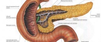
Primary digestion in the stomach
The stomach also has peculiar muscles on its walls, the contraction of which causes the expulsion of chyme.
After chewing and passing through the canals, the food enters a pouch-like expansion, which can change its size as needed. The stomach is located immediately after the esophagus, which determines a large number of diseases associated with an improper diet.
The connection with the esophagus occurs through the cardinal foramen, and with the first section of the small intestine through the pylorus. The internal cavity of this organ is covered with a mucous membrane, which has a huge number of different glands. Through them mucus and various enzymes, hydrochloric acid and other substances are produced. If we consider the structure and purpose of the stomach, we can say that its work is associated with mixing and partial digestion of food. Gastric juice has the primary chemical effect.
Gastric juice deserves special attention. It is produced by special glands that are located in the mucous membrane. The composition of gastric juice includes:
- The enzyme pepsin.
- Hydrochloric acid.
These substances break down the incoming food mass into simpler elements. Gastric juice has a greater effect on protein, since under its action this component is broken down. Under the influence of acid and enzymes, food is digested and becomes a liquid substance, which is subsequently subjected to major processing. In medicine, it is customary to call the substance in question chyme. The stomach also has peculiar muscles on its walls, the contraction of which causes the expulsion of chyme.
The role of nerves in food digestion
Under conditions of parasymptomatic and symptomatic innervation, the activity of the digestive section of the body is regulated. The anatomy of the distribution of nerves leads to the fact that in the first case there is an increase in the functioning of the gastrointestinal tract, and in the second there is a decrease in the level of digestion. In this case, the nerve signal conduction circuit involves 2-3 neurons, which are responsible for stimulating or inhibiting the digestion process.
Disturbances in one system that controls the process leads to disruption of digestive functions.
Major processing in the small intestine
If a disease occurs in one department, the entire digestive process may be disrupted.
This is due to the fact that all organs of the digestive system are interconnected. The length of the small intestine is up to 4.5 meters. It is located in the form of loops. In this case, the area of the internal cavity expands due to the presence of a huge number of villi. The juice of the human intestinal tract is secreted in this section, since the internal cavity is filled with glands. People who assume that food digestion takes place in the stomach are wrong - the main work of excreting and separating nutrients takes place in the gastrointestinal tract. The villi are responsible for transferring beneficial microelements into the blood and lymph, which transport them throughout the body.
The small intestine is divided into:
1. The duodenum is the initial section that receives chyme from the stomach.
2. Jejunum - middle section. This name was given because after the death of a person, when the abdominal cavity is opened, it always turns out to be empty.
3. The ileum is the last section, which many do not separate from the previous one, but there are still significant differences - it is wider, the walls are thicker.
It is in these departments that the main processing of incoming food occurs, the separation of useful components from unnecessary ones, as well as the absorption of necessary elements into the blood and lymph.
Main diseases of the digestive tract
Gastritis is a common disease of the digestive system.
Different parts of the gastrointestinal tract react differently to irritants, so there are many different pathologies, common among which are:
- Gastritis of various origins. A healthy stomach has a solid layer of mucous membrane, which begins to collapse under the influence of irritants.
- Colitis. The disease affects the intestinal area and leads to inflammation of its walls. If the pathology is not treated, heavy bleeding may develop and pose a threat to human life.
- Viral type hepatitis. The affected area is the filter of the human body, i.e. the liver. They are usually provoked by various viruses that penetrate the body.
- Cirrhosis of the liver. A chronic type of disease that leads to the inevitable death of the patient.
- Ulcer. The integrity of the tissues of the gastrointestinal tract is compromised, which can lead to life-threatening complications.
- Dysbacteriosis. In this case, there is a change in the normal intestinal microflora, which leads to problems in the functioning of the gastrointestinal tract.
- Cholecystitis. The gallbladder is affected, which leads to painful symptoms, nausea, and vomiting.
- Appendicitis. The vermiform appendix of the cecum becomes inflamed, and the symptoms depend on the form of the disease.
The structure of the intestinal wall
For those who want to know everything about the intestines, its structure, function and anatomical structure, it is important to understand what the structure of the intestinal wall is. The anatomy of the intestine influences the functioning of the digestive system.
Another article on this topic: Causes and treatment of gas formation in the intestines in women
The intestine consists of 4 layers, each of which has a huge number of capillaries and arteries. These layers are arranged in order as follows:
- The first layer is the mucosa with an epithelial layer. It contains the Luberkühn glands, which look like small villi with crypts. The muscle plate is also located here.
- Next is the submucosal part . Everything on its surface is connective tissue, where nerves and blood vessels are located. This layer has a complex structure of collagen fibers, nerve plexus, and connective reticular fibers.
- The third section contains the muscular layer. Between it and the submucosa is the Auerbach nerve plexus.
- The last layer consists of connective tissue. This is a serous layer that tightly covers the epithelium, like a protective membrane.
Having figured out what the intestinal wall is made of, you can understand how the intestines are structured, what they look like and how they work. On which side is it vulnerable, and on which side is it more protected from external influences?
What causes a digestive system disorder?
Frequent overeating can cause digestive disorders.
The normal functioning of the human gastrointestinal tract can be disrupted due to the following factors:
- Unhealthy diet. Frequent overeating or fasting, unsystematic eating, rapid rhythm of food absorption, as well as imbalance of foods lead to the development of various problems in the digestive system.
- Low level of ecology. Directly affects the quality of consumed products and water, which irritates the mucous membrane of the digestive system.
- Addictions. Nicotine and alcohol do not help maintain healthy microflora in the body.
- Medicines. The drugs must be taken with caution, since they directly affect the gastrointestinal tract.
- Genetics. If relatives have abnormalities in digestion, then you need to pay special attention to your health.
- Poor level of sanitation. Cooking food requires compliance with a number of rules that help avoid the entry of harmful microorganisms into the body.
- Endocrine disorders. They can also lead to serious gastrointestinal disorders.
Structure of the small intestine
The longest organ is the small intestine. It starts from the stomach and ends at the beginning of the large intestine. The physiology of the work of this organ lies in various digestive processes. The intestine has a mesentery, which has two parts. It connects the intestines to the abdominal cavity.
This part includes 3 sections, which do not have any partitions between them. Each department has its own zones that perform their functions.
Department of the duodenum
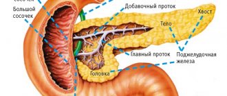
The section of this intestine is the beginning of the organ. The length of the organ is about 30 cm. It is located in the pancreas area. It includes the bile and pancreatic ducts. Therefore, this department is responsible for the high-quality digestion of food consumed by humans .
Bile and gastric juice react, affecting food, which breaks down into elements, begins to be absorbed and supplies all the necessary substances.
Section of the jejunum
This section is located at the very top of the small intestine . It got its name because it is always empty, regardless of food intake. Its shell consists of two layers of smooth muscle tissue.
Most often, this section of the small intestine suffers from ascariasis and enteritis. This is where cancerous tumors form.
Another article on this topic: Intestinal obstruction: symptoms and treatment
Aerial part of the intestine
The aerial part of the small intestine is located in the lower half and is completely covered by the peritoneum. The average length is more than 2.5 m. With a large number of vessels and capillaries. For women it may be slightly less. After death, it stretches almost 2 times.
The walls of the aerial section have 2 layers. They actively contract, so they are responsible for peristalsis. The main feature of this department is the production of neurotensin, which affects the drinking and eating reflex.
Symptoms characteristic of gastrointestinal disorders
Heartburn is a common symptom of increased acidity in the digestive system.
Unpleasant manifestations develop quite clearly, so it will not be difficult to suspect abnormalities in the functioning of the digestive system. A general range of signs includes:
- Pain. It can develop as a result of increased intestinal work or with the development of an ulcer.
- Heartburn. It is one of the most common symptoms of high acidity. The sensation spreads down the esophagus and is accompanied by a lump or a bursting sensation.
- Chest pain. They indicate problems in the gastrointestinal tract, and are also similar to symptoms of cardiovascular abnormalities.
- Belching. If a person has a sick stomach or duodenum, then unpleasant gas emissions are felt.
- Dysphagia. The swallowing reflex does not work properly, causing food to enter the nose or trachea.
- Nausea. Often observed with the development of gastritis or ulcers, as well as with poor bowel function.
- Flatulence. Heaviness and swelling are observed in the peritoneum, which are accompanied by attacks of pain.
- Problematic chair. This includes problematic bowel movements and diarrhea, indicating the development of dysbiosis, ulcers or pancreatitis.
Diseases of the gastrointestinal tract[ | ]
Stomatitis[ | ]
Main article: Stomatitis
Stomatitis
(from ancient Greek στόμα - mouth + -itis - inflammation) - the most common lesion of the oral mucosa. The causes of stomatitis have not yet been fully elucidated; most likely, its development is associated with the reaction of the immune system to irritants. With this disease, the oral mucosa becomes swollen, painful, hyperemic, and may be covered with a white or yellow coating. Hypersalivation (increased salivation) is noted. Bleeding gums and bad breath may occur. Diseases of the gastrointestinal tract, such as gastritis, duodenitis, colitis, as well as helminthic infestation, can cause catarrhal stomatitis.
Esophagitis[ | ]
Main article: Esophagitis
Esophagitis
(novolat. oesophagitis from ancient Greek οἰσοφάγος - esophagus + -itis - inflammation) - a disease of the esophagus, accompanied by inflammation of its mucous membrane. Causes of esophagitis:
- the most common cause is gastroesophageal reflux, which leads to damage to the esophageal mucosa due to the effects of the acid-peptic factor. In case esophagitis is caused by reflux, it is called reflux esophagitis
; - infections (most often Candida fungi, herpes simplex virus, cytomegalovirus). These infections most often occur in patients with reduced immunity, in particular in those suffering from AIDS or receiving immunosuppressive therapy, glucocorticoids, and antitumor chemotherapy;
- a chemical burn with an alkali or acid, a solvent (for example, gasoline, acetone), or a strong oxidizing agent such as potassium permanganate can also cause esophagitis. Such esophagitis is usually observed in children after an accidental test or in adults after a suicide attempt using an alkali, acid, solvent or oxidizing agent. Often observed in alcoholics - in this case, the damaging factor is ethyl alcohol;
- Physical damage to the esophagus due to radiation therapy or tube insertion can also cause esophagitis.
Reflux esophagitis[ | ]
Main article: Gastroesophageal reflux disease
Gastroesophageal reflux disease
(
reflux esophagitis
) is a chronic relapsing disease caused by spontaneous, regularly repeated reflux of gastric and/or duodenal contents into the esophagus, leading to damage to the lower esophagus. The development of gastroesophageal reflux disease is facilitated by lifestyle features (stress, work associated with an inclined position of the body, obesity), pregnancy, smoking, nutritional factors (fatty foods, chocolate, coffee, fruit juices, alcohol, spicy foods), as well as taking peripheral concentration of dopamine drugs (phenamine, pervitin, other phenylethylamine derivatives). The following reasons contribute to the development of gastroesophageal reflux disease:
- decreased tone of the lower esophageal sphincter;
- decreased ability of the esophagus to cleanse itself;
- the damaging properties of the refluxant, that is, the contents of the stomach and/or duodenum thrown into the esophagus;
- inability of the mucous membrane to resist the damaging effects of the refluxant;
- impaired gastric emptying;
- increased intra-abdominal pressure.
Heartburn[ | ]
Main article: Heartburn
Heartburn
- a feeling of discomfort or burning behind the sternum, spreading upward from the epigastric (epigastric) region, sometimes extending to the neck. Popular patient literature states that heartburn is the result of gastric acid affecting the lining of the esophagus from the stomach as a result of gastroesophageal reflux or regurgitation[4]. Other sources note that, in addition to hydrochloric acid, pepsin, bile acids, lysolecithin, and pancreatic enzymes also play a damaging role [5].
Chronic gastritis[ | ]
Main article: Gastritis
Chronic gastritis
(Latin gastritis, from ancient Greek γαστήρ (gaster) “stomach” + -itis inflammatory or inflammatory-dystrophic changes in the mucous membrane) is a long-term recurrent inflammatory lesion of the gastric mucosa, occurring with its structural restructuring and disruption of the functions of the stomach. Chronic gastritis often develops asymptomatically[6].
There are two main forms of chronic disease: superficial and atrophic gastritis. These terms, based on the results of endoscopic studies of the gastric mucosa, were first proposed in 1948 by the German surgeon R. Schindler
). These terms have received universal recognition and are reflected in the classification of gastritis according to ICD-10. The division is based on the factor of preservation or loss of normal glands, which has obvious functional and prognostic significance [7]. In addition to the two main forms, there are also special forms of chronic gastritis: atrophic-hyperplastic gastritis (or polypous, “warty”), hypertrophic gastritis, giant hypertrophic gastritis (Menetrier’s disease), lymphocytic, granulomatous (Crohn’s disease, sarcoidosis, Wegener’s granulomatosis of gastric localization) , collagen, eosinophilic (synonym for allergic), radiation, infectious (gastrospirillum, cytomegalovirus, yeast-like fungi Candida) [8].
Chronic duodenitis[ | ]
Main article: Duodenitis
Chronic duodenitis
(duodenitis; anat. duodenum duodenum + -itis) is an inflammatory disease of the duodenum, most often only the mucous membrane. The occurrence of chronic duodenitis is promoted by irregular nutrition with frequent consumption of spicy, irritating, too hot food. Secondary chronic duodenitis is observed in chronic gastritis, gastric and duodenal ulcers, chronic pancreatitis, giardiasis, food allergies, uremia. In addition to the direct effect of the irritating agent on the mucous membrane of the duodenum, the proteolytic effect of active gastric juice on it (with trophic disorders, dyskinesias) is important in the pathogenesis of chronic duodenitis.
Gastroduodenitis[ | ]
Main article: Gastroduodenitis
Chronic gastroduodenitis
- (Latin gastroduodenitis; other Greek γαστήρ stomach + duodenum duodenum + -itis - inflammation) - inflammatory disease of the mucous membrane of the duodenum and pyloric zone of the stomach. The causes of gastroduodenitis are divided into endogenous (internal) and exogenous (external).
Among endogenous
The causes of gastroduodenitis are given great importance to increased acid formation, decreased mucus formation, and disruption of hormonal regulation of secretion. In addition, diseases of the liver and biliary tract, and endocrine pathology predispose to the development of gastroduodenitis.
Among exogenous
etiological factors include physical ones, such as eating spicy, cold or hot food, and chemical ones (exposure to pesticides). The most important factor is the entry of the bacterium Helicobacter pylori into the digestive tract.
Duodenogastric reflux[ | ]
Main article: Duodenogastric reflux
Duodenogastric reflux
- reflux of the contents of the duodenum into the stomach. The cause of reflux is insufficiency of the pyloric closure function, chronic duodenitis and increased pressure in the duodenum[9]. Duodenogastric reflux leads to damage to the gastric mucosa, mainly adjacent to the duodenum of the antrum of the stomach, by bile acids, their salts, pancreatic enzymes, lysolecithin and other components of duodenal contents [9].
Regarding duodenogastric reflux in healthy people, gastroenterologists have slightly different opinions, which are expressed in formulations. So, some believe that duodenogastric reflux occurs
in healthy people[10], others write more specifically: duodenogastric reflux
is constantly present in healthy people
, takes up about 40% of the time of day and intensifies at night[11].
Enteritis[ | ]
Main article: Enteritis
Chronic enteritis
(from ancient Greek ἔντερον - intestine) - inflammation of the small intestine. Chronic enteritis can be the result of poor nutrition (systematic eating disorders, abuse of spicy foods, strong alcoholic drinks, etc.), helminthiasis, giardiasis, geotrichosis, chronic intoxication with certain industrial poisons (for example, lead compounds), long-term uncontrolled use of drugs (for example, saline laxatives, broad-spectrum antibiotics), some congenital diseases characterized by impaired synthesis of certain enzymes in the intestines, and so on. Atrophy of the mucous membrane gradually develops, its villi are smoothed out, the production of intestinal enzymes decreases, and absorption is impaired. Patients are concerned about rumbling in the intestines, mild pain in the umbilical region, nausea, weakness, diarrhea (mainly with enterocolitis). Due to malabsorption in the intestine, various nutritional disorders can occur. Recognition of enteritis is helped by studies of feces, cavity and parietal digestion, and others.
Colitis[ | ]
Main article: Colitis
Chronic colitis
- inflammatory disease of the inner lining of the colon. Chronic colitis is considered as a fading of the symptoms of acute colitis, with periodic exacerbations.
Proctitis[ | ]
Main article: Proctitis
Proctitis
- is an inflammation of the mucous membrane of the rectum and sigmoid colon. It is a consequence of an untreated acute disease or has a specific nature - tuberculosis, syphilitic, gonorrheal, due to helminthic infestation or another. Clinically, it is manifested by a periodic feeling of discomfort in the rectum, a feeling of incomplete emptying, periodic exacerbations, accompanied by increased frequency of stools mixed with mucus and sometimes blood, and a painful urge to defecate. Chronic inflammatory processes can lead to the development of ulcers on the intestinal mucosa and the formation of fistulas[12].
How is diagnosis carried out?
FEGDS allows you to identify pathologies of the mucous membrane of the digestive organs.
If unpleasant symptoms develop, a person is advised to seek help from a specialist in order to identify abnormalities at an early stage of development. The functions of the gastrointestinal tract are checked using the following methods:
- FEGDS. Allows you to study the mucous membrane of the esophagus, stomach and features of the duodenum. The technique is capable of identifying pathologies such as esophagitis, gastritis, and ulcers.
- Colonoscopy. It is used to determine the causes that provoked problems in the colon.
- Radiography. It is usually performed after injecting a barium solution into the esophagus, which does not affect the patient's health.
- Capsule endoscopy. The patient swallows a capsule that contains a chamber and moves throughout the digestive system. The device allows you to identify abnormalities of the stomach and other parts of the gastrointestinal tract without discomfort.
How does the digestive system work?
It has a complex structure. Each organ in a healthy body functions in a certain sequence, without any failures, which guarantees high-quality food processing and human well-being. This is due to the characteristic structure of the elements and the functions they perform.
The digestive system is represented by the following organs:
- salivary glands;
- liver;
- gallbladder;
- pancreas;
- stomach and other parts of the gastrointestinal tract.
The salivary glands are located in the oral cavity. Their structure allows the production of a certain amount of secretion necessary for the normal formation of a food bolus and its further movement. The liver is a kind of filter; it helps to release beneficial substances and eliminate toxins from the body. The gallbladder produces bile, which is directly involved in the digestion process. The stomach is responsible for processing incoming food and its further movement to the intestines. The pancreas secretes special enzymes involved in the digestion process.
Each of the presented elements of the digestive structure performs its own specific job and is responsible for the normal movement, breakdown and processing of incoming products. It is difficult to imagine human life without the normal functioning of the digestive system.
Treatment of gastrointestinal pathologies
Gastrointestinal diseases are different, so the therapeutic method is selected individually, after conducting a series of examinations and making an accurate diagnosis. Most often, a conservative method helps to cure the problem, sometimes surgical intervention is provided. The course is a medicinal therapy, which is supported by folk decoctions that can soothe the irritated mucous membrane and relieve nervous tension provoked by uncomfortable sensations. Maintaining a special diet helps restore intestinal function, which turns into proper nutrition after the end of treatment, which helps strengthen the intestinal structure.
Therapy is selected individually by the doctor based on the diagnostic results.
Methods for preventing pathologies of the gastrointestinal tract
Smoked products irritate the mucous membrane of the digestive tract.
The normal functioning of the human gastrointestinal tract directly depends on compliance with a number of rules:
- Healthy food. It is recommended to exclude fatty, fried, spicy and smoked foods, as they irritate the mucous membrane of the digestive system.
- Fractionality of nutrition. Meals should be divided into 5-6 approaches throughout the day and include small portions.
- Thermal balance. To maintain the health of various parts of the gastrointestinal tract, you need to avoid excessively cold or hot foods and eat food at room temperature.
- Simple carbohydrates. Products with the highest content should be excluded from the menu to prevent intestinal problems.
- Psycho-emotional balance. Disorders of the gastrointestinal tract functions can provoke prolonged stay in a tense state. Therefore, it is important to provide a comfortable environment and eliminate sources of anxiety.
The work of the gastrointestinal tract is a well-coordinated mechanism, each section of which plays an important role in the process of digestion and absorption of food. Therefore, when the work of one of its departments is disrupted, it affects the condition of the entire organism. At the first unpleasant manifestations, you should immediately consult a doctor who will diagnose and prescribe appropriate treatment.
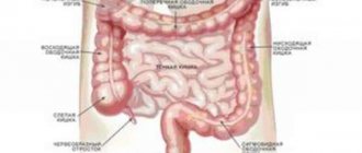
Structure of the large intestine
The large intestine is the end of the gastrointestinal tract. Its length is approximately 2 m, and its diameter is 4-10 cm. Its dimensions can be very clearly seen in the form of a three-dimensional image, which is shown in the picture during diagnosis. The task of this organ is to digest food, absorb water and form feces.
The intestines include:
Blind intestine
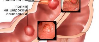
This is a worm-like outgrowth, i.e. appendix . Despite the common belief among ordinary people that the appendix does not play any role in a person’s life, it is a vitally important department. Thanks to it, the level of activity and development of pathogenic microorganisms decreases. It affects the development of beneficial bacteria in the large intestine. The appendix is directly related to the proper functioning of the human immune system, protecting the body from infections and diseases. This is an important part of the large intestine. It is located on the right side of the abdominal cavity.
Its mucous layer contains the Luberkühn gland, which plays an important role in the human body. When it becomes inflamed, a person is diagnosed with appendicitis or typhlitis. If inflammation lasts for a long time, rapid aging of a person occurs. The roots of malignant tumors also develop here.
Colon
The colon is the main part of the small intestine. It does not participate in the work of digestion, in the assimilation, digestion and movement of food. Despite this, it is important for the human body. It is in this section that maximum absorption of water and liquids occurs. If liquid food from the small and large intestines is not completely digested, it enters the colon. From a liquid state it stops in feces.
The following description of characteristics will help you understand the operation. The entire length of this section is 1.5 meters. The diameter, due to the individual characteristics of the body, can be 8 cm. This department consists of subdivisions:
- Ascending, about 20 cm long.
- Transverse colon with a maximum length of up to 56 cm;
- Descending, with a length of up to 22 cm.
Another article on this topic: What to do if you have a prolapsed stomach? Symptoms, causes and treatment
Damage to these areas by bacteria and infections leads to diseases such as:
- constipation;
- diarrhea;
- colitis;
- intussusception.
Sigmoid colon
The sigmoid colon is an important part of the gastrointestinal tract, since the entire functioning of the large intestine depends on its proper functioning. Any ailment can provoke diseases of the entire gastrointestinal tract. The intestine is located in the right hypochondrium, between the descending and colon. It reaches 70 cm in length and 4 cm in diameter. This section is involved in digestion. It can be compared to a large sponge that absorbs liquids and then distributes them throughout the vital systems.
Rectum
Its scientific description is rectum. It is located in the pelvis and ends at the anus. It is small in size: 14-16 cm. In the area of the anus, the diameter is approximately 4 cm, and higher up the intestines it increases to 7.5 cm. The length of the anal canal ranges from 3-5 cm.
The rectum is a kind of reservoir in which processed food and feces accumulate. Then, with the help of the intestinal muscles, they come out. An important component of this section of the intestine is the diaphragm muscles. They do not allow feces to pass out constantly, keeping them inside the intestine until maximum accumulation.