The possibilities of ultrasound examination are very wide. Ultrasound of the abdominal cavity is especially informative, in which vital human organs are located - the abdominal aorta, liver, spleen, gallbladder, pancreas, etc. Where to do an ultrasound?
Thanks to a routine preventive examination, recommended once a year, there is a high probability of diagnosing and preventing further development of the disease at an early stage.
As a rule, patients agree to this type of study because it is painless and does not pose any health risks. Ultrasound allows you to determine with very high accuracy the size, shape, location and structure of the abdominal organs, the condition of the vessels and ducts.
Focal formations are easily visualized by ultrasound, such as pancreatic cancer, liver cancer, tumor metastasis, cysts, abscesses, hematomas, adenomas, and stones. But confirmation of the diagnosis, especially oncopathology, is possible only after histological examination of the tissue. Even small amounts of free fluid (from 100 ml) in the abdominal cavity are diagnosed by ultrasound. We will talk about what is included in an abdominal ultrasound, how to properly prepare for it and how the study is done in our article.
Symptoms for which abdominal ultrasound is indicated
- bitterness in the mouth, hypersalivation
- heaviness in the right hypochondrium
- bursting and nagging pain in the epigastrium after eating
- pain in the abdominal area of various types
- increased gas formation
Diseases for which ultrasound of the PD is necessary
Ultrasound is indicated in the presence or suspicion of the following diseases:
- pancreatitis
- hepatitis
- cholecystitis
- cirrhosis of the liver
- cysts in PD organs
- kidney and gallstones
- neoplasms of PD organs
Preparing for an abdominal ultrasound - detailed recommendations
If you are scheduled for an abdominal ultrasound, preparation is no less important than the examination itself, because this directly affects the information content of the procedure.
3 days before the ultrasound: | The evening before the ultrasound: | Day of ultrasound: |
| Eating a small amount of food every 3-4 hours, about 4-5 times a day. Fluid intake is about one and a half liters daily. | A light dinner is allowed and must be eaten before 20.00. | If the study is carried out in the morning, breakfast is excluded. |
| Products that increase gas formation are completely excluded from the diet: brown bread, baked goods, fruits and vegetables, fatty meat and fish, alcohol, soda, milk, juices, legumes, etc. | Meat and fish products, even dietary ones, should not be included in dinner. | If the study is scheduled after 15:00, a light breakfast is allowed and should be eaten before 11:00. |
|
| 2 hours before the ultrasound, take 5 - 10 tablets of activated carbon or simethicone (2 capsules or 2 teaspoons of emulsion) |
| Adsorbents may be prescribed if the patient has a tendency to flatulence: activated carbon, enterosgel, espumisan, etc. | If laxatives are poorly tolerated, it is recommended to insert a Besacodyl suppository into the rectum (see suppositories for constipation). | If you are prone to flatulence, a cleansing enema may be prescribed in the morning, before the procedure. |
| Enzyme preparations can be prescribed to improve digestion and prevent gas formation: mezim, festal, pancreatin, creon, etc. | If laxatives are ineffective, a cleansing enema is prescribed 12 hours before the ultrasound. | Before an ultrasound, you should not chew gum, suck on lollipops, smoke, or take antispasmodics. |
Ultrasound of the abdominal cavity - preparing the child
- Infants under 1 year of age are recommended not to feed for 2-4 hours and not to drink for about 1 hour before the ultrasound.
- Children 1-3 years old - do not feed for 4 hours and do not drink 1 hour before the ultrasound,
- Children over 3 years old - preparation before an ultrasound is more strict; you cannot eat for about 6-8 hours and drink liquid 1 hour before the examination.
How to prepare for research
Proper preparation for an abdominal ultrasound is the key to a reliable result!
This research method is prescribed by a specialist for the following patient complaints:
- Nausea and vomiting
- Feeling of bitterness in the mouth
- Pain of varying nature and intensity in the abdominal area
- Increased gas formation
- Increased abdominal volume
Ultrasound of the abdominal cavity is also prescribed if tumor processes or infectious and inflammatory diseases are suspected.
To obtain accurate research results, you need to know the specifics of preparing for diagnosis. To this end, you must adhere to the following rules:
- Do not eat or drink liquids 4-5 hours before the ultrasound. You can drink water before diagnosis only if the patient needs to have his kidneys examined or if his gallbladder has been removed.
- To cleanse the intestines, it is advisable to do an enema before undergoing diagnostics. You can also do without an enema - then the specialist will advise you to take medications intended to cleanse the organ. These are drugs such as Senadexin, Fortrans, Prelaxan, Senade, Dufalak, Normaze, which have a laxative effect. You can take adsorbents, for example, Espumisan or Activated Carbon, for two days.
- It is advisable not to drink alcoholic beverages several days before the procedure. It is also not recommended to smoke two hours before the ultrasound.
- It is best to carry out the procedure on an empty stomach in the morning.
- A few hours before the test, you should not chew gum or hard candies.
- After the x-ray, an ultrasound examination is recommended to be performed three days later.
- If the patient took any painkillers the day before, this must be reported to the specialist.
Before diagnosing the abdominal cavity, it is recommended to follow a diet for three days that excludes the following foods from the diet:
- Carbonated drinks
- Milk
- Raw fruits and vegetables
- Fat meat
- Confectionery
- Fruit juices
- Black bread
In addition, you should not eat fatty, spicy or fried foods these days. Limiting such products helps to significantly reduce gas formation in the intestines, which will allow the specialist to better examine the abdominal cavity. It is advisable that the meals be fractional, every three hours.
Executing the procedure
Abdominal ultrasound procedure
In order to carry out the diagnosis, the patient is placed in a supine position on his back. The specialist may ask you to take deep breaths or stop breathing and turn to the side for an accurate check.
A special product in the form of a gel, which is a contrast agent, is applied to the abdominal area. An ultrasound probe is then passed over the areas of the abdomen where the internal organs are located.
The study is carried out using high-frequency ultrasonic waves (from 2.5 to 3.5 MHz). They allow you to obtain a three-dimensional or two-dimensional picture of organs.
A special sensor receives readings and sends them to a computer monitor.
During the ultrasound procedure, the following organs located in the abdominal cavity are examined:
- Pancreas
- Liver
- Gallbladder and its ducts
- Spleen
In addition, with this procedure, you can view the kidneys located behind the abdominal cavity. It is possible to study using ultrasound of the stomach and intestines. However, in this case, it is important to say that the result is not always accurate, since there is an accumulation of air in these organs. Colonoscopy is considered an effective method for diagnosing these organs.
When should you not perform an ultrasound?
- After fluoroscopy of the gastrointestinal tract using contrast (irrigoscopy, gastrography).
- After endoscopy of the gastrointestinal tract (fibrogastroduodenoscopy, colonoscopy).
- After laparoscopy and pneumoperitoneum.
In the first and second cases, a delay of 2 days is made, in the last - 3-5 days. Preparation for abdominal ultrasound in these cases is the same as described above.
The mechanism of action of ultrasound - how it is done
Ultrasound waves easily pass through the skin, are repelled from the organ being studied and display the result on the monitor in black and white. For better contact with the sensor, a special gel-like substance is applied to the patient’s skin.
How often can an abdominal ultrasound be done? If for a routine examination, then once a year, for existing indications - as much as you like, no harm from ultrasound was identified, although studies of this are being carried out all the time. Ultrasound can be destructive, but at much higher powers and exposure time to tissue.
How is ultrasound performed?
Ultrasound of the PD, as a rule, includes a mandatory examination of the gallbladder, liver, retroperitoneum, spleen, pancreas and blood vessels. The remaining organs are optional for examination and are examined according to indications.
The standard study protocol includes:
- determining the location and size of organs
- study of organ structure
- determination of free fluid in the abdominal space (more precisely, confirmation of its absence)
- exclusion of formations, cysts, stones, etc.
Description of the procedure
How is an abdominal ultrasound done? The procedure itself takes on average 15-20 minutes. An ultrasound is performed by an ultrasound specialist, who is assisted by a nurse, filling out a research protocol. There are no painful or uncomfortable sensations during the procedure. A special conductive gel is applied to the contact sensor.
The examination is carried out in a supine position; if necessary, the doctor may ask you to turn over on your side and hold your breath for a few seconds. Using a sensor connected to the monitor of an ultrasound machine, the doctor moves along the skin of the anterior abdominal wall, moving down and to the sides. During the procedure, the doctor will name numbers and medical terms that the nurse will enter into the protocol. Immediately after the ultrasound, you can eat and lead a normal lifestyle without restrictions.
Preparing for a pelvic examination
This ultrasound can be performed on any day of the cycle. The exception is the days of menstruation. To clarify the diagnosis, a repeat ultrasound is performed on the day of the cycle, which is prescribed by a specialist. The location of the pelvic organs is the abdominal cavity. They are located deep, making them difficult to explore.
In order to examine the pelvis (uterus, ovaries, fallopian tubes), a special vaginal sensor is used. This research method is called transvaginal. If the girl is a virgin, then this method is not acceptable for her. Transvaginal ultrasound does not require preliminary preparation and is performed through the anterior abdominal wall. Before starting the study, you need to go to the toilet and empty your bladder.
An hour before the appointed time, virgins drink 3-4 glasses of non-carbonated liquid.
What is included in the examination - the organs examined and ultrasound capabilities
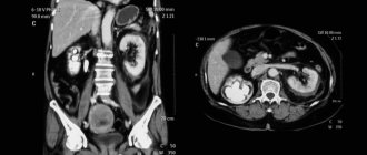
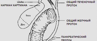
- Liver. Inspected first. Hepatosis, cirrhosis, cysts, and tumors can be diagnosed.
- Gallbladder and ducts. The patency of the ducts, the presence of polyps, gallstones, and the condition of the organ wall are assessed.
- Stomach. Inspected at the time of exclusion of formations.
- Pancreas. Where possible, all shares are valued. Pancreatitis, tumor and pancreatic necrosis may be detected.
- Spleen. The structure, location and size of the organ are assessed. Neoplasms, cysts, and inflammations are excluded.
- Intestines. Most often, only the large intestine is examined. If formations and polyps are detected, the patient is sent for a narrow examination.
- Kidneys. Localization and relative position, dimensions are assessed. Inflammatory changes, conglomerates, tumors and cysts may be detected.
- Bladder. The shape, size, condition of the walls, and contents are assessed.
- Vessels. The abdominal aorta and large vessels feeding the organs must be evaluated. Blood flow and the condition of the vascular wall are determined.
- The lymph nodes. Their size is assessed (an increase is typical for oncological pathology).
- The uterus in women and the prostate gland in men. These organs are located in the pelvis, however, they can be examined. Tumors and inflammatory processes can be detected.
Features of abdominal ultrasound in children
Ultrasound in infants is of particular interest, since in children one year and older, ultrasound is virtually no different from that in adults. Young children are referred for an abdominal ultrasound if:
- the presence of congenital pathologies;
- abdominal injuries;
- abdominal pain and fever of unknown origin;
- routine screening, which is mandatory during the neonatal period.
Ultrasound allows you to assess the condition of the digestive and excretory systems, namely: liver, bladder and ureters, kidneys, gall bladder, pancreas, stomach, intestines. The retroperitoneal space, adrenal glands, arteries, veins and nerve plexuses must be examined.
The procedure is carried out according to the same principle as the examination of an adult, but in the presence of one of the parents, who helps to hold the baby.
This study is necessary to exclude (or confirm) congenital pathologies, confirm the normal condition and functioning of organs according to age standards.
Ultrasound can reveal:
- congenital organ pathologies
- tumors, cysts and polyps of organs
- reactive pancreatitis
- kinks and constrictions of the gallbladder
- hyperplasia, liver cirrhosis and hepatitis
- enlarged spleen
- enlargement of regional lymph nodes
- blood flow disorders
Rules for preparing for diagnostic studies
Rules for preparing for diagnostic studies
For the most accurate diagnosis of diseases, the most modern laboratory equipment is not enough. The accuracy of the results depends not only on the reagents and equipment used, but also on the time and correctness of collection of the test material. If the basic rules for preparing for analysis are not followed, their results can be significantly distorted.
Rules for preparing patients for laboratory tests.
1.Blood test:
All blood tests are taken before radiography, ultrasound and physiotherapeutic procedures.
If the patient is dizzy or weak, warn the treatment nurse about this - your blood will be taken in a lying position.
A general blood test, determination of blood group, Rh factor, biochemical tests are taken on an empty stomach, no less than 12 hours after the last meal.
1-2 days before the examination, exclude fatty and fried foods from the diet.
On the eve of the examination, light dinner and good rest.
On the day of the examination, you cannot have breakfast.
(including drinking tea, coffee or juice), avoid physical activity, take medications, and refrain from smoking.
If you have difficulty stopping medications, you should definitely consult with your doctor.
Drinking water does not affect blood counts, so you can drink water.
We recommend that all tests be taken in the morning, due to the fact that blood counts change significantly during the day and the standards are calculated for this period of the day.
2 days before the examination, you must give up alcohol, fatty and fried foods.
Do not smoke 1-2 hours before blood sampling.
- Before a blood test, you should reduce physical activity as much as possible and avoid emotional excitement. You need to rest for 10-15 minutes. Before donating blood, you need to calm down in order to avoid an unmotivated release of hormones into the blood and an increase in their levels.
- You cannot donate blood immediately after physiotherapeutic procedures, ultrasound and x-ray examinations, massage and reflexology.
- Before a hormonal blood test in women of reproductive age, you should follow the recommendations of your doctor about the day of the menstrual cycle on which you need to donate blood, since the result of the analysis is influenced by physiological factors of the phase of the menstrual cycle.
How to prepare for a tumor marker test?
To ensure that the results of the tumor marker analysis are reliable, be sure to first consult with your doctor .
and follow his recommendations.
Basic rules for preparing for blood tests for tumor markers:
- Blood is given strictly in the morning on an empty stomach, i.e. At least 8–12 hours should pass after the last meal.
- 3 days before the test, you should not drink alcoholic beverages or fatty foods.
- Cancel all physical activity.
- On the day of the test, refrain from smoking.
- Do not use medications.
- When testing for PSA, you must abstain from sexual intercourse for a week.
Patients undergoing treatment for cancer are strongly recommended to undergo testing several times a year.
2.Urine analysis
General clinical urine analysis:
-only morning urine taken in the middle of urination is collected; -morning urine sample: collection is done immediately after getting out of bed, before drinking morning coffee or tea; – previous urination was no later than 2 am; – before collecting a urine test, a thorough toilet of the external genitalia is performed; – 10 ml of urine is collected in a special container with a lid, provided with directions, and the collected urine is immediately sent to the laboratory; – storing urine in the refrigerator is allowed at 2-4 C, but not more than 1.5 hours; -Women should not donate urine during menstruation.
Collection of daily urine:
- the patient collects urine for 24 hours with normal drinking regimen (about 1.5 liters per day); – in the morning at 6-8 o’clock he empties the bladder and pours out this portion, then during the day he collects all the urine in a clean wide-necked dark glass vessel with a lid with a capacity of at least 2 liters; – the last portion is taken at the same time when the collection began the day before, the start and end times of the collection are noted; — the container is stored in a cool place (preferably in the refrigerator on the bottom shelf), freezing is not allowed; – at the end of urine collection, its volume is measured, the urine is thoroughly shaken and 50-100 ml is poured into a special container in which it will be delivered to the laboratory; – be sure to indicate the volume of daily urine.
Collection of urine for microbiological examination (urine culture)
-morning urine is collected in a sterile laboratory container with a lid; – the first 15 ml of urine are not used for analysis, the next 5-10 ml are taken; – collected urine is delivered to the laboratory within 1.5 – 2 hours after collection; – urine can be stored in the refrigerator, but not more than 3-4 hours; – urine collection is carried out before the start of drug treatment; – if it is necessary to evaluate the effect of the therapy, then urine culture is performed at the end of the course of treatment.
3.Analysis in gynecology, urology
For women:
- you cannot urinate for 3 hours before taking a test (smear, culture); – it is not recommended to have sexual intercourse 36 hours before, especially with the use of contraceptives, which can distort the result, as they have an antibacterial effect; – the day before you should not wash yourself with antibacterial soap and douche; – antibiotics cannot be used internally; – you cannot take tests during menstruation.
For men:
- you cannot go to the toilet 3 hours before taking the test; – do not take uroseptics or antibiotics internally; – apply externally solutions with a disinfectant effect, soap with an antibacterial effect; – It is not recommended to have sexual intercourse 36 hours before taking tests.
Sputum analysis
— the analysis is collected in a sterile laboratory container; – before collecting sputum, you need to brush your teeth, rinse your mouth and throat.
4.Ultrasound examinations
Preparation of ultrasound examination of the abdominal cavity and kidneys
- 2-3 days before the examination, it is recommended to switch to a slag-free diet, exclude from the diet foods that increase gas formation in the intestines (raw vegetables rich in plant fiber, whole milk, brown bread, legumes, carbonated drinks, as well as high-calorie confectionery products - pastries, cakes );
- For patients with problems with the gastrointestinal tract (constipation), it is advisable to take enzyme preparations and enterosorbents (for example, festal, mezim-forte, activated carbon or espumizan, 1 tablet 3 times a day) during this period of time, which will help reduce the manifestations of flatulence;
- Ultrasound of the abdominal organs must be performed on an empty stomach; if the study cannot be performed in the morning, a light breakfast is allowed;
- If you are taking medications, notify the ultrasound doctor;
- You cannot conduct research after gastro- and colonoscopy, as well as R-studies of the gastrointestinal tract.
Preparation of ultrasound examination of the pelvic organs (bladder, uterus, appendages in women)
- The examination is carried out with a full bladder, so it is necessary not to urinate for 3-4 hours before the examination and drink 1 liter of non-carbonated liquid 1 hour before the procedure.
- No special preparation is required for transvaginal ultrasound (TVS). If the patient has problems with the gastrointestinal tract, it is necessary to perform a cleansing enema the night before.
Preparation of ultrasound examination of the bladder and prostate in men
- The examination is carried out with a full bladder, so it is necessary not to urinate for 3-4 hours before the examination and drink 1 liter of non-carbonated liquid 1 hour before the procedure.
- Before transrectal examination of the prostate (TRUS), it is necessary to do a cleansing enema.
Preparing for breast ultrasound
- It is advisable to carry out examination of the mammary glands in the first 7-10 days of the menstrual cycle (phase 1 of the cycle).
Ultrasound of the thyroid gland, lymph nodes and kidneys
– do not require special preparation of the patient.
The patient must have with him:
- data from previous ultrasound studies (to determine the dynamics of the disease);
- referral for an ultrasound examination (purpose of the study, presence of concomitant diseases);
- a large towel or diaper.
5. Functional diagnostics.
Functional methods for studying the heart:
Echocardiography (ultrasound of the heart):
- The study is carried out after a 10-15 minute rest.
- Before examinations, it is not recommended to eat a large meal, strong tea, coffee, as well as to carry out after taking medications, physiotherapeutic procedures, physical therapy and other examinations that contribute to the patient’s fatigue (X-ray, radioisotope).
- Know the exact weight.
Studies of wall tone and vascular patency:
Rheocephalography (REG), rheovasography (RVG of the extremities), ultrasound Dopplerography of the vessels of the brachiocephalic region and lower extremities, USDG-BCA, transcranial Dopplerography.
- All these studies do not require special preparation. They are carried out before therapeutic exercises, physiotherapeutic procedures, or taking medications.
6.Endoscopic studies
Fibrogastroduodenoscopy : how to prepare correctly:
- arrive at least 5 minutes before the appointed time;
in the morning on the day of the study before FGDS it is PROHIBITED:
- have breakfast and eat any food, even if the study takes place in the afternoon
in the morning on the day of the study before FGDS IT IS NOT RECOMMENDED:
- smoke
- take medications in tablets (capsules) orally
in the morning on the day of the study before the FGDS is ALLOWED:
- brush your teeth
- do an ultrasound of the abdominal cavity and other organs
- 2-4 hours before drinking water, weak tea with sugar (no bread, jam, sweets...)
- take medications that can be dissolved in the mouth without swallowing or taken with you
- give injections if you do not need to eat after the injection and it is not possible to do it after FGDS
- Before the examination, you need to remove removable dentures, glasses, and a tie.
The night before: easy-to-digest (no salads!) dinner until 18.00.
No special diet is required before FGS (FGDS), but:
- exclude chocolate (chocolate candies), seeds, nuts, spicy foods and alcohol for 2 days;
- when examining from 11 a.m. and later, preferably in the morning and 2-3 hours before the procedure, drink in small sips one glass of still water or weak tea (without jam, sweets, cookies, bread, etc.);
It is important that:
a) the clothes were loose, the collar and belt were unbuttoned;
b) you did not use perfume or cologne;
You promptly warned the doctor about the presence of drug, food or other allergies.
The patient must have with him:
- regularly taken medications (take after examination, and under the tongue or spray for ischemic heart disease, bronchial asthma.. - before examination!);
- data from previous FGDS studies (to determine the dynamics of the disease) and biopsy (to clarify the indications for repeat biopsy);
- referral for an FGDS study (purpose of the study, presence of concomitant diseases...);
- a towel that absorbs liquid or diaper well.
If you are unable to show up at the appointed time, please call the doctor in advance or wherever you made an appointment!!!
Respect yourself and save your doctor’s time!
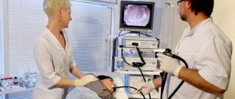
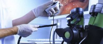
Colonoscopy
How to prepare properly:
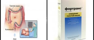
Preparation for colonoscopy using the drug "Fortrans"
Two days before the test:
Recommended diet: boiled white fish, chicken, eggs, cheese, white bread, butter, cookies, potatoes
It is recommended to drink a sufficient amount of fluid - up to 2.5 liters per day (if you do not have diseases for which heavy drinking is contraindicated, consult your doctor about this)
It is not recommended to eat: fruits and berries with seeds, red meat, vegetables, cereals, salad, mushrooms, nuts, grain bread, sweets
The day before the test:
In the morning - a light breakfast of the products recommended above. After breakfast, until the end of the study, you cannot eat solid food, you are only allowed to drink
After breakfast, before 17-00, it is recommended to drink a sufficient amount of liquid to cleanse the intestines - up to 2 liters (you can drink water, low-fat broths, fruit drinks, juices without pulp, tea with sugar or honey, compotes without berries). It is not recommended to take milk, jelly, kefir
At 17:00 you need to prepare a Fortrans solution
For this:
Dilute 1 packet of Fortrans in 1.0 liter of boiled water at room temperature.
The prepared Fortrans solution must be drunk within two hours (from 17:00 to 19:00). Fortrans should be taken in small portions, 1 glass every 15 minutes, in small sips.
At 19.00, drink the second packet of Fortrans using the same method.
1-3 hours after starting to take the Fortrans solution, you should have copious, frequent, loose stools, which will facilitate complete cleansing of the intestines.
If loose stools do not appear 4 hours after the start of treatment or signs of an allergic reaction appear, you should contact medical personnel and refrain from taking the drug again.
On the day of the study:
In the morning at 7.00 it is necessary to repeat taking Fortrans to completely cleanse the intestines of contents (1 packet of Fortrans).
Drink the resulting solution in separate small portions within 1 hour (07-00 to 08-00). You will again have loose stools, which should last until the intestines are completely empty and cleansed.
By 12-00 you will be ready for research. When preparing for a study with Fortrans, enemas are not required!
You need to have with you:
Referral for colonoscopy (if you were referred from another medical institution), conclusions and protocols of previously performed endoscopic studies, ECG (if you have cardiovascular diseases)
The key to a successful colonoscopy is proper preparation of the patient. Preparation for an intestinal examination begins 2-3 days before the scheduled date of the examination. Additional products used to prepare the bowel for examination are recommended.
To reduce the likelihood of unpleasant sensations during and after the examination, an intestinal antispasmodic (a drug that relieves intestinal spasms) Dicetel 50 mg (1 tablet) 3 times a day the day before the examination and 50 mg immediately before the colonoscopy is prescribed. No-shpa, baralgin, spasmalgon and other similar drugs are ineffective.
How to behave after the study?
Immediately after the procedure, you can drink and eat. If the feeling of fullness of the abdomen with gases persists and the intestines are not emptied of remaining air naturally, you can take 8-10 tablets of finely crushed activated carbon, stirring it in 1/2 cup of warm boiled water. It is better to lie on your stomach for several hours after the examination.
7. PREPARATION OF PATIENTS FOR COMPUTED TOMOGRAPHY
You need to know that a computed tomography scan of the abdominal organs after examining the stomach and intestines using a barium suspension can be performed no earlier than 3 days later.
STUDIES THAT DO NOT REQUIRE SPECIAL PREPARATION
Brain
The study usually begins without contrast. The question of the use of intravenous contrast is decided by a radiologist.
Chest organs
Examined without contrast. The question of the use of intravenous contrast is decided by the radiologist and, if necessary, it is administered intravenously by the attending physician directly on the tomograph table.
Liver
The study is performed on an empty stomach, without contrast. The question of the use of intravenous contrast of the liver parenchyma of vessels and ducts is decided by a radiologist.
Liver parenchyma
To contrast the liver parenchyma and its vessels, it is injected intravenously by the attending physician on the tomograph table.
Bile ducts
To contrast the bile ducts, a contrast agent is injected intravenously. The injection is carried out by the attending physician on the tomograph table.
Gallbladder
The study is performed on an empty stomach, usually without contrast. The question of using intravenous contrast for the gallbladder is decided by a radiologist.
Pancreas
The study is performed on an empty stomach. Before the examination, the patient drinks 200 ml of mineral or boiled water, as well as a special mixture, which is prepared by an x-ray technician in the computed tomography room immediately before the examination.
Kidneys
The study is carried out without contrast. The issue of using intravenous contrast for the parenchyma, pelvis, and ureters is decided by a radiologist. If necessary, intravenous jet administration is carried out by a doctor directly on the tomograph table.
Abdominal aorta and inferior vena cava
The study is carried out without contrast. The question of the use of intravenous vascular contrast is decided by a radiologist. Intravenous fluid is carried out by a doctor directly on the tomograph table.
AREAS OF STUDY REQUIRING SPECIAL TRAINING
Retroperitoneal lymph nodes
In the hospital, you need to drink two glasses of water 2 hours before the test. 1 hour before the examination and immediately before the examination (in the computed tomography room), the patient drinks one glass of the mixture prepared by the x-ray technician.
Bladder
5 hours before the test for 30 minutes. You must drink 1 liter of mineral water and a medication prescribed by your doctor (if necessary). Before the examination in the computed tomography room, the bladder is emptied through a catheter, after which 150 ml of oxygen is injected into the bladder through the catheter. The catheter, clamped with a clamp, remains in the bladder for the entire period of the study. All preparatory operations are performed by a urologist.
Female pelvic organs (uterus, appendages)
5 hours before the test for 30 minutes. drink 1 liter of mineral (without gases) or boiled water, if necessary with a mixture of a contrast agent prescribed by a doctor. Breakfast in the morning. Immediately before the examination, the bladder is emptied through the catheter, followed by the introduction into the bladder of a mixture consisting of 50 ml of distilled water and a contrast agent (if necessary). A gauze pad is inserted into the vagina to the level of the cervix. The question of the use of intravenous contrast is decided by a radiologist. All preparatory manipulations are carried out by a gynecologist.
Male pelvic organs
5 hours before the test for 30 minutes. drink a mixture prepared from 1 liter of mineral or boiled water and, if necessary, a contrast agent specified by the doctor. The study is carried out with a full bladder. The question of the use of intravenous contrast is decided by a radiologist. All preparatory procedures are carried out by a urologist.
A CT scan of the abdominal cavity is done on an empty stomach for the reasons that many organs change their shape and volume after eating or drinking a large amount of water, especially those containing gas-forming substances. The information turns out to be somewhat distorted and it is very difficult to describe the resulting picture. Before an abdominal CT scan, you must avoid eating foods that cause gas!
8. RULES FOR PREPARATION FOR DIAGNOSTIC STUDIES IN THE DEPARTMENT OF RADIOISOTOPIC DIAGNOSTICS (Scintigraphy of the kidneys, skeleton)
Dynamic renal scintigraphy and isotope renography are performed after a meal and 2 glasses of liquid (no coffee)
-skeletal bone scintigraphy is performed no earlier than 3 months after radiation and chemotherapy
Contraindications for research in the RDL: relative – high temperature, exacerbation of chronic diseases, breastfeeding, cachexia, children under 1 year of age; absolute contraindication: pregnancy.
9.Preparation of x-ray studies.
X-ray examination of the skull, cervical spine, paranasal sinuses - remove jewelry (chain, earrings, hairpins, piercings).
X-ray examination of hands – remove jewelry (rings, bracelets, watches)
X-ray examination of the pelvis, SIJ, lumbar spine - do an enema.
X-ray examination of the stomach and esophagus in the evening, light dinner in the morning, do not eat or drink. X-ray examination of the intestines (irrigoscopy, irrigography) - light dinner no later than 19.00, the night before and in the morning a cleansing enema is done until the waters are clear. Avoid gas-forming foods (brown bread, vegetables, fruits, carbonated drinks, sour dairy products)
When prescribing survey and excretory urography, careful preparation is required; For 2-3 days, follow a diet to exclude gas-forming foods (brown bread, vegetables, fruits, carbonated drinks, sour dairy products). On the eve of the study, in the evening and in the morning, a cleansing enema until the waters are clear. Light dinner, no later than 19.00.
Survey radiography of the abdominal organs will be performed without preparation, while standing.
Ultrasound of the abdominal cavity - how to evaluate the results of the study
As a rule, at the end of the study, the ultrasound doctor provides a short summary that is understandable to the patient, which may sound like “You are doing well” - the most expected and reassuring phrase. But there may be another conclusion, for example: “You need to see a gastroenterologist (urologist, etc.) for consultation.” But this should not be scary; any, even the most serious, pathologies can be treated, the main thing is not to delay it.
A research protocol is issued, which contains standard columns describing each organ, as well as a doctor’s conclusion. The conclusion indicates all identified pathologies or suspicions of them. If the patient is healthy, the conclusion will sound something like this: The abdominal organs are unremarkable.
Study results - what ultrasound shows
When examining the abdominal organs, their size, location, shape, width of the ducts, and the condition of the external walls are assessed.
Also an integral part of the examination is the examination of blood vessels, including the inferior vena cava, portal and splenic veins. During ultrasound, it is possible to identify areas of tissue change, the presence of foreign objects and stones.
At the end of the examination, the diagnostician issues a protocol to the patient. In some cases, an echogram (photograph) is attached to the ultrasound report. It does not contain vital information and is optional.
Ultrasound signs of some diseases
Disease | Ultrasound sign | |
| Fatty liver |
| |
| Cirrhosis of the liver | Direct signs:
| Indirect signs:
|
| Congestive liver circulatory failure |
| |
| Focal liver pathologies: | Cysts, abscess, area of tumor necrotization, hematoma | An area devoid of echostructure |
| Cancer metastases (poorly differentiated), hepatocellular cancer, malignant lymphoma, sarcoma, adenoma, hemangioma, hematoma, abscess | Area with reduced echostructure | |
| Metastases of cancer (highly differentiated), adenoma, hepatoma, hemangioma, scars, foci of calcification | Area with enhanced echo structure | |
| Malignant liver tumor | Strengthening the echostructure in the center of the area and decreasing the echostructure along the edge of the space-occupying formation | |
| Acute cholecystitis |
| |
| Chronic cholecystitis |
| |
| Cholelithiasis | Direct signs:
| Indirect signs:
|
| Acute pancreatitis | Significant reduction in echostructure | |
| Chronic pancreatitis |
| |
| Pancreatic tumor |
| |
Author:
Sabuk Tatyana Leonidovna hygienist, epidemiologist