Author
Baryshnikov Vladimir Leonidovich
Leading doctor
Candidate of Medical Sciences
Radiologist
COVID-19
Family doctor diagnoses COVID-19 Read more All promotions
MSCT of the abdominal cavity and retroperitoneum
– a modern diagnostic method that provides a high degree of detail and makes visualization of internal organs as convenient as possible. Multislice computed tomography (as the abbreviation MSCT stands for) is based on the use of X-ray radiation. Tissues with different densities transmit X-rays differently. This information is read using special equipment - a tomograph. Multislice tomographs used in “Family Doctor” make it possible to obtain 64 slices with a thickness of 0.625 mm. for one revolution of the gantry (the working part of the tomograph). This information is processed using powerful computer software that reconstructs images of the area of interest in any plane, as well as in 3D (3D). MSCT examination of the abdominal cavity and retroperitoneal space is actively used to assess the condition of organs related to this area, differential diagnosis and detection of diseases in the early stages.
What it is?
Multislice computed tomography (MSCT) is an X-ray method for visualizing the patient’s body. The cola apparatus contains several detectors of radiation, which is supplied in such a way as to completely and evenly cover a certain area of the body. A series of pictures are taken and automatically processed by the software. As a result, the doctor receives a three-dimensional image on the screen.
However, some pathological processes (inflammation, tumors, blood clots in blood vessels) are difficult to detect using simple MSCT, especially if they are small in diameter (2-4 mm). Therefore, to increase the information content of the examination, the patient is injected with a contrast agent, which accumulates differently in different types of tissue. This can significantly increase the information content of diagnostics, which is especially important in oncology or emergency surgery.
Important
Contrast can be administered intravenously (in the vast majority of cases), orally (through the mouth), or rectally (into the rectum). The choice of technique remains with the attending physician.
Indications and contraindications for MSCT of the abdominal cavity
MSCT creates a three-dimensional image of the abdominal cavity and retroperitoneal space
MSCT is a modern non-surgical X-ray diagnostic method, one of the types of computed tomography. It is distinguished by short-term irradiation of organs with a small amount of radiation. Scanning with ultrathin sections makes it possible to detect pathological changes up to several millimeters in size. The data is transformed into a three-dimensional model of the abdominal organs.
Multislice computed tomography is used when diagnosis is difficult, questionable CT results, or the presence of unclear X-ray images. Most organs are hollow, so a contrast agent is used for better visualization. Direct indications for examination are the following situations:
- suspected tumor of the stomach, liver and biliary system;
- inflammatory bowel disease - Crohn's disease, ulcerative colitis;
- pancreatic diseases;
- spleen injury;
- inflammation of the kidneys of various origins;
- urolithiasis disease;
- preparation for surgery.
Indications for MSCT of the abdominal cavity with contrast may include renal vascular disease or existing blood clots in the hepatic arteries and veins. The method is suitable for planning surgery and follow-up. The procedure is also carried out to monitor dynamics during complex courses of treatment.
Multislice tomography with contrast has some contraindications:
- pregnant women, especially in the last trimester;
- nursing women;
- children under 14 years of age;
- allergy to contrast;
- diabetes;
- body weight more than 120 kg;
- severe kidney disease.
The procedure is not recommended for people with metal implants or pacemakers. They give away the background and distort the picture of the indicators.
Indications for use
Typically, MSCT with contrast is prescribed by a medical or surgical doctor after examining the patient. Sometimes it is required in a situation where conventional CT, ultrasound, FGDS or colonoscopy could not find the cause of the patient’s deterioration in well-being. Among the symptoms that are the reason for prescribing diagnostics, it is necessary to highlight:
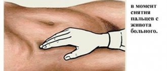
abdominal pain of any location, of varying nature and intensity, which intensifies when pressed;- the presence of tension in the muscles of the anterior abdominal wall;
- detection of blood impurities in the stool (visually or laboratory);
- nausea, frequent or periodic vomiting with bile, mucus or blood;
- marked decrease in appetite in the patient
- traumatic injury to the abdomen (open or closed);
- tendency to constipation;
- decrease in the patient’s body weight (especially against the background of unchanged appetite, diet and physical activity);
- severe weakness, which is accompanied by an increase in body temperature;
- enlarged lymph nodes in the abdominal cavity (detected during palpation or ultrasound examination);
- the presence of signs of hemorrhagic shock (low blood pressure, increased heart rate) without external blood loss.
Appointment for examination
Having determined the need for contrast, we can talk about the reasons that require instrumental diagnostics. So, in what cases is it necessary to perform MRI or MSCT:
MRI: indications for use
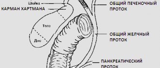
- diseases of the liver or gall bladder: cirrhosis, hepatosis, changes in the parameters of internal organs, parenchymal tissue damage, cholelithiasis;
- Abdominal abnormalities, congenital or acquired, including signs of infiltration;
- non-tumor neoplasms, for example, cysts or hematomas;
- vascular lesions, such as portal hypertension;
- all forms of pancreatitis;
- tumors of varying localization and severity.
MSCT with contrast
- preparation for abdominal surgery;
- injuries in the abdominal area;
- pathological processes in the liver;
- suspicion of urolithiasis or its diagnosis;
- biliary dyskinesia;
- inflammatory diseases of the abdominal organs;
- identification of benign or malignant neoplasms;
- pathologies of blood vessels and heart muscle;
- internal bleeding;
- spinal injuries;
- joint diseases, such as arthrosis or arthritis.
It should be noted here that MSCT of the abdominal organs with contrast allows one to determine not only the presence of tumors in the body, but also to identify the cause of its development. It is also possible to determine the degree of malignancy of the tumor. Moreover, MRI can only establish the causes of the disease and the current state of important body systems.
Contraindications
Multislice computed tomography has a specific contraindication, which is due to the fact that MSCT is an x-ray technique. The examination should not be carried out during pregnancy , as this may affect the development of the fetus. Many devices also have a maximum patient weight limit (120-160 kg), which must be clarified when making an appointment.
It is also necessary to take into account that iodine preparations, which are used as a contrast, can accumulate in certain organs and affect their functioning. Therefore, the examination cannot be done if the patient has:
- chronic renal failure (glomerular filtration rate below 15 ml/min);
- the period of breastfeeding (it must be suspended);
- pathologies of the thyroid gland (toxic goiter, adenoma, cancer) with clinical manifestations of thyrotoxicosis;
- intolerance to iodine drugs;
- severe heart failure (for example, in the acute phase of myocardial infarction, in the presence of life-threatening arrhythmias).
What is multislice CT?
MSCT is a multispiral computed tomography that shows a “slice” of each organ during the examination. Most often, such a study is necessary for those who have diseases associated with the gastrointestinal tract. Thanks to it, the stomach and intestines are better viewed.
The MSCT device produces almost no radiation. At the same time, the pictures are of high quality, which allows you to see the most invisible details. Thanks to MSCT images, you can see very thin sections of the abdominal organs less than 1 mm thick. Thanks to the three-dimensional representation of the stomach, the treating specialist can accurately diagnose the disease.
What does a contrast study of organs show?
MSCT allows you to diagnose various diseases of the digestive system and other organs located in the abdominal cavity:
developmental abnormalities of the stomach or intestines;- benign and malignant neoplasms of the digestive tract;
- nonspecific ulcerative colitis and Crohn's disease;
- acute mesenteric thrombosis;
- intestinal obstruction;
- increased pressure in the hepatic portal vein system;
- cirrhosis;
- liver cancer;
- steatohepatosis (deposition of fatty tissue in the liver);
- appendicitis and diverticulitis;
- violation of the integrity of the intestinal wall;
- internal bleeding in the abdominal cavity;
- metastases of tumors from another location;
- traumatic damage to the wall of the stomach or organs in its cavity;
- Budd-Chiari syndrome (hepatic vein thrombosis);
- aneurysm and dissection of the aortic wall;
- hematological pathologies;
- cholelithiasis;
- acute cholecystitis or cholangitis;
- splenomegaly (enlarged spleen);
- ascites (accumulation of fluid in the abdominal cavity).
FAQ
This section contains the most common questions that visitors to our website have about MSCT.
Which is better - MSCT or MRI?
Despite the fact that MSCT and MRI are almost equally informative for diseases of the digestive system, specialists more often insist on performing MSCT. The presented type of diagnostics is capable of identifying microscopic tumors and vascular pathologies, which MRI is not able to provide. However, MSCT also has disadvantages such as higher cost and duration of the procedure.
How often can MSCT be done?
The frequency of MSCT depends on the patient’s age and general health. Typically, experts recommend conducting such an examination no more than twice a year. At the same time, for acute indications, the number of sessions can be increased to three or four.
How does MSCT differ from CT?
Today, standard CT is practically not performed. All such devices have been replaced by spiral ones, which scan the body using sensors located on a rotating tube.
MSCT is distinguished by the fact that specialized detectors are arranged in several rows, due to which scanning is carried out without interruptions. It is due to this that significantly more information can be obtained during a tomography, and the examination itself takes less time. Pictures can be taken in a variety of planes: transverse, lateral, frontal, oblique. In this regard, MSCT is much more preferable compared to CT.
Similar articles:
How do they do it?
To conduct the examination, the patient comes to the CT room at the appointed time. First, passport data is recorded, after which the doctor quickly examines the patient and studies medical documentation (referral, medical history). It is necessary to remove outer clothing and shoes. The patient lies down on the tomograph table in a supine position. The nurse places a catheter in the antecubital vein, which is connected to an infusion pump (a device that contains a syringe with contrast agent). The doctor sets the position of the table so as to maximally cover the abdominal area during the examination.
Next, all medical workers and accompanying persons go to a special protected room with transparent glass. First, a classic MSCT of the abdominal organs is performed. Then the administration of the contrast agent begins, and several more series of images are taken at short intervals. This allows you to record all phases of drug intake and accumulation.
note
During the entire diagnostic process, the patient remains under the control of medical personnel. If general health deteriorates, the procedure is immediately stopped and first aid is provided.
In oncological practice, it is important how long the drug will remain in the tissues. Sometimes this allows you to detect a small tumor or metastasis. Therefore, such patients are given another series of images after 10-20 minutes.
If a contrast based on Barium sulfate is used, which must be drunk, then the study begins one minute after the end of drinking. Subsequent images are taken after 15, 30 and 60 minutes (if necessary).
Barium sulfate
After completing the diagnosis with intravenous or oral contrast, the doctor will advise the patient to drink a large volume of fluid (1-1.5 liters). This promotes faster removal of the medication from the body.
When contrast is administered rectally, the examination is done immediately. After its completion, it is necessary to do a cleansing enema to ensure complete removal of the drug.
Price
The price of MSCT of the abdominal organs, depending on the chosen clinic, will vary, but on average it starts from 6,000 rubles. The use of contrast also increases the cost of the study by approximately 1000 rubles. It is worth noting that in some clinics, delivery of the results in person is also paid separately - it is better to clarify this point before concluding an agreement for the examination.
The price for an MRI of the abdominal cavity is within similar limits, but an ultrasound will cost significantly less - 800-1000 rubles.
Attention! Some studies require special preparation: visit plan.
Kinds
In modern practice, contrasts based on iodine salts (Vizipak, Urografin, Omnipak, Ultravist, Telebrix) are most often used. They are introduced into the patient's body intravenously and have a number of advantages: they are safe, quickly accumulate and are eliminated from the body while maintaining renal function. Some of them (Urografin) are used primarily for vascular diagnostics (CT angiography), while others (Omnipaque, Visipaque) are well suited for screening oncological pathology (tumor and its metastases).
The second group of contrasts are preparations based on Barium sulfate . They are dissolved in water and either given to the patient to drink or injected into the rectum. They make it possible to clearly visualize the mucous membrane of the digestive tract and disorders of its structure. Today this method is used less frequently.
There are several main types of contrasting:
First, the patient is slowly injected intravenously with a contrast agent, after which the examination is performed. The main disadvantage is the inability to detect some vascular pathologies, but visualization of the accumulation of contrast in the abdominal organs is good.
Classic technique.- Bolus contrast enhancement of organs. The drug is administered slowly intravenously using a special device (infusion pump) or a dropper. Diagnostics begins almost immediately after the start of the infusion. Allows you to visualize the passage of contrast through the aorta and its branches (vascular phase) and gradual accumulation in organs (parenchymal phase). This is the most informative technique for conducting MSCT, which is the gold standard for examination.
- Administration of a double dose of contrast. In oncological practice it is sometimes used to search for metastases in lymph nodes and distant organs.
- Constant administration of contrast using a dropper. Used to visualize the aorta and its branches.
- Rectal - a contrast agent is injected into the rectum by enema, after which the diagnosis begins. Used in proctological practice.
- Oral. The patient drinks a contrast agent, and then after a certain period of time he undergoes MSCT.
Using Contrast
As previously written, the use of contrast accompanies almost any tomography of the abdominal cavity. With the help of a special contrast agent, it becomes possible to highlight exactly the organ that needs to be examined in the greatest detail. This substance is absolutely safe and leaves the body naturally in about a day. Also, depending on the organ being examined, the types of contrasts and methods of introducing the illuminating substance into the body vary. For example, to study the condition of the liver, an iodine-based contrast agent is injected into the artery that provides direct blood flow to the organ. Thanks to this, the contrast immediately reaches the desired organ and the MSCT procedure is immediately possible. If it is necessary to highlight the organs of the gastrointestinal tract, then another barium-based contrast agent is used, which the subject simply drinks before the tomography procedure.
It is worth noting that when contrast is administered, discomfort may occur, for example, in the case of intravenous contrast, a metallic taste may be felt in the mouth, or the body will feel a surge of warmth, as with a hot injection. If you use an oral contrast agent, you may experience stomach or headache pain. In any of the above cases, you must immediately tell the specialist conducting the diagnosis.
Preparation
Before diagnosis, special preparation is recommended, which will help increase the information content of the study. Therefore, during a planned study, a special diet is pre-prescribed, which helps reduce intestinal gas formation. It is also recommended to refrain from eating for 6-8 hours before MSCT.
Before the study, an assessment of kidney function is also carried out (blood test for creatinine), and, if necessary, an assessment of the hormonal status of the thyroid gland.
Important
If MSCT with contrast is urgently required, then most of the preparation principles can be neglected (except for a blood test for creatinine).
Preparing and conducting analysis
MSCT of the abdominal cavity with contrast requires compliance with certain recommendations. Patients should begin preparing approximately two days before the test.
First of all, it is necessary to exclude bloating and flatulence. The accumulation of excess gases in the lumen of the stomach and intestines is the main factor that complicates the visualization of the contours and structure of the internal organs.
Therefore, 2 days before the procedure, it is recommended to refrain from eating foods that cause fermentation, rotting and the accumulation of excess gases in the intestines:
- cabbage;
- beans, peas and other legumes;
- black varieties of bread;
- sodas;
- kvass;
- flour products, baked goods;
- alcohol;
- milk, fermented milk products;
- apples
Experts strongly recommend waiting an eight-hour fasting interval before the test.
If the patient suffers from diabetes, the last meal may be 4 hours before the test.
MSCT of the abdominal organs involves examination using contrast. Therefore, it is important to evaluate renal function before undergoing it.
With a pronounced decrease in glomerular filtration rate (GFR), they speak of renal failure. If there is stage 3b-4 renal failure, it is dangerous to administer contrast. To determine GFR, a blood test is taken to measure the concentration of creatinine and urea.
Another problem that can be encountered when performing MSCT with contrast is allergic reactions to contrast compounds. To prevent them, it is important to collect an allergic history as carefully as possible, as well as conduct a test. A small amount of iodine-containing contrast is injected into the vein of the elbow. Within 5-10 minutes. the patient's condition is assessed.
If necessary, assistance is provided, so the office always has a kit for assistance with anaphylactic shock and other allergic reactions. In the absence of adverse reactions, contrast is administered entirely based on body weight.
It is best to come to the examination in light, non-restrictive clothing that can be easily unbuttoned and removed if necessary. This is relevant for elderly patients, as well as for people who have serious joint problems, or have paresis or paralysis, which makes movement and self-care difficult. It is better to remove jewelry and decorations.
You should notify your doctor about the presence of prostheses and implants.
The doctor who will interpret the results of the study must have before him the results of previously conducted studies for dynamics and comparison. They need to be demonstrated.
If there is a pronounced fear of enclosed spaces, premedication in the form of sedatives and sedatives is indicated. Sometimes ordinary atropine and relanium are enough.
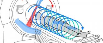
This completes the preparation stage. The hardware table is located in a horizontal plane, and the patient is placed on it. After it is conveniently fixed, the table falls into a ring element, where X-ray sources move in a spiral, invisible to the patient.
The study time with contrast can be up to half an hour. As soon as the patient begins to feel discomfort, he presses a special button to call medical personnel.
Price
In Moscow, you can undergo MSCT in many public or private medical institutions.
The cost of an abdominal examination ranges from 4,000 (Research Institute of Rheumatology) to 12,000 (Pokrovsky Gates) rubles. Almost similar prices in St. Petersburg. MSCT with contrast in the abdominal area will cost from 5,500 to 10,500 rubles.
In Kazan, the examination can be completed for 7,500 (“Expert+”), in Yekaterinburg for 5,500 (“Diagnostics Plus”), in Krasnodar – 5,200 rubles (“WTM”).
In many public or private clinics, examination can be done under the compulsory medical insurance policy . To do this, you need to come for an examination with a referral from your attending physician (no older than 3 months), a passport (for children - a birth certificate), a compulsory medical insurance policy, the results of a blood test for creatinine, and for some patients - with the minutes of the meeting of the medical commission. It is necessary to clarify all the nuances when registering for examinations.
What does MSCT OBP show?
Using MSCT of the abdominal cavity and retroperitoneal space, the doctor examines the organs and structures located in this area. Unlike computer scanning, this diagnostic method is used when it is necessary to examine hollow organs. If there are symptoms indicating violations, the use of a contrast solution is indicated (most often this is the drug Trazograph with triiodide or barium solution).
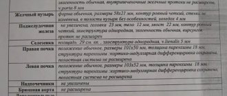
Examination using MSCT with contrast establishes the position (including mutual), condition, structure of organs located in the abdomen. Diagnostics reveals:
- OBP dysfunction;
- pathological changes (inflammation, necrotic processes and infectious foci);
- injuries;
- tumor processes, including at stage 1.
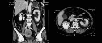
It is important to understand that MSCT shows abnormalities in the form of a black and white image that can only be read by a radiologist.