What it is?
Echogenicity is a technical term used in echography to refer to the ability of organs to reflect sound waves.
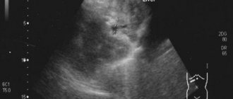
The ultrasound machine converts sound waves reflected from tissues with different acoustic densities into a picture that is visible on the screen during the examination. Knowing the exact data on the echogenicity of each organ, the doctor states an increase or decrease. Deviation from generally accepted parameters means that negative factors provoked diffuse changes in the structures and functioning of internal organs: kidneys, pancreas, intestines, spleen, stomach and liver. Ultrasound makes it possible to visualize organs, identify the disease and monitor its dynamics.
When the echogenicity of the parenchyma of an organ is increased, it means that at the moment its tissues are different from healthy ones. If echogenicity increases or decreases, or the homogeneity of the structure or contours of an organ changes, a targeted examination of the questionable area is carried out. Interpretation of ultrasound gives a clear idea of the condition and diffuse changes in the liver parenchyma and the entire digestive system. The manipulation allows the doctor to clarify the following questions:
what is the density and size of the organ; homogeneous or heterogeneous structure; are scars or nodes present; what is the concentration of metabolic products; infection with worms; are there tumor formations; condition (expansion or narrowing) of blood vessels and bile ducts; formation of stones and obstruction of veins; is it accompanied by increased echogenicity and enlarged lymph nodes.
Liver imaging with computed tomography
The liver is the largest organ of the human body, visualized by computed tomography as a homogeneous structure with a density (without contrast) within +55...+70 Hounsfield scale units.
A decrease in liver density on CT below +55 units indicates fatty hepatosis (infiltration of liver tissue with adipose tissue cells), more than +70 units indicates metallosis (deposition of metal salts in the parenchyma - this is how, for example, hemosiderosis manifests itself). Observations (CT of the liver) demonstrating pronounced fatty infiltration of the hepatic parenchyma before contrast administration (left). It can be seen that the parenchyma density is only -8 units on the Hounsfield scale, while normally it should be at least +55. The image in the middle shows that even after the administration of contrast, the parenchymal density increased slightly and is only -4 HU. Compare the image data with the norm (on the far right liver scan).
In the structure of the liver, it is customary to distinguish two lobes (main) - right and left, as well as the so-called. the caudate lobe is smaller in size. For more accurate localization of pathological foci, the liver is usually divided into segments. The blood supply to the liver is carried out by its own hepatic artery, which is a branch of the celiac trunk; venous outflow is carried out through the hepatic vein into the inferior vena cava system. In addition, at the porta hepatis, CT scan can visualize another large vessel that collects venous blood from the stomach, large and small intestines, pancreas, and spleen - the portal vein. Normally, the width of the portal vein on computed tomography of the liver is 10-20 mm (such a wide variation is mainly due to differences in body size). It is also necessary to evaluate the width of the splenic and superior mesenteric veins, which form the portal vein, and also carefully examine their lumen for the presence of filling defects caused by blood clots, as well as pathological changes in the lumen.
Contrast in computed tomography of the liver. Blue arrows indicate the portal and splenic vein.
Arterial and venous vessels during CT angiography of the liver: on the left, the abdominal part of the aorta is marked with number 4, the celiac trunk with number 5, and the hepatic artery with number 6. The blue numbers mark the veins: 1 – portal, 2 – splenic, 7 – superior mesenteric, 3 – inferior vena cava.
The portal vein system (left) and the vessels of the celiac trunk (right). Number 1 on the left indicates the portal vein, 2 – splenic vein, 3 – superior mesenteric vein, on the right, red numbers mark the arterial vessels of the liver (CT angiography): 1 – celiac trunk, 2 – hepatic artery, 3 – splenic artery, 4 – abdominal aortic section.
MRI of the liver (for comparison with CT and radiographs).
Another example of liver imaging with MRI.
Liver on radiographs. With X-rays, you can only evaluate the edges of the liver, approximately its size (as in the image on the left), as well as the presence of high-density objects (as in the X-ray of the liver on the right - metal staples after endoscopic cholecystectomy are circled).
Another point that you need to pay attention to when interpreting a computed tomography scan of the liver is the presence of lymph nodes with an altered structure and shape in its gates, which can be observed with hematogenous metastasis of gastrointestinal tumors to the liver. It is also necessary to carefully evaluate the structure of the liver, highlighting areas of different density in its parenchyma - cystic or solid in nature.
Normal liver structure
Diagram of the liver and its structure.
The liver is an exocrine gland, the most important unpaired organ of the human body, performing more than 500 functions. In this unique “laboratory” the most complex processes are carried out. It actively participates in digestion, producing the required volume of bile, cleanses the blood of toxins and other toxic substances accumulated by the body due to unfavorable ecology, poor nutrition and alcohol abuse.
Normally, the liver parenchyma is a homogeneous structure, penetrated by many vessels and bile ducts.
The echostructure of the tissue is generally fine-mesh and uniform. The anatomical location of the liver allows you to effectively practice ultrasound and collect the necessary data that allows you to draw conclusions about normal function or pathological abnormalities. It is located on the right side, weighs from 1.2–1.5 kg, and has a dark red color.
Reasons for increased echogenicity of the liver
Deviations in echogenicity are a signal indicating problems with the liver, which should not be ignored, since everything in the body is interconnected. Impaired functioning of one organ can lead to disruption of the functioning of other individual organs and subsequently to an unfavorable outcome in general. The reasons for increased echogenicity are summarized in the table:
| Chronic hepatitis | The structure of the liver is homogeneous, echogenicity is moderately increased. |
| Cirrhosis | At an early stage of the disease, the liver is enlarged. In later stages, dystrophy appears with a decrease in size. Heterogeneous, mosaic-type structure. Mixed echogenicity, depending on the location of the lesion. |
| Dystrophy and steatosis (fatty infiltration) | Moderate enlargement of the liver. Echogenicity increases with increasing intensity of reflection of sound waves from fatty inclusions into the liver cells. |
| Chronic cholangitis | There is a high echogenicity (hyperechogenicity), manifested in the rich reflection of sound waves from the walls of the dilated bile ducts. |
| Alveococcosis, opisthorchiasis (helminthic infestation) | The screen image shows diffuse increased echogenicity, blurring of invasive and healthy tissue, and a mesh-like structure. |
| Liver abscess | The initial stage of the incipient inflammatory process is represented by a small segment of reduced echogenicity, but as the abscess develops, heterogeneous echodensity is observed - either low or too high. |
Diabetes mellitus can impair organ function.
Other factors:
Echoic formation (hematoma, hemangioma, adenoma). Obesity or sudden weight loss. Alcoholic fibrosis and sclerosis. Diabetes mellitus. Intensive use of medications.
Prevention of liver diseases
To prevent the development of parenchymal changes in various liver diseases, you must follow simple rules. Such prevention will be useful for the normal functioning of not only the liver, but also other internal organs.
The main recommendations of experts are:
- Eat right. Every day your diet should include lean meats or fish, vegetables, and fruits. Avoid fatty, fried foods. Products with a high fiber content are also useful: cereals, whole grain bread, vegetables.
- Watch your weight. Overeating and, as a result, obesity have a negative effect on the liver. If a person constantly eats a lot, it simply does not have time to break down all the enzymes. However, if you need to lose weight, avoid strict diets, it is better to adhere to the principles of proper nutrition and exercise.
- Don't abuse alcohol. If there are no liver diseases yet, you must adhere to the established permissible alcohol limits. For women – 12 g of ethanol per day, for men – 24 g. If you already have chronic diseases, you must completely give up alcohol.
- Avoid stress. Due to constant stress and adrenaline production, pressure on the liver increases. Try to live a measured life, if necessary, consult a psychotherapist.
Thus, the liver parenchyma or its tissue is the first thing that reacts to the onset of disease development. If diffuse or focal changes begin, it is necessary to diagnose the organ, establish the cause and select appropriate treatment.
Symptoms of liver pathology, accompanied by increased echogenicity
Increased echogenicity has external signs or certain symptoms that indicate an unfavorable condition of the liver and require immediate consultation with a doctor to determine the causes of the ailment. Some symptoms are usually characteristic of those diseases that cause changes in echogenicity:
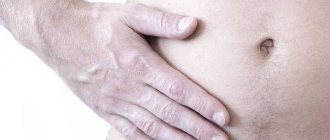
frequent pain, tingling, pain on the right side below the chest; causeless nausea or vomiting; yellowness of the skin; digestive problems; changes in the liver (enlargement, deformation) upon palpation; overweight or obesity; emerging heart problems; decreased immunity.
Diagnosis and treatment
If ultrasound cannot immediately accurately diagnose liver disease, then each subsequent study is prescribed by a specialist based on the data from previous procedures.
A biochemical blood test will help detect markers of hepatitis and HIV.
When, based on the results of ultrasound, the doctor sees that the echogenicity of the liver parenchyma is increased and a diffusely heterogeneous structure is observed, additional diagnostic methods will be proposed. This will allow us to find out what caused the anomaly. Held:
Blood chemistry. Needed to clarify data about processes occurring inside the liver, or to detect markers of hepatitis or HIV. Computer or magnetic resonance imaging. Confirm that the echogenicity of the liver is increased. Biopsy. Allows you to identify or exclude neoplasms in a separate area if local heterogeneity of echogenicity is observed.
The final diagnosis is made based on a combination of data from a medical examination, general tests, patient complaints, and liver ultrasound. Therapy is aimed at eliminating the disease that caused the increase in echogenicity. When treating, doctors use a method to relieve symptoms:
severe pain is relieved with antispasmodics; congestion of the hepatic ducts is removed with choleretic drugs; for excess accumulations in the abdominal cavity, diuretics are prescribed.
To normalize and protect liver cells, hepatoprotectors such as Essentiale and Hepa-Merz are used. To restore the normal functioning of blood vessels and the flow of nutrients into the body, antiplatelet agents are prescribed. If inflammation is present, a course of antibiotics is required. When hepatitis or cirrhosis is diagnosed, the patient undergoes treatment, selected by the doctor for each person individually.
Diet for liver problems
When liver problems begin, diet is a mandatory addition to the main treatment. The doctor will advise you to limit the use of fats and prescribe a complex of vitamins or medications with essential phospholipids that restore damaged liver cell membranes. In case of serious complications, doctors prescribe therapeutic diet No. 5. The healing menu includes:
raw, boiled or baked vegetables; milk or vegetarian vegetable soups; steamed or baked chicken, turkey, beef; milk, kefir, yogurt, cottage cheese; boiled or baked low-fat fish; cereal porridge and pasta; sauerkraut (not very sour) ); compote, jelly; honey and jam; tea with lemon, fresh juices from vegetables and fruits.
It is recommended to avoid the consumption of alcohol, tobacco, fatty meat and fish, smoked meats, pickles, legumes, fried foods, chocolate, coffee, etc. Take medications only as prescribed by a doctor. You need to know that antibiotics, antivirals, antihistamines and some diuretics can have negative side effects.
The results of an ultrasound of the liver make it possible to determine the contours of the organ, the size of the lobes, echogenicity and examine individual structures. This diagnostic method reveals cavities, compactions, the presence of tumors and metastases, and measures the pressure in the portal vein. Sometimes a sign such as increased echogenicity of the liver is determined.
Types of changes
In total, there are two types of parenchymal changes:
- focal – affect certain areas of tissue;
- diffuse - the lesion affects the entire organ.
The most common are diffuse changes, and they are also the most dangerous. With focal inflammation, the entire liver parenchyma is not affected, but this is usually only temporary. For example, these include cysts and abscesses, but often immediately against the background of the appearance of these focal changes, diffuse changes appear that affect the entire organ.
Diffuse liver changes
Pathological changes in the liver parenchyma, called diffuse, are of several types:
- Hypertrophy. It implies an enlargement of the organ, in which the natural liver tissue dies and fibrous tissue grows in its place. Often hypertrophy is one of the signs of hepatitis of various forms. An increase in fibrous tissue is called hepatomegaly.
- Heterogeneous fabric structure. The evenness of the contours of the parenchyma gradually changes, and at first it is difficult to notice this on ultrasound. The diffusely heterogeneous structure is clearly visible with other diagnostic methods - CT, MRI.
- Dystrophic and other structural changes. The liver decreases in size and its functionality suffers. The contours of the organ become uneven. This condition increases the risk of developing chronic liver failure. Dystrophy can be congenital or acquired.
- Fatty parenchymal degeneration. This condition means that there is an excessive amount of fat in the liver tissue. This is often associated with lipid metabolism disorders.
- Reactive changes. They are a secondary disease that occurs against the background of inflammation of other organs of the gastrointestinal tract, poisoning by toxins, malfunctions of the endocrine system, etc.
Normally, the structure of the parenchyma is homogeneous and of low density. Any deviations from this condition mean changes in the organ, which requires urgent diagnosis.
One of the complications of many diffuse changes is cirrhosis, a disease in which liver cells simply do not work.
Causes
The main causes of diffuse changes in the parenchyma are incorrect human habits. In more than 80% of cases of liver disease in young people, their lifestyle is taken into account. Among the possible reasons, the most common are:
- Poor nutrition. The main problems with choosing a diet are eating semi-finished products, fast foods, fried, fatty, and smoked foods. Non-compliance with food intake, overeating, and incorrect diets for weight loss also play an important role. If a child has had such nutritional problems since school age, then by the age of 30-35, if not earlier, the liver will begin to show the first alarming signs.
- Alcohol. Drinking alcohol in large quantities harms the liver. She has to process alcohol, it is broken down by her enzymes. Harmful compounds are produced, as a result of which parenchyma cells die and fat cells appear in their place.
- Ecology. Environmental pollution negatively affects the functioning of many internal organs, among which the liver is especially affected. All toxic substances that enter the body enter the liver through the bloodstream. People living in large cities are most susceptible to negative environmental impacts.
- Side effects of medications. Some medications contain potent components that are difficult to process by the liver. Danger arises when they are used incorrectly or excessively.
- Stress. When a person experiences a strong emotional shock, adrenaline is released. The liver must dissolve it, but for it this hormone is toxic. One-time experiences are not so dangerous, but if a person lives in constant stress, this will affect the functioning of the liver.
Some factors that provoke the development of diffuse changes do not depend on a person’s lifestyle, but they are less common. These include heredity and congenital pathologies.
Symptoms of diffuse changes
Damage to the liver tissue does not have pronounced signs, which creates difficulties with the timely diagnosis of diseases. Typically, patients complain about the functioning of the gastrointestinal tract, without even suspecting the presence of liver problems.
The main symptoms of diffuse changes are:
- pain in the right hypochondrium;
- yellowing of the tongue, bad breath;
- feeling of heaviness after eating food, especially fatty or fried food;
- frequent nausea;
- feeling of weakness;
- fast fatiguability;
- headache.
What is increased echogenicity
The ultrasound technique is similar to echolocation used by some animals (bats), fishing boats and the military. An ultrasound machine generates ultrasonic waves that cannot be detected by human hearing. They pass through tissue and are reflected at boundaries with different acoustic densities. Each fabric reflects waves differently. This property is called echogenicity. The device picks up reflected waves, which makes it possible to judge the condition of organs.
The speed of propagation of ultrasound depends on the density and elasticity of the medium. The more air or liquid contained in the tissue, the slower the wave speed. The ability to reflect, absorb, and scatter ultrasound is also taken into account. On the monitor this is shown by dark and light areas.
How violations are detected
Ultrasound examination is the safest and most informative. The data allows us to confirm or refute the presence of structural changes in the organ, impaired blood flow, the presence of stones and other formations.
Echogenicity reflects the density of the object being studied. Media represented by liquid are homogeneous, freely transmit waves and do not reflect them. Such objects are called echo-negative. Examples include gallbladder and cysts. They are dark on the screen.
Dense tissues reflect ultrasound in full, therefore they are always echo-positive and displayed in white. For example, bone, stones.
An organ with medium echogenicity is a healthy liver.
Causes of increased echogenicity
An increase in echogenicity occurs with the development of such diseases and conditions:
alcohol poisoning; cirrhosis of the liver; chronic hepatitis; chronic disease in a state of relapse; endocrine diseases, including diabetes mellitus; excess body weight; fatty degeneration of the liver; long-term use of medications.
An increase in echogenicity may be accompanied by a change in the size of the organ, unevenness of its edges, and disruption of the shape of the corners. Such an indicator detected on ultrasound, supported by symptoms of the disease, changes in laboratory data, requires appropriate treatment.
Changes in the liver parenchyma, treatment and prevention
If the examination reveals a disease that has caused changes in the liver tissue, then it is necessary to undergo treatment as soon as possible aimed at removing toxic substances from the liver and restoring liver cells. It is necessary to take medications that improve the functioning of the digestive system (enzymes to restore intestinal flora), anti-inflammatory drugs. An important role is played by the intake of choleretic agents and antispasmodics, which can facilitate the passage of bile and pancreatic stones.
It is necessary to cleanse the liver of toxins, including using folk remedies - using decoctions of medicinal herbs or infusions of medicinal plants. In some cases, it is necessary to take painkillers and vitamins (especially group B).
Before you start taking medications, you need to quit smoking, completely forget about alcoholic beverages, and also be sure to follow a diet. Food should be low in calories, without pepper, spices and salt. You can only eat boiled or steamed dishes and low-fat dairy products. Sweet and sour foods should also be excluded from the diet. Fruits are very healthy, but only ripe and not sour. We need to fight obesity. Diet and the fight against bad habits play a therapeutic as well as a preventive role in improving the health of the liver parenchyma.
Symptoms of increased conductivity
Detection of increased echogenicity usually occurs in the presence of health complaints. Symptoms of the disease may include the following:
pain in the right hypochondrium; bitterness in the mouth, belching, nausea, periodic vomiting; indigestion of food, loose stools; brown coating on the tongue; skin itching; yellowing of the skin; enlarged liver, its edge protrudes from under the ribs; decreased immunity; development ascites, edema; unjustified weight gain; gynecomastia in men, menstrual irregularities in women; palmar erythema - redness of the palms.
The following changes are observed in biochemical analysis:
increase in bilirubin, ALT, AST, alkaline phosphatase; lipid imbalance; decrease in total protein levels, imbalance in the ratio of protein fractions; increase in glucose.
Diagnosis of echogenicity. How to check the diagnosis is correct
Changes in echogenicity are determined using ultrasound. But this sign in itself is not informative. To confirm the disease, additional methods are used.
Patient complaints. Examination of the skin, oral mucosa, sclera of the eyes, palpation of the liver, abdomen. Laboratory blood tests for sugar, bilirubin, liver enzymes, hepatitis viruses, HIV. Computed tomography and magnetic resonance imaging will help clarify the localization of objects with increased echogenicity. If there is a suspicion of cancer, a liver biopsy is performed to clarify cirrhotic changes.
Diagnosis of organ parenchyma
An increase in echogenicity in the liver may be accompanied by changes in the parenchyma of other organs. Calcifications can form at the site of inflammatory processes or foci of infection. Examination of the kidneys and pancreas can reveal similar processes.
Tumors and metastases are located not only in the liver parenchyma, but also in other organs. Additional testing may be required.
Types of Changes
So what are “parenchymal pathologies” and how do they manifest themselves? How do they differ from each other, and why do they arise at all? Several types of changes can occur in liver tissue: in density, shape and composition.
These deviations are divided into the following types:
- Diffuse – changes throughout the parenchyma.
- Local - damage to one part of the parenchyma.
- Focal - compaction or one small lesion.
Focal changes
Focal abnormalities in the liver parenchyma can be detected using echography. As a result of this procedure, the density of the parenchyma and the lesions are determined, divided into areas with weak, mixed, strong echostructure, or its complete absence.
Focal lesions can be multiple, merging or single. As the patient's condition worsens, changes in the echogenicity of the affected area are observed.
Echography also makes it possible to determine the development of numerous or single cases of calcification in the parenchyma, i.e. seals. They occur less frequently in children than in adults.
The following ailments usually contribute to this:
- cirrhosis;
- chronic hepatitis;
- tuberculosis;
- echinococcosis;
- hypomotor dyskinesia of the biliary ducts;
- bile stagnation in the liver;
- malaria;
- sepsis;
- parasitic infection.
Lesions whose echostructure has not been identified are parenchymal cysts. Using an echogram, they can be determined only when they reach 3-5 mm in diameter. Cyst formations are divided into several types, differing in the cause of their development (acquired or congenital) and the methods of appearance (parasitic, non-parasitic, false and true).
Diffuse type deviations
Diffuse changes are acute and chronic hepatitis, cirrhosis, fat accumulation and other structural disorders due to diseases. When a person has hepatitis, the size of the liver increases, but its structure remains unchanged.
Damage to the surface of the parenchyma occurs if inflammation begins to grow. In this case, the thin wall of the liver thickens. When carrying out echography, low echogenicity of the organ is determined, but on the contrary, increased sound conductivity.
In the presence of hepatitis, unexpressed inflammation in the parenchyma causes a heterogeneous level of echogenicity. If a patient is diagnosed with cirrhosis, then lesions with impaired echogenicity begin to grow in number, because the homogeneous structure of the liver is quickly destroyed.
The parameters of such foci can be 0.5-2 cm. The structure of the parenchyma becomes heterogeneous as a result of metabolic disorders, bile stagnation and fatty degeneration.
How to treat liver echogenicity
Therapy is aimed at eliminating the causes of changes in the liver, rather than combating symptoms. Additional examination methods help determine the cause of the disease and prescribe appropriate treatment.
Any liver pathology involves changing your lifestyle and following a diet. Meals are needed in small portions, 5-6 times a day. The last meal is optimal 2 hours before bedtime.
Dishes are prepared in a double boiler, stewed, boiled, baked. You should not eat fried, fatty foods, fast food, alcoholic drinks, or sweet carbonated waters. Spicy, smoked, salty foods, sour fruits, heavy foods (legumes, mushrooms, duck, lamb, fatty fish) will lead to an increase in the symptoms of the disease.
Fermented milk products, cereal porridges, vegetables, dietary rabbit meat, turkey, and lean fish have a beneficial effect.
Once a week it is recommended to arrange fasting days.
Dietary nutrition is supplemented with drug treatment. It is selected by the doctor depending on the diagnosed disease. Drugs are prescribed to improve liver function - hepatoprotectors, essential phospholipids. For pain, use antispasmodics and compresses with a warm heating pad. Ascites and edema of other localizations are eliminated with the help of diuretics. Inflammatory diseases are treated with antibiotics.
Viral hepatitis types B and C require antiviral medications. Treatment of cancer is carried out with surgery, chemotherapy drugs and radiation therapy.
The development of severe complications requires treatment in the intensive care unit.
How to be treated
The largest gland in our body is a vital organ, so it is necessary to make a diagnosis as quickly as possible and begin the right treatment. Depending on the nature of the disease, the necessary treatment tactics are chosen.
Is your liver healthy? Take the free test!
- Small abscesses are eliminated with doses of new generation antibiotics or antiparasitic drugs. Large ulcers are cleaned by aspiration or puncture drainage with the introduction of antibacterial and antiseptic solutions inside.
- Elimination of Caroli syndrome with medications is theoretically possible, but usually ineffective. Therefore, in case of recurrent bleeding, endoscopic ligation (suturing) is performed on esophageal varices.
- Ischemic hepatitis (heart attack) is eliminated when the causes that caused it are eliminated (in 50% this is a shock state).
- Extensive subcapsular hemorrhage with a hematoma requires emergency surgery to stop blood loss.
- Cysts that are not prone to growth are subject to dynamic observation; if they grow, they are removed. For invasive nodes with parasites, an echinococotomy is performed (opening and cleansing the bladder or removing it entirely).
- Treatment of parenchymal dystrophy is aimed at getting rid of the main pathology. This is mainly weight loss, alcohol coding, taking antiviral and antidiabetic drugs.
- The decision on methods of treating benign tumors is made separately in each case. To prevent degeneration into a malignant form, according to indications, part of the organ is removed: segmentectomy or sectionectomy.
- Taking into account the stage of cancer, surgery is performed in combination with chemotherapy. In the absence of metastases to other tissues and irreversible changes in the cellular structure, transplantation is performed.
You can contact the hepatologist in the comments. Don't hesitate to ask!
This article was last updated: 05/23/2020
Didn't find what you were looking for?
Try using search
doctor or administrator.
Read the dictionary of terms.
Expert author: gastroenterologist Daniela Sergeevna Purgina