What it is?
The essence of an X-ray examination of the gastrointestinal tract with a contrast agent is the introduction of a special substance (with a high absorption coefficient of X-rays) into the patient’s body and subsequent visualization of the organ using ionizing radiation. It includes fluoroscopy and taking pictures under its control in the required projections.
As a contrast, barium sulfate is most often used , less often - water-soluble contrast agents (verografin, urografin).
Barium sulfate is a white powder, almost tasteless, resembling chalk. It is absolutely harmless to the patient and is excreted from the gastrointestinal tract unchanged, but can cause the development of short-term discomfort. If the integrity of the walls of the stomach or intestines is damaged, barium can escape into the tissue, which is unacceptable (danger of peritonitis).
In order to increase the astringent properties, this contrast is used in the form of an aqueous suspension; if necessary, tanning and foam-reducing substances are added to it. There are also ready-made barium sulfate preparations that have high contrast, viscosity and fluidity.
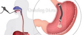
Contrast can be introduced into the body in two ways:
- orally (the person is asked to drink a specially prepared liquid);
- through a probe.
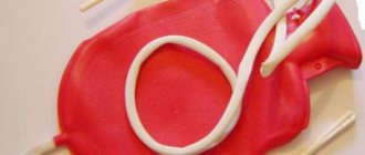
Filling the stomach with barium
What can you see during the examination?
This method of examining the gastrointestinal tract allows, using X-rays, to obtain a complete picture of the examined organs with all pathological changes in their shape, structure, and even evaluate them over time. But it is possible to take an x-ray of the stomach and other organs only under certain conditions that the patient will definitely need to fulfill in preparation for the study.
Due to the structural features of the digestive organs - the esophagus, stomach, small and large intestines, which have a hollow shape that does not block X-ray radiation, contrast is used to successfully carry out the procedure. As a contrast agent, a drug made from barium is used, diluted with water to the consistency of a kind of suspension.
Barium envelops the walls of the esophagus, which makes it possible to detect their defects.
In some cases, in order to straighten the folds of the mucous surface of the gastrointestinal tract, immediately before the start of the study, the patient is asked to drink a solution of crystalline soda, which increases visualization. Sometimes, for the same purposes, air is introduced under pressure or using a perforated tube, through which the patient simultaneously receives a barium suspension.
X-ray of the stomach in a direct projection shows:
- relief of the antrum of the stomach and esophagus after taking the first sips of a contrast agent;
- the condition of the folds and grooves of the upper stomach and body;
- How does the gastro-rectal junction function?
- with the first sip of barium, you can see what the structure of the folds of the duodenum is.
X-ray of the stomach in oblique and lateral projection shows:
- pneumorelief of the antrum;
- how does a gatekeeper function?
- displacement;
- gastroesophageal reflux.
The Trendelenburg position during radiography helps to detect a hiatal hernia (the stomach can prolapse there if the muscle fibers are weakened).
With the patient lying supine, the dimensions of the lesser curvature and elasticity are assessed.
Kinds
To diagnose stomach diseases in medicine, 2 main types of X-ray contrast studies are used.
- Normal contrast.
- Double contrast method.
In the first case, for the study it is sufficient to ingest an aqueous barium suspension and visualize the organ on an X-ray.
If the data obtained is not enough, then additional gas is injected into the stomach cavity. This method is called “double contrast” and is used for radiography of the stomach and duodenum. The role of contrast is played by two components at once - a suspension of barium particles and gas. If indicated, this procedure can be planned in advance. In such cases, it is customary to talk about primary double contrast. For this, a barium mixture with special characteristics is used:
- higher density;
- homogeneity;
- good adhesive properties (adhesion to the inner lining of the stomach).
After its introduction into the body, the patient is offered to drink a special gas-forming powder or gas is administered through a probe. The method makes it possible to study the structure of the organ in more detail.
Indications
Indications for performing an X-ray examination of the stomach with barium are very wide. They correspond to the symptoms of various diseases of this part of the digestive system:
pain or discomfort in the abdomen after eating;- night hunger pain in the stomach;
- persistent nausea, vomiting of eaten food or sour stomach contents;
- bloating;
- swallowing disorder;
- hidden blood in the stool or its darkening;
- weight loss with lack of appetite;
- selectivity in eating food (for example, aversion to meat or bread);
- the presence of a palpable (to the touch) tumor-like formation in the area of the stomach;
- long-term anemia of unknown origin
- achilia (lack of hydrochloric acid and enzymes in gastric juice).
Contraindications
There are limitations to barium X-ray examination of the stomach. It is contraindicated:
- pregnant women;
- persons with individual intolerance to contrast;
- patients with impaired renal function;
- if you suspect a perforation of the wall of the digestive tract, intestinal obstruction or ongoing gastric bleeding.
In some cases, the procedure is impossible due to the general serious condition of the patient.
Reference
If contraindications are caused by the introduction of barium into the body, then it can be replaced with another contrast (water-soluble).
How does the procedure work and how long does it take?
Features of the procedure for X-ray of the stomach with barium:
- On the day of the study, you should refrain from drinking and eating.
- Immediately before the x-ray, you need to get rid of various jewelry and removable dentures.
- Next, a survey X-ray of the chest and abdominal cavity is performed.
- The patient is given a contrast agent (barium sulfate). It has a rich chalk flavor and a dense consistency.
- The specialist takes the first x-ray, determining the relief of the walls of the esophagus.
- The patient takes various positions: lying on his back, on his side, and also using the Trendelenburg method.
On average, the procedure takes up to 40 minutes. If there is a need to take x-rays of the lower intestines, this may take up to a day.
Photo gallery
X-ray of the stomach with barium sulfate Violation of the lumen of the stomach Gastric ulcer on an x-ray
Preparation
It is necessary to prepare for the study in advance, since its results and their reliability depend on the quality of the preparation.
How to prepare:
- limit food and water intake (the procedure is carried out on an empty stomach);
- follow a diet;
- take medications (if necessary);
- to refuse from bad habits.
More information about this and some other features of the preparatory stage can be found in a separate article.
Preparation for the procedure
You should not drink water if the test is to be done with barium.
X-ray of the esophagus and stomach does not require special preparation for those patients who have obvious symptoms indicating dysfunction of the gastrointestinal tract. Patients with this pathology come for examination on an empty stomach. Also, the patient who is scheduled for the procedure must notify the doctor in advance about taking medications and, if necessary, limit the dosage. The specialist will also need information about chronic pathologies. Before taking x-rays for older people, 24 hours before the procedure it is better to switch to boiled food with a minimum of spices. Preparation for fluoroscopy of the stomach is especially important if it is planned to study the gastric cavity and intestines with barium: if the rules are not followed, X-ray diagnostics will not show contours. For example, you should not drink large quantities of water, as during an ultrasound.
How do they do it?
The patient comes to the X-ray room on an empty stomach. The further course of action depends on what procedure is planned. But, there is a rule - every x-ray examination of the stomach always begins with a survey x-ray of the abdominal organs, because various diseases of the digestive system can cause a reaction in the surrounding tissues. This provides the opportunity to:
- assess the situation;
- identify signs of the formation of through defects in the wall of a hollow organ, intestinal obstruction or fluid accumulation in the sloping parts of the abdomen.
If no contraindications to the study are identified, then it is continued.
In a routine X-ray of the stomach, a contrast agent is administered orally, in other words, the patient is given a few sips of barium to drink. At this time, the specialist monitors the progress of the contrast through the esophagus using fluoroscopy. Reaching the stomach, it is distributed along its walls, and the radiologist takes a series of photographs in which the relief of the internal surface of the organ is visible. Then the subject takes another 100-150 ml of barium orally, and the doctor determines in which projections and positions it is better to take radiographs to obtain the maximum amount of information about the condition of the organ.- When performing primary double contrast, the study begins with premedication (administration of anticholinergics, such as metacin), which allows to reduce the tone and motor function of the stomach for the entire period of the study. Then the patient drinks 2-3 sips of the barium mixture. While it drains into the stomach, the person is in an upright position and the doctor assesses the condition of the esophagus. After taking 50-70 ml of contrast, he is recommended to drink gas-forming powder. As a result of a chemical reaction, gas is formed in the stomach, fills its lumen and the hollow organ swells, and barium is evenly distributed throughout its inner lining. The study continues in a horizontal position. Under fluoroscopy control, the most optimal projections for taking pictures are selected. Usually these are 2-3 anterior (straight and oblique) and the same number of posterior projections.
The results of the study are interpreted by a radiologist using a series of radiographs.
X-ray of the stomach and esophagus with barium - what shows
X-ray examination (hereinafter referred to as RI) is a projection of human organs, resulting from the passage of special rays through them, which their inventor Konrad Roentgen himself called X-rays. The study is based on the ability of body tissues to absorb x-rays to one degree or another.
Accordingly, hard tissues (bones) are clearly visible in the image, while soft tissues appear as dark structures. Therefore, to study organs such as the stomach, esophagus and others, they came up with a special method - x-ray using the substance barium, which absorbs radiation well.
Indications
RI of the esophagus and stomach can also be carried out as part of the study of other organs of the gastrointestinal tract directly related to them.
Contraindications
In these situations, alternative examination methods are prescribed, such as fibrogastroscopy or MRI.
Preparation
As with any type of medical examination of internal organs, it is also necessary to prepare in advance for fluoroscopy.
X-ray radiation is one of the types of gamma radiation, and there are standards that can be given to the patient in one procedure.
The pause before the next study must be maintained, so if the patient comes unprepared, the doctor will take a picture, but may not see anything there. This is bad for the patient himself, as time for treatment is missed. Therefore, it is necessary to follow some rules:
- Before RI, it is forbidden to consume foods that increase gas formation (rye bread, legumes, vegetables and others containing fiber), since distension of the intestines can block access to parts of other organs. 3 days before the study, it is advisable to start taking drugs that reduce gas formation - Espumisan, activated carbon, etc.
- The stomach should be empty during the examination, since some of the food on the wall of the stomach will not allow the contrast mixture to flow around the mucous membrane, and this can be interpreted in the image as a tumor. Therefore, in order to avoid an erroneous diagnosis, you need to fast in the evening and come to the examination with a completely empty stomach.
- Immediately before the procedure, it is necessary to remove all metal jewelry from the neck and hair from the back of the head.
- Before examining the stomach and esophagus directly, a survey fluoroscopy of the internal organs is performed, which allows one to judge their condition and the presence of gas accumulations in the surrounding tissues.
- The examination of the esophagus and stomach itself can be carried out using the following methods:
- traditional;
- according to Trendelenburg;
- double contrast.
As barium suspension (barium sulfate solution) passes through the gastrointestinal tract, the radiologist will have an idea of defects in the mucous membranes, and the time of its movement will make it possible to judge the narrowing or obstruction of the organ.
Traditional method
The contours of its shadow are a reflection of the inner surface. In order to achieve uniform distribution of the mixture and examine all the walls of the organ, the study is also carried out with the patient in a horizontal position.
The mixture does not remain in the esophagus for long - when you exhale, the esophageal sphincter relaxes and the barium passes into the stomach. The contrast mass is distributed over the mucous membrane and fills the gaps between the folds. For better distribution, the contrast agent is “smeared” by palpation.
After examining the relief of the mucous membrane, the patient is given another 150-200 ml of contrast agent to drink. At the same time, the shape, position and size of the stomach are also determined in different projections, and the course of its emptying is examined.
According to Trendelenburg
The Trendelenburg study involves a special position of the patient - the pelvis is raised at an angle of 45 degrees relative to the head. This position is used to detect a hernia of the diaphragm - the organs are displaced towards the diaphragm, and thanks to the contrast agent, the doctor can see whether they prolapse into the chest cavity.
The Trendelenburg study is contraindicated in the elderly, with cardiovascular diseases, sclerosis and limited respiratory function.
Many people are interested in the procedure of X-ray of the stomach and esophagus with barium, and it will not be superfluous to find out what it shows in adults and children.
Stomach diseases are very common, and now not only adults, but also children, starting from primary school age, suffer from them. The most common problems are gastritis and peptic ulcers.
The easiest way to find out what the condition of the gastric mucosa is, whether there are erosions, ulcers, bleeding or neoplasms, is gastroscopy.
However, this diagnostic method is not suitable for everyone: it is not used in children, as well as in people suffering from mental disorders. Sometimes it is impossible to carry out it purely technically - a person does not allow the gastroscope to be inserted due to panic, pain, or it is not feasible due to the severity of the patient’s condition.
But the patient still needs to be examined, since he needs adequate treatment. In this case, an x-ray of the gastrointestinal tract comes to the rescue.
Many people believe that X-rays only examine the condition of the lungs or bones if a fracture is suspected. It's true: these two situations are the most common reasons to send a person to the x-ray room. However, they are not the only ones.
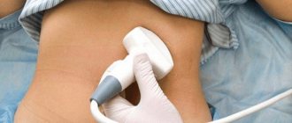
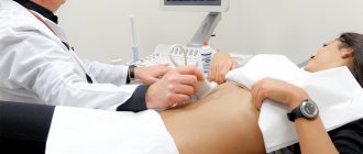
X-ray examination of the stomach is a non-invasive research method in which the condition of the organ is assessed by visual inspection of X-ray images (radiography) or in real time (fluoroscopy).
A huge advantage of this diagnostic method is that it causes virtually no inconvenience to the patient: it is painless, so it can be performed on a person in any condition (including bedridden patients and newborns). It is used both independently, if endoscopy is impossible, and in addition to it, if the doctor still has questions regarding making the correct diagnosis and prescribing treatment.
While during gastroscopy only the esophagus, stomach and the initial part of the duodenum are examined, and during recto- and colonoscopy - the rectum and colon. These are three separate procedures, each of which requires special preparation.
There are several ways to assess the condition of the stomach using x-rays. Among them are the main four.
- If, with the help of this study, the doctor can clearly assess the condition of the lungs and bone tissue, since their density in the first case is very low, and in the second, on the contrary, it is high, then the abdominal organs in such a picture are only approximately visible.
- The density of the stomach, intestines and other organs is not very different from each other, so the image is not clear.
- For this reason, this study as an independent diagnostic method is not very informative; it only allows the doctor to make certain assumptions regarding the condition of the abdominal organs.
Unlike the previous method, before taking a picture, the doctor asks the patient to drink a special liquid - an X-ray contrast agent (it most often contains barium). After ingestion, it gradually moves down the digestive tube, filling all sections.
- As a result, the doctor sees a clear outline of the organ in the image and can assess its boundaries, the presence of a foreign body, obstruction, tumor, perforation, motor skills, etc.
- Despite the fact that the specialist does not see the gastric mucosa itself, using this method one can obtain very important information regarding the state of human health.
- Before starting the study, be sure to take a simple overview photo without contrast.
This diagnostic method involves the use of two types of contrast - an X-ray contrast agent (barium, iodine) and air.
This method is more informative than the previous one, since it allows you to see very minor formations on the gastric mucosa and suspect cancer at its very beginning.
X-ray
In this case, the doctor does not evaluate the result obtained in the images, but looks at the dynamics of the progress of the contrast along the digestive tube in real time. The study is recorded and can be reviewed again if necessary.
X-ray contrast examination of the stomach is a procedure for which it is necessary to properly prepare. If this is not taken care of, the doctor will receive an unclear image: the contrast agent will be mixed with food bolus, feces, mucus, and as a result, it will be almost impossible to evaluate the study.
Therefore, 2-3 days before the date when the procedure is planned, you need to start following a special diet. It is necessary to exclude all foods that cause fermentation - yeast baked goods, fruits, juices, vegetables, milk and dairy products.
These days you should eat meat broths, fish, durum wheat bread, water porridge and cheese. 10-12 hours before the test, you need to completely abstain from food and drink only water.
The need for an enema is decided individually. It will be needed if a person has a tendency to constipation.
In general, an enema is advisable for everyone - the evening before the examination, to be sure that the digestive tube will be completely free of food and will not interfere with the procedure.
After special training, the person approaches the office at the specified time. First, he is given a plain X-ray of the abdominal cavity. Next, they give you a glass of contrast agent to drink. After this, they begin to take a series of photographs to assess the dynamics of its progress.
Then the liquid fills the stomach, and the doctor examines its condition, decides whether more pictures are needed, and in what position it is best to take them. When performing fluoroscopy, all this is visible in real time.
With double contrasting, a probe is inserted into the patient's stomach and a contrast agent is already supplied through it, or air is pumped in. In some cases, preliminary administration of antispasmodic drugs may be required.
As in any other case, this type of research is clearly prohibited for pregnant women.
The issue regarding its implementation in children is controversial. If possible, it should be avoided, but sometimes it turns out to be indispensable if anatomical defects of the digestive tube are suspected in newborns.
What does esophageal examination show?
A barium X-ray of the stomach allows the doctor to obtain the following information about the condition of the organ:
- position;
- displacement and relationship with nearby structures;
- form;
- size;
- tone and peristalsis;
- relief of the inner shell.
Also during the study, you can evaluate the condition of the esophagus and duodenum.
Taking into account these characteristics and identifying radiological symptoms, the specialist makes a conclusion about the presence (or absence) of various stomach diseases in the patient.
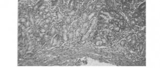
(only a biopsy can confirm the diagnosis): disruption of the relief of the mucous membrane, compaction of folds, minor erosions, functional disorders (changes in motor activity).
Chronic gastritis- Peptic ulcer disease : ulcerative niches (a shadow of a contrasting mass that fills a defect in the mucous membrane), erosion in the form of round-shaped clearings with an accumulation of barium in the center, signs of gastritis, gross deformations of the organ wall (scars).
- Congenital malformations of the stomach : diverticulum (blindly ending protrusion of the organ wall in the form of a process), congenital pyloric stenosis (narrowing of its outlet part), dextrogastria (change in the position of the organ with a shift to the right), duplication of the stomach.
- Stomach cancer (X-ray changes depend on the type of malignant neoplasm, the nature of its growth and the stage of the process): filling defect, niche in it, deformation of the organ, narrowing of its lumen, change in the relief of the inner lining, rigidity of the stomach walls and lack of peristalsis in the affected area.
- Benign tumors (the x-ray picture is also determined by the type of tumor and the stage of its development).
- Complications after gastric surgery are afferent loop syndrome (the flow of contrast from the stump of the stomach into the expanded afferent loop, where barium suspension accumulates) and dumping syndrome (accelerated emptying of the remaining part of the stomach and the rapid movement of barium through the intestinal loops).
Possible consequences
Modern X-ray examinations are considered quite safe. They are no longer what they were before. Using the latest X-ray equipment and digital equipment, the radiation dose that the patient receives is reduced several times (by 5 or more). The latter can also be achieved by reducing the exposure time.
The radiation exposure received during an X-ray of the stomach with barium does not exceed that of other types of X-ray examination and is not dangerous to humans.
- The average radiation exposure during an X-ray of the stomach (1 image) is 6-8 mSv. This dose corresponds to exposure to radiation under natural conditions for 3 years (solar radiation, soil and landscape background, cosmic radiation).
- The X-ray method involves visual inspection of the organ on a monitor screen. It involves significantly less radiation. But due to the increase in the duration of the procedure, the total dose may increase. So, during 15 minutes of the study, the patient receives a dose of 2-6 mSv.
note
The dose that can cause damage to body tissue (radiation sickness) under the influence of radiation is more than 3 Sv for humans. It is impossible to obtain it with conventional x-ray diagnostics (even with a combination of several research methods).
Negative consequences during the study are mainly associated with the use of contrast. It can be:
- constipation;
- bloating;
- feeling of discomfort;
- abdominal pain (intestinal cramps).
But they occur only in some patients, are short-term and quickly disappear after barium is removed from the gastrointestinal tract. To prevent them, it is recommended to drink more fluids and eat foods rich in fiber.
How to prepare for a barium x-ray
The patient is advised to follow standard requirements, which confirm that special preparation for the x-ray is not necessary.
- Three days before the procedure, avoid fatty, spicy, highly salty foods and alcohol intake. You should not eat foods that can cause flatulence (brown bread, legumes, fresh fruits and vegetables). You can - boiled meat, fish (steamed), porridge (cooked in water).
- Ten hours before the examination, eating is prohibited.
- To obtain high-quality images, it is advisable to rinse the stomach and colon with an enema (when it is impossible to cleanse the intestines naturally, it is necessary to perform cleansing using a probe).
- When lying down for installation, you need to remove your dentures and all metal objects (jewelry, accessories, clothes with fasteners).
If you prepare correctly for the procedure, it will be carried out accurately. Subsequently, the doctor will have the opportunity to make the correct diagnosis and develop an effective treatment regimen.
( 1 ratings, average: 5.00 out of 5)
Price
Pricing policies in different regions of the country have some differences. It is determined by the prestige of the clinic, its location, equipment, and qualifications of the staff.
- The highest price for an x-ray of the esophagus in Moscow and St. Petersburg is from 4,500 rubles.
- In the regions, prices are slightly lower: in Nizhny Novgorod and Novosibirsk - from 3000, in Voronezh - from 2000, in Krasnodar and Ufa - from 1000 rubles.
The official websites of clinics do not always have accurate data on the cost of the study. Often, when a client contacts him personally, it increases due to various reasons (for example, overestimation, outdated data). Moreover, the price may not be indicated in full for the entire procedure, but only for fluoroscopy or 1 x-ray. But it is also possible for the price to decrease during promotions or discounts. Therefore, when choosing a clinic, you should remain vigilant and clarify financial issues before the procedure begins.
Doctors provide consultations in the following clinics:
"SM-Clinic" on Volgogradsky Prospekt (metro station "Textilshchiki")
- Atyasova Elena Viktorovna
- Babaeva Afelia Mirzoevna
- Bikineeva Dina Ilyinichna
- Blinova Elena Nikolaevna
- Koksharov Dmitry Vladimirovich
- Muratkhodzhaeva Malika Farkhadovna
- Tebieva Svetlana Temurovna
"SM-Clinic" on the street. Yaroslavskaya (metro station "VDNH")
- Babaeva Afelia Mirzoevna
- Krasnykh Maxim Alexandrovich
- Krinina Inna Vladimirovna
- Mansimov Ramin Magomedalievich
- Muratkhodzhaeva Malika Farkhadovna
- Timashkov Ivan Alexandrovich
- Fedina Oksana Nikolaevna
"SM-Clinic" on the street. Novocheremushkinskaya (metro station “New Cheryomushki”)
- Bikineeva Dina Ilyinichna
- Krasnykh Maxim Alexandrovich
- Povytchikova Natalya Vladimirovna
"SM-Clinic" on the street. Clara Zetkin (metro station "Voikovskaya")
- Azaryan Samvel Amirkhanovich
- Dubrova Sofya Erikovna
- Ikonnikova Nina Yurievna
- Karachenkova Inna Alexandrovna
- Muratkhodzhaeva Malika Farkhadovna
- Romanova Olga Ivanovna
- Rudaya Anna Ivanovna
- Chekaida Natalya Vladimirovna
"SM-Clinic" in 2nd Syromyatnichesky lane. (metro station "Kurskaya")
- Babaeva Afelia Mirzoevna
- Mukhidinova Diana Magomedovna
- Sevastyanov Sergey Yurievich
"SM-Clinic" on Simferopol Boulevard (metro station "Sevastopolskaya")
- Atyasova Elena Viktorovna
- Gubina Svetlana Vasilievna
- Ikonnikova Nina Yurievna
- Kim Elena Georgievna
- Makarov Artem Mikhailovich
- Morozova Natalya Mikhailovna
- Tebieva Svetlana Temurovna
"SM-Clinic" on the street. Marshal Timoshenko (metro station "Krylatskoe")
- Minkina (Shcherbakova) Natalya Igorevna
- Ochirov Bembya Yurievich
- Timashkov Ivan Alexandrovich
"SM-Clinic" on the street. Yartsevskaya (metro station “Molodezhnaya”)
- Gubina Svetlana Vasilievna
“SM-Clinic” in Staropetrovsky Proezd (metro station “Voikovskaya”)
- Garashchenko Natalya Viktorovna
- Nazarova Anna Anatolyevna
- Sevastyanov Sergey Yurievich
"SM-Clinic" in Solnechnogorsk, st. Red
- Gavrilov Kirill Viktorovich
- Levin Roman Eduardovich
- Mineeva Irina Vasilievna
"SM-Clinic" in Solnechnogorsk, microdistrict. Requinzo
- Gavrilov Kirill Viktorovich
Children's clinic "SM-Doctor" in Maryina Roshcha (m. Maryina Roshcha)
- Mukaeva Evgenia Ivanovna
- Musinova Ksenia Olegovna
- Ryabchenko Mikhail Mikhailovich
- Sevastyanov Sergey Yurievich
- Kharlamova (Grabovskaya) Tatyana Vladimirovna
Children's department on Volgogradsky Prospekt (Textilshchiki metro station)
- Atyasova Elena Viktorovna
- Babaeva Afelia Mirzoevna
- Bikineeva Dina Ilyinichna
- Blinova Elena Nikolaevna
- Koksharov Dmitry Vladimirovich
- Muratkhodzhaeva Malika Farkhadovna
- Tebieva Svetlana Temurovna
Children's department on the street. Novocheremushkinskaya (metro station “New Cheryomushki”)
- Bikineeva Dina Ilyinichna
Children's department on Simferopol Boulevard (metro station Sevastopolskaya)
- Atyasova Elena Viktorovna
- Gubina Svetlana Vasilievna
- Ikonnikova Nina Yurievna
- Kim Elena Georgievna
- Makarov Artem Mikhailovich
- Morozova Natalya Mikhailovna
- Tebieva Svetlana Temurovna
Children's department on the street. Yartsevskaya (metro station “Molodezhnaya”)
- Gubina Svetlana Vasilievna
Children's department on the street. Yaroslavskaya (metro station "VDNH")
- Babaeva Afelia Mirzoevna
- Krinina Inna Vladimirovna
- Mansimov Ramin Magomedalievich
- Timashkov Ivan Alexandrovich
Children's department in Solnechnogorsk, st. Red
- Gavrilov Kirill Viktorovich
- Levin Roman Eduardovich
- Mineeva Irina Vasilievna
Children's department in the city of Solnechnogorsk, microdistrict. Requinzo
- Gavrilov Kirill Viktorovich