Superficial bulbitis is one of the mildest forms of a dangerous inflammatory process that occurs in the duodenal bulb. The symptoms of this disease are considered nonspecific; they accompany a number of pathologies of the gastrointestinal tract. Basically, signs of superficial bulbitis appear, such as severe flatulence, nausea, vomiting, pain in the epigastric region on an empty stomach and after eating, as well as various phenomena accompanying dyspeptic syndrome.
In diagnosis, radiography, esophagogastroduodenoscopy, and antroduodenal manometry play an important role. In the treatment of superficial bulbitis, many medications are used: anticholinergics, antispasmodics, anti-Helicobacter, antacid drugs, as well as antiparasitic drugs.
Causes of the disease
Typically, bulbitis appears along with gastritis. Or gastritis comes later. Factors contributing to the development of the disease:
- infection with the bacterium Helicobacter pylori;
- disruption of the motor activity of the duodenum, as a result of which food stagnates in the bulb, and hydrochloric acid disrupts the integrity of the mucous membrane;
- unhealthy diet (eating fried, salted, smoked foods, fast food), as well as alcohol and smoking;
- constant nervous tension and anxiety contribute to disruption of blood microcirculation in the intestinal walls;
- parasitic infestations (ascariasis, enterobiasis);
- stomach and intestinal injuries.
Immunity disorders and severe concomitant diseases also lead to bulbitis.
Causes of the disease
Almost the entire adult population has acute or chronic gastritis.
But in order for it to move from the stomach to the duodenum, a person must eat improperly, eat a lot of fatty and fried foods, indulge in alcoholic beverages, smoke a lot, and not maintain immunity at the proper level.
Doctors highlight several main points that contribute to the fact that people develop superficial bulbitis and gastritis of the stomach. They are:
- Improper nervous and humoral regulation of digestion, which occurs in the stomach and intestines.
- The presence of Giardia, opisthorchiasis, Helicobacter and Campylobacter in the human body.
- Taking certain medications that lead to damage to the mucous membranes of the duodenum.
- Improper and irregular nutrition.
- Nicotine abuse.
- Hormonal disbalance.
- Regular stress.
- Gastritis of the stomach with high acidity.
- Treatment with painkillers.
Only a qualified specialist can determine the true cause of the disease after a series of procedures and a complete examination of the body.
Symptoms of superficial bulbitis
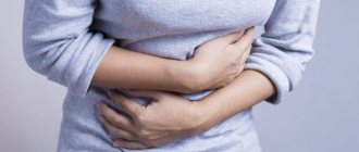
The main manifestation is pain. The pain appears an hour and a half after eating. It is aching and localized just above the navel. It may radiate to the right hypochondrium and back. In the acute stage of the disease, the pain intensifies and requires the use of antispasmodics. Additionally, the presence of bulbitis is indicated by:
- belching is bitter, often with an unpleasant odor;
- nausea after eating;
- vomiting mixed with mucus and bile.
As with any disease, the general condition suffers: headaches, palpitations, drowsiness and irritability.
Diagnosis of the disease
If you have at least several of the listed symptoms, you should consult a doctor. Having carefully examined your complaints, the doctor will conduct an external examination (when palpating the abdomen, pain is felt in the epigastric or navel area) and prescribe a full examination.
If focal bulbitis is suspected, an FGDS (fibrogastroduodenoscopy) is performed. During this method, a camera at the end of a special hose enters the duodenal bulb and the doctor is able to examine the organ from the inside. Inflammation will be indicated by redness of the mucous membrane, the presence of contact bleeding, and an increase in size of this section of the intestine. There will also be stagnant food masses there.
Additionally, for superficial bulbitis, X-rays of the stomach and duodenum with contrast are prescribed. The photographs will show erratic intestinal peristalsis and a tendency for contents to flow back into the stomach.
To clarify the severity of the disease, a biochemical blood test is prescribed. Due to malabsorption of food, it will have low hemoglobin and protein.
Diagnostics
Catarrhal bulbitis is diagnosed based on the patient’s complaints, his condition and symptoms. Afterwards, an examination is performed by palpation, which helps to establish the source of the inflammatory process. When pressing on the inflamed area, the patient feels unbearable pain.
An important step for correct diagnosis is instrumental examination. Prescribed:
- Fibrogastroduodenoscopy. The procedure allows you to detect inflammation in the stomach and the upper zone of the 12 intestinal rings;
- CT AND ultrasound;
- X-ray of the stomach;
- Esophagogastroduodenoscopy. Gives the opportunity to visually assess the condition of the mucous membrane of the stomach, 12 duodenum and, in particular, the bulbar zone;
- Probing.
To obtain a general clinical picture, the doctor also prescribes laboratory tests:
- Analysis of gastric contents;
- Blood analysis;
- Breath test;
- Enzyme immunoassay;
- PCR diagnostics of Helicobacter.
Currently reading: What is duodenal bulbitis - symptoms, treatment and diet
Treatment of superficial bulbitis
For successful therapy, it is necessary to correctly determine the cause of the disease. If it is helminthic infestation or Helicobacter pylori infection, then these factors are primarily affected. And then the symptoms of the disease are treated. In the treatment of bulbitis, there are three areas: diet, treatment with medications and folk remedies.
Diet for bulbitis
In principle, the diet for superficial bulbitis is the same as for any lesion of the gastrointestinal tract. Avoid foods that stimulate the secretion of hydrochloric acid in the stomach and bile. These are spices, pickles, smoked meat, soda, coffee, chocolate, kvass, horseradish. Suitable for use:
- lean boiled meat, fish (can be baked);
- porridges cooked in milk or water (except millet, pearl barley and corn);
- seasonal vegetables, steamed or boiled in water (exclude cabbage, turnips, sorrel, cucumbers and mushrooms in any form);
- flour products made from premium flour, biscuits;
- scrambled eggs;
- butter and vegetable oil.
There should be at least 5-6 small meals per day. Food should not be too cold or hot. Table salt is limited whenever possible, as in any diet.
Drug therapy
Drugs are selected depending on the severity of the disease and the financial capabilities of the patient. Treatment usually lasts 2-3 weeks, and repeated courses may be prescribed. Most often used:
- antibiotics for irradiation of Helicobacter pylori in the stomach - Amoxicillin, Clarithromycin, Tetracycline and Metronidazole (an antimicrobial drug, not an antibiotic);
- proton pump inhibitors - Omeprazole, Rabeprazole, Esomeprazole - reduce gastric acidity;
- antacids - neutralize hydrochloric acid (Rennie, Gastal, Maalox, Almagel, Taltsid);
- antispasmodics - to relieve pain and relax the muscles of the duodenum and stomach (Drotaverine or No-shpa, Papaverine);
- light sedatives (valerian or motherwort extract);
- agents that increase the formation of mucus to heal erosions (Biogastron, Liquiriton).
Treatment with folk remedies
Herbal medicine for superficial bulbitis is based on the use of infusions containing celandine, St. John's wort, chamomile, and yarrow. Mix all these herbs in equal quantities. Pour boiling water (500 ml) over two tablespoons of the mixture and leave for 3-4 hours. The resulting product is drunk half a glass before each meal. Treatment takes approximately 10 days. Then you definitely need to take a break.
You can also make an infusion from oak bark; it has an antimicrobial and astringent effect. Pour two tablespoons of bark into a glass of boiling water and leave for 2 hours. Drink 2 tablespoons before meals.
Drinking mineral waters “Essentuki” No. 4 and 17, “Borjomi” gives a good effect. 2 glasses a day (in the morning on an empty stomach and at night before bed). You can drink them for a long time, 2-3 weeks.
If you consult a doctor for help in a timely manner and correctly prescribed therapy, it is not difficult to cope with superficial bulbitis. But you need to strictly follow all recommendations and be careful in treating with folk remedies.
If you have been diagnosed with this disease at least once, you will have to stick to a diet for life and say goodbye to bad habits. Don't forget to devote at least two hours a week to sports activities and maintain good personal hygiene. Then you will forever forget what a superficial bulbite is.
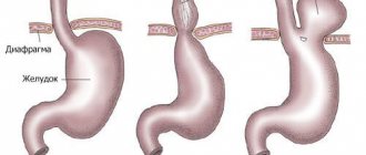
Superficial bulbitis (focal) is an inflammatory process in the area of the duodenal bulb, located at the outlet of the stomach. It is into this department that the mass of semi-digested food enters for subsequent processing and penetration into the large intestine.
Superficial bulbitis is classified as a type of duodenitis.
Treatment and prevention of bulbitis
In order to cure a superficial problematic bulbitis, the cause of its occurrence should be identified.
If the examination reveals a Helicobacter or parasitic infection, then specific therapy is prescribed.
In order for the symptoms of acute bulbitis to disappear, you need to adhere to diet No. 1 and take antispasmodics with anticholinergics.
If earlier doctors prescribed astringent and enveloping drugs, today the effectiveness of such treatment is in question.
Chronic bulbitis requires adherence to a diet throughout life.
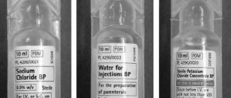
During periods of exacerbation, doctors recommend symptomatic treatment using vitamin therapy and parenteral administration of a protein hydrolyser.
In the treatment of giardiasis, Macmiror shows good results. But in the treatment of opisthorchiasis it is customary to use Praziquantel.
Duodenal bulbitis caused by Helicobacter or Campylobacter can be cured with broad-spectrum antibiotics.
It is noteworthy that the Helicobacter bacterium is usually treated with several types of antibiotics at the same time.
In addition, it is appropriate to take acid secretion suppressors: Omeprazole or Nolpaza. The drug De-Nol, which contains bismuth, has proven itself well.
To increase the tone of the pyloric sphincter, it is customary to prescribe trimedate or domperidone - broad-spectrum prokinetics.
Bulbitis of the duodenum often provokes disturbances in the excretion of bile. You can get rid of the problem with the help of a choleretic or antispasmodic drug.
With timely diagnosis and treatment, the prognosis for the pathology in question is quite favorable.
Preventive measures include maintaining a healthy lifestyle, eating the right foods, and treating chronic gastrointestinal diseases.
What is surface bulbite
A feature of superficial bulbitis is inflammation of the duodenal bulb, which occurs in a mild form and can be cured without much difficulty. However, it is much more difficult to control the running process.
The disease is characterized by swelling of the mucous membrane of this section, which causes stagnation of bile and enzymes, which are necessary for the complete digestion and absorption of food.
Ducts from the pancreas and gallbladder exit into the bulb. Due to such anatomical features, the inflammatory process can spread to the pancreas or duodenum. Often this phenomenon precedes a peptic ulcer.
When the duodenal bulb is inflamed, the specialist diagnoses “Superficial gastritis bulbitis.”
The following forms of superficial bulbitis are distinguished: acute and chronic. In the first case, the inflammatory process develops in response to nutritional disorders, intoxication of the body, or damage to the intestinal mucosa by a foreign object.
The chronic form can be primary or secondary. In the primary version, the pathology develops due to stress and poor nutrition, in the secondary - against the background of inflammatory diseases of the gastrointestinal tract.
How is pathology diagnosed?
People who experience superficial intestinal bulbitis should definitely consult a gastroenterologist to clarify the diagnosis and prescribe adequate treatment.
Only those patients who are experiencing severe pain, have a stomach ulcer and hypoproteinemia in their blood biochemistry are hospitalized in the department.
There is a standard examination scheme for patients complaining of bulbitis. She is like this:
- Visit to an endoscopist for esophagogastroduodenoscopy. The procedure allows us to detect degeneration of epithelial cells, swelling of intermediate tissues, and filling of the mucous membrane with lymphocytes.
- X-ray of the stomach and duodenum. Such diagnostics are required to determine discoordination of peristalsis and its spasms, retrograde peristalsis, and an increase in the speed of movement of food through the digestive canal.
- Gastric impedancemetry is a procedure that allows you to make a final diagnosis.
The specialist is faced with the important task of differentiating bulbitis and superficial gastritis, ulcers, pancreatitis, cholecystitis. In addition, the pathology in question is similar to cancer and hiatal hernia.
Causes of the disease
Superficial bulbitis of the stomach develops as a result of factors such as:
- complicated family history;
- penetration of foreign bodies into the intestines and their traumatic effect on the bulb;
- violation of the diet, predominance of fried, spicy and fatty foods in the diet;
- helminthic infestations (enterobiasis, ascariasis);
- eating unwashed vegetables and fruits, which causes infection;
- smoking, alcohol abuse;
- eating too hot food and drinks;
- decreased protective functions of the body;
- chronic disease of the gastrointestinal tract characterized by inflammation (Crohn's disease);
- gastritis.
The causes of the disease can be different. To get rid of inflammation, it is necessary to find out what triggered it, based on diagnostic measures.
Forms and stages
Catarrhal bulbitis can have different forms. Each form of pathology is expressed by certain specifics.
The following types of bulbite are distinguished:
- Surface. It is a mild form of the disease. In the superficial form, only the outer zones of the mucous membrane are affected. This type can occur hidden, the patient is unaware of the problem with the digestive tract for a long time. When the disease is neglected, there is a risk of developing an erosive form of pathology. The erosive type of bulbitis is expressed by deep damage to the layers of the epithelium.
- Erosive. Manifested by nausea and vomiting, mainly after eating. In complex cases, this type can cause ulcerative lesions.
- Follicular. This form of catarrhal bulbitis is rare. The pathology is accompanied by serious disorders of the digestive tract, which negatively affects the patient’s quality of life.
- Moderate. Most often, the pathology occurs hidden. Patients may experience headaches, nausea and vomiting. Such symptoms are recorded when the disease relapses.
- Focal. Characteristic of this form is the defeat of individual sections of the 12th bulb - the rings of the intestine. This species is quite dangerous to health; timely treatment prevents the development of serious complications.
- Atrophic. This form of the disease is expressed by impaired functioning of the duodenum. As a result, the patient experiences frequent vomiting. The patient with this type experiences rapid weight loss.
In accordance with the morphological characteristics, bulbite is classified as follows:
- First degree. Defined as weak. The preservation of the structure of the mucous membrane is recorded, infiltration with lymphocytes and plasma cells is observed;
- Second degree or moderate bulbitis. Damage to the surface layer and structural changes in the microvilli of the mucous membrane are recorded;
- Third degree or severe. It is expressed by deformations of the villi, lymphoplasmacytic infiltration is observed, and there is a possibility of the appearance of erosive formations.
Chronic catarrhal bulbitis
The chronic type of the disease develops as a result of neglect of catarrhal bulbitis, manifested by the duration of the disease, periods of remission and exacerbation, the body's immunity decreases, headaches and muscle weakness occur. Lesions of the intestinal lining may be microscopic. Chronic gastric bulbitis has certain symptoms. Basically, the pathology is accompanied by pain of varying degrees of intensity; the presence of ulcers is excluded.
Focal catarrhal bulbitis
Against the background of hormonal disorders, focal bulbitis often occurs. The inflammatory process covers various zones of the mucous membrane of the bulb, located between the stomach and duodenum. The lesions are not localized; they are located in different parts of the affected organ. In severe cases, inflammation spreads, moving towards the intestines and stomach.
Pain with focal bulbitis is similar to the symptoms of a peptic ulcer. The patient has an intestinal disorder, in the morning the patient feels nausea, after eating he often experiences sour belching, bloating, and a feeling of heaviness that turns into pain in the epigastric region.
Exacerbation occurs against the background of the development of vitamin deficiency, alcohol abuse, and prolonged fasting. The duration of catarrhal focal bulbitis is about 7 days, in some cases up to 2 months. Stomach acidity may remain normal. With focal bulbitis there is a risk of bleeding.
Erosive
Erosive bulbitis is characterized by the formation of erosive foci on the walls of the duodenum, which can cause destruction of the walls of the stomach. This type of inflammation is provoked by irregular diet, constant stress and a genetic predisposition to gastrointestinal diseases. Advanced erosive bulbitis is dangerous due to the risk of gastric bleeding, the appearance of ulcers, and various complications in the functioning of the gastrointestinal tract.
Follicular
Follicular bulbitis is one of the rare types of disease. This type belongs to the varieties of gastroduodenitis and is often a harbinger of its development. Factors that negatively affect the mucous membrane of the bulbus lead to a malfunction of the motility of this zone and an increase in the reaction of lymphoid tissue in this part of the intestine. An increase in the amount of gastric juice produced, as well as a decrease in the protective functions of the bicarbonate layer, enhances the aggressive influence of HCL in this area of the gastrointestinal tract and the growth of lymphoid follicles. This leads to the formation of white vesicles on the mucous membrane of the bulbus, which indicate the development of follicular bulbitis.
Currently reading: What is acute bulbitis of the stomach and duodenum - what it looks like, symptoms and treatment
Surface
With superficial bulbitis, the upper part of the mucous membrane becomes inflamed. This form of the disease is the initial stage of the disease. In most cases there is no severe pain or vomiting. The patient feels heartburn, heaviness and slight pain. Most often, this form of bulbitis occurs due to poor nutrition, smoking, and alcohol abuse. If neglected, the disease can progress.
Symptoms of pathology
Characteristic symptoms of superficial gastritis bulbitis are:
- constant rumbling in the stomach;
- attacks of nausea ending in vomiting, after which a bitter taste appears in the mouth;
- constant feeling of hunger, even after eating enough;
- pain in the stomach;
- bad breath;
- abdominal pain localized in the navel area and appearing 1-1.5 hours after eating;
- general weakness;
- headache;
- increased irritability;
- constipation
In the superficial form of this pathology, pain is not the leading symptom. Typically, severe pain occurs already in advanced stages, as well as in the case of chronic inflammation .
Symptoms
The inflammatory process of the duodenal bulb is accompanied by symptoms that are similar to other gastrointestinal disorders. The patient complains of:
- Hunger pain if the stomach is empty. At night, aching pain often appears. Pain is recorded in the area of the navel or shoulder blade;
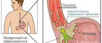
- Frequent belching and heartburn associated with acidic content entering the esophagus. Some of the foods eaten with acid from the stomach enter the esophagus, discomfort and a burning sensation appear, which are often localized in the chest and throat;
- Presence of bitterness in the mouth. Appears as a result of solidification of a food coma in the follicle of the stomach;
- Vomiting and nausea after eating. In most cases, after this the pain begins to decrease. Before an attack, the patient experiences pallor of the skin, weakness and tremors in the muscle tissue;
- Chronic constipation.
Treatment of superficial bulbitis
Treatment of superficial bulbitis requires not only taking medications, but also following diet rules.
Drug treatment
Medicines are used only during periods of exacerbation of the disease.
The doctor selects the medicine based on what factor provoked the development of the pathology:
- If Helicobacter pylori (a bacteria that provokes the development of ulcers, gastroenteritis, duodenitis, and stomach cancer) is present in the gastrointestinal tract, antibiotics are prescribed. Amoxicillin and Clarithromycin are usually prescribed.
- Antacids are prescribed to neutralize hydrochloric acid. These are Maalox, Reni, Gastal.
- In order to stimulate the process of mucus formation, which helps heal erosion of the membranes, medications such as Liquiriton, Biogastron are prescribed.
- For severe pain, taking No-Shpa and Drotaverine is indicated.
Diet for bulbitis and gastritis
Proper treatment of superficial gastritis bulbitis involves following a diet. Food should be light and in no case irritate the mucous membranes of the gastrointestinal tract.
The patient's meals should be divided: you can eat up to 5-6 times a day, but in small portions.
- lean fish and meat, boiled or baked;
- vegetables cooked steamed or in water (except cabbage, mushrooms, sorrel);
- cereal side dishes with milk or water (excluding corn and pearl barley);
- soft-boiled eggs.
Traditional medicine recipes
Herbal treatment is another way to correct the patient’s condition. It must be taken into account that traditional medicine is purely auxiliary in nature and can only complement the main course of therapy.
For superficial bulbitis, the following folk recipes are useful:
- Plant composition. To prepare, mix chamomile, St. John's wort, yarrow, celandine in equal proportions, take 2 tablespoons of this raw material. Pour the mixture into 0.5 liters of boiling water and let it brew for 3-4 hours. You need to drink 100 ml of this composition before each meal.
- Plantain-based medicine. You will need 3 tablespoons of fresh plantain juice and a tablespoon of honey. The components need to be mixed. Before meals, eat a teaspoon of the prepared product.
- Infusion based on propolis. To prepare, 60 g of raw material is poured with a glass of alcohol and left in a dark place for a week. Take 1-2 times a day, adding 5 ml of infusion to a glass of warm water.
Superficial bulbitis is a pathological process in which one of the parts of the stomach becomes inflamed. This process can be easily treated, but in advanced cases the situation gets worse.
Treatment of bulbitis involves taking medications during the period of exacerbation, as well as following a special diet.
Minimal inflammation of the mucous membrane in the duodenal bulb is called superficial bulbitis. The isolated development of this pathology is unlikely; damage to the duodenum is often combined with gastritis.
Diet
When bulbitis worsens, it is necessary to follow a special diet, which is an important part of therapeutic therapy. The diet during the period of exacerbation of pathology should include products (meals) that prevent the release of excessive secretion. Basically, a strict diet is prescribed for a month.
Currently reading: What is chronic bulbitis of the stomach and duodenum - prognosis and treatment
The patient is advised to eat steamed or boiled foods. Food should be warm, but not hot, you need to eat in limited portions, 5 times a day, at the same hours.
It is necessary to completely abandon salty, spicy and fatty foods; coffee, carbonated drinks, spices and seasonings, saltiness, and canned foods should be excluded from the diet. You need to quit smoking and give up alcoholic beverages.
Pureed porridges cooked in water or milk, low-fat cottage cheese, homemade yoghurts, jelly made from non-acidic fruits and berries, and natural jelly are useful. The diet should include lean meats (fish, veal, rabbit, poultry).
Food must be steamed. Instead of fats, you need to consume oils of vegetable origin; it is recommended to give preference to olive oil, since the product has an enveloping effect and facilitates the movement of food through the esophagus.
Mechanism of disease development
The main factors contributing to inflammation of the mucous membrane at this level of the digestive system include:
- Helicobacter pylori contamination;
- helminthic infestations;
- dysfunctional state of the pyloric sphincter, leading to uncoordinated reflux of acidic gastric contents into the duodenal lumen;
- smoking and systematic abuse of alcoholic beverages;
- improper organization of nutrition;
- increased stress background.
Various external influences lead to an imbalance between aggressive components and mechanisms that prevent destructive changes in the internal lining of the duodenum. Reserves of protective cells are sent to the site of damage, and a significant amount of biologically active substances is supplied and locally produced. A nonspecific inflammatory reaction is formed.
How to suspect the manifestation of bulbitis?
Inflammation of the duodenal bulb is accompanied by pain in the upper abdomen. The disease is characterized by late or hunger pain, which is associated with the time of chyme from the stomach entering the duodenum (2-3 hours after eating) or the response of the irritated intestinal mucosa to a long break in food. The painful sensations have a pressing, squeezing nature and can go away either independently or after taking antacid medications or be weakened by the action of antispasmodics.
Patients note manifestations of dyspepsia: belching, nausea, vomiting bile. General health is often disturbed: patients complain of increased fatigue, weakness, drowsiness, and other manifestations of asthenia.
How to identify a superficial, just beginning bulbitis?
The pathology at the initial stages has mild clinical symptoms. The presence of the above complaints may correspond to several diseases of the digestive system. In order to verify the diagnosis, the patient is comprehensively examined.
The doctor prescribes blood and urine tests and an ECG. The main method of examination is fibrogastroduodenoscopy (FGDS).
The condition of the mucous membrane of the esophagus, stomach, and duodenum is visually assessed, after which the doctor writes a conclusion based on the endoscopic picture.
What is superficial bulbitis from an endoscopy point of view?
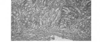
Examination of the mucous membrane of the duodenum on FGDS in the superficial form of the disease reveals a heterogeneous state of the tissues. Inflammation appears as swollen areas and alternates with intact intestinal lining. Foci of plethora (hyperemia) up to 0.2-0.3 cm in size appear above the altered areas of the mucosa. Physiological folds at the level of inflammation are slightly increased in volume.
To clarify the nature of the pathology, the endoscopist takes the material for analysis. Detection of H. pylori confirms the bacterial etiology of inflammation of the duodenum and helps determine treatment tactics.
The choice of therapy largely depends on the nature of the disease. In most cases, the prescription of antibacterial agents is required. Taking medications from the omeprazole group, bismuth preparations, and H2 blockers helps reduce acid production and restore the mucous membrane of the stomach and intestines. Prokinetics are indicated in the presence of impaired motor function, as well as in order to reduce the discoordinated entry of gastric juice into the lumen of the duodenum.
For emergency help, they resort to antacids and antispasmodics. But the patient can get rid of bulbitis only by following a diet and medication prescriptions.
Fried, spicy, and fatty foods are excluded from the diet. Organize 5-6 meals a day in small portions. Food is prepared steamed or boiled. During the period of exacerbation, the diet is observed as strictly as possible.
Following the recommendations of the attending physician helps to bring the disease into stable remission and prevent the progression of the pathology with the development of more severe forms.
Superficial bulbitis is a common phenomenon among diseases of the gastrointestinal tract. As a rule, this disease occurs in conjunction with other gastrointestinal pathologies: gastritis, gastric ulcer, pancreatitis, cholecystitis. But in practice there are isolated cases of the disease.
Superficial gastric bulbitis - what is it?
This is primarily an inflammatory process that is localized in the mucous layer of the duodenal bulb. To understand the mechanism of formation and progression of pathology, you need to have an idea of the anatomical and physiological characteristics of this organ.
The duodenum is the initial section of the small intestine, and at the same time, it is a continuation of the stomach. It consists of an upper sector - an onion, about 5 cm in size; descending, horizontal and ascending parts. The peculiarity of this organ is that the major and minor papilla open in the thickness of its mucous membrane.
They, at one time, are the final part of the bile duct and pancreatic duct. That is, all the main enzymatic digestion processes originate in this organ. If the mucous wall is involved in inflammation, then these processes will be disrupted.
There are 2 main courses of bulbitis: acute and chronic. In terms of severity, it can be mild, moderate and severe.
Causes of the disease
Isolated superficial bulbitis occurs due to the following factors:
- Non-compliance with daily routine and nutrition. Appears when eating spicy, over-salted, irritating, scalding, sour foods, fast food. In addition, the development of this disease is facilitated by: overeating, a sudden change in diet to a large meal, rare meals during the day (1-2 times), overeating at night before bed.
- Infectious agent – Helicobacter pylori. Infection occurs from a carrier or a patient suffering from the disease.
- Exposure to excess gastric juice containing abundant amounts of hydrochloric acid.
- Sustained stress. In addition to emotional disturbances, stressful situations for the body are: constant lack of sleep, spending a lot of time at the computer or in front of the TV, sedentary lifestyle, obesity, physical inactivity.
- Taking pharmacological drugs: non-steroidal anti-inflammatory drugs (painkillers and antipyretics), use of hormones, antibacterial agents, chemotherapy and radiation therapy.
- Bad habits: smoking, drinking alcohol, psychotropic and narcotic drugs.
- Hazardous working conditions and environmental pollution: work in mines, in the chemical industry, with paint and varnish products, repair work.
- Concomitant diseases: diabetes mellitus, renal failure, pathology of the cardiovascular and endocrine systems.
In combination with other pathologies, it can occur against the background of:
- Various forms of gastritis.
- Peptic ulcer disease.
- Gastric bleeding.
- Pancreatitis.
- Cholecystitis.
- Hepatitis of both viral and toxic etiology.
- Oncological processes of the digestive system.
- Food poisoning, toxic infectious diseases, gastroenteritis.
Prevention
To prevent the development of bulbitis, it is necessary to promptly treat gastritis and other gastrointestinal pathologies. After treatment of gastritis, it is necessary to conduct special tests that will confirm the eradication of the pathogen.
The patient should undergo regular examinations by specialists. You should take medications prescribed only by your doctor.
It is important to follow a diet, adjust your diet, eat meals at specific times with small portions. You need to completely give up bad habits (smoking, alcohol, coffee), avoid stress and overexertion, promptly treat inflammation and infections of the oral cavity, and observe good hygiene.
Symptoms and treatment of superficial gastric bulbitis
The symptoms of this disease are similar to the manifestations of various forms of gastritis or gastric ulcer. In each patient, due to the individual characteristics of the body, the disease may develop differently, but common complaints and signs are noted. These include:
- Unpleasant pain in the upper abdominal cavity. The pain is constant, aching or pulling, not associated with eating. Basically, patients note that pain occurs due to errors in diet and nutrition. Sometimes this symptom may be diffuse in nature, and its localization changes, discomfort is noted in the umbilical region, left or right hypochondrium.
- Appetite disorder. Patients complain of a decreased desire to eat food or a sophistication of taste. Many people claim that they are giving up their usual food and starting to eat something they have never eaten before. Lack of appetite subsequently leads to weight loss.
- Phenomena of changes in stool. Due to the fact that enzymatic activity is disrupted during inflammatory processes, the phenomenon of alternating constipation and diarrhea occurs. More often, patients note a mushy stool consistency.
- Nausea, vomiting, coating on the tongue.
- Symptoms of general intoxication. These include general weakness, weakness, decreased ability to work, and apathy. Slight rises in temperature are often observed.
- Signs of deficiency of vitamins and macroelements - hypovitaminosis. Manifested by dry skin, brittle nails, hair loss, dizziness.
- Abdominal bloating, increased gas formation.
- With insufficient therapy or its absence, the disease can progress, and complications will appear in the form of gastrointestinal bleeding and malignancy.
This disease is diagnosed using endoscopic methods - fibrogastroduodenoscopy, radiological methods - radiography of the abdominal organs with contrast, ultrasound - ultrasound of the abdominal cavity.
They may resort to computed tomography or magnetic resonance imaging if necessary. In extreme cases, they resort to surgical intervention - diagnostic laparoscopy.
Diet as the basis of therapy
The fundamental factor in the treatment of pathology is compliance with the daily routine and diet.
The diet should be balanced and correctly divided into portions throughout the day. Nutrition must satisfy physiological and energy needs, contain the required amount of proteins, fats, carbohydrates, macro- and microelements.
There should be at least six to seven doses per day in small portions.
Breakfast should not be the densest, but also not the lowest in calories. It is useful to eat non-dairy cereals: semolina, oatmeal, oatmeal, rice. You can supplement it with low-fat yoghurts.
There must be a second breakfast, which may contain a boiled egg, cottage cheese or a light vegetable salad.
Lunch should be the most saturated in terms of calories. It may consist of several dishes. For example, vegetable soup, then boiled potatoes with chicken breast and green salad as a side dish. Don’t forget about dessert; you don’t need to completely eliminate sweet dishes.
The patient chooses the afternoon snack and snacks before dinner. It should be something easily digestible and not rough. For dinner you can eat buckwheat porridge with steamed fish and fresh vegetables. Drinks include jelly, chamomile or green tea.
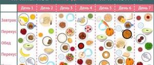
Here is an example of a proper balanced diet for the day:
The most important thing is not to eat rough, hot, fried, fatty, spicy, or solid foods. It is healthy to eat a lot of greens, protein-rich poultry, cereals, soups, fish, cottage cheese, low-fat dairy products, vegetables, and fruits.
Drug therapy
The goals of therapy depend on what caused the disease. Medicines should be carefully selected and prescribed only by a qualified physician.
If the disease arose due to improper consumption of food, then you should discuss it with your doctor during treatment and in the future and choose the right and healthy food for your diet.
In such cases, proton pump inhibitors are prescribed, which reduce the secretion of hydrochloric acid. These include drugs such as Omeprozole, Omez, Esomeprazole and others.
If the cause is an infectious agent - Helicobacter pylori, then its eradication is carried out. They use regimens with antibiotics and proton pump inhibitors. Antibacterial agents are used: metronidazole, clarithromycin, tetracycline drugs.
To relieve pain, antispasmodics are prescribed: drotaverine, duspatalin.
If the disease occurs against the background of other pathological processes, then the original cause is treated directly. Enzymes are prescribed: pancreatin, creon; hepatoprotectors: heptral, antimicrobials: nitrofurans, nifuroxazide.
To restore normal microflora and the functioning of the gastrointestinal tract, probiotics are recommended: Linex, Enterol.
Features of the diet for bulbitis
A therapeutic diet is one of the most effective and mandatory conditions for carrying out therapeutic activities.
Bulbit, like gastritis of the stomach, requires control of its course and adherence to the correct diet.
Specialists must select a strict diet that will have to be followed for some time.
But it is worth considering that throughout his life a person will have to limit himself to eating fried, sour, spicy and salty foods, even if the symptoms of the disease do not manifest themselves.
With the next exacerbation of the pathology, the following foods should be excluded from the diet:
- Black tea.
- Coffee.
- Alcohol.
- Smoked meats.
- Salt.
- Tough vegetables.
- Bread with yeast.
- Carbonated drinks.
Such food leads to the fact that bulbitis and gastritis worsen, and the person suffers from new attacks. After the onset of pain, doctors recommend eating liquid food, which is quickly absorbed.
Salt intake should be strictly limited. The permissible dose is 5 grams per day. This also applies to sugar - 50 grams per day.
The following foods are allowed to be eaten:
- Omelette.
- Scrambled eggs.
- Various cereals.
- Chicken fillet.
- Kissel, compote.
- Milk soup.
- Apples baked in the oven.
- Boiled or steamed meat.
A few days after the exacerbation, patients are allowed yesterday's bread, sour cream with a small percentage of fat, cottage cheese products, pasta, and biscuits.
You can drink weak tea. In addition, nutritionists strongly recommend drinking a tablespoon of olive oil half an hour before meals.
It is very important to avoid stressful situations and worry less. You need to change your harmful work and get proper rest at night.
If you adhere to all of these recommendations, then the pathology in question will not make itself felt for a long time.
Folk remedies for superficial bulbitis
Herbal medicines are often prescribed in the complex therapy of bulbitis.
For example, a decoction of chamomile flowers helps relieve inflammation and pain.
A decoction of the following herbs has medicinal properties:
- liquorice root,
- marshmallow,
- St. John's wort,
- chamomile inflorescences.
Mix all the herbs in equal proportions and pour boiling water over them. Let it brew. Use cooled 5-6 times a day, 5-7 sips.