The proper functioning of the digestive tract plays a vital role in people's daily lives. Chronic diseases lead to a decrease in quality of life, productivity and cause neuropsychiatric disorders. Megacolon is a severe pathology of the large intestine, which is characterized by thickening of its walls, a change in length, which causes a disruption of the blood supply, and the development of an inflammatory process up to atrophy. All this leads to disruption of peristalsis and passage of intestinal contents.
Definition and classification
Megacolon is said to exist if the x-ray shows a pathological increase in the diameter of the colon:
- blind – more than 12 cm;
- ascending – 8 cm;
- descending and sigmoid - 6.5 cm.
In clinical practice, there are three types of megacolon:
- spicy,
- chronic,
- toxic.
Acute megacolon (Ogilvie's syndrome) is an enlargement of the colon that occurs in patients with severe surgical diseases. The chronic version lasts for several months or more. It is caused by persistent intestinal dysfunction, such as in Hirschsprung's disease or muscular dystrophies. Toxic megacolon is provoked by various infections: bacterial or pseudomembranous colitis, Chagas disease, etc.
Causes
Depending on the etiology, megacolon is divided into primary (congenital) and secondary (acquired). The primary form is Hirschsprung's disease. This is a congenital pathology, which is based on the absence of intramural (inside the intestinal wall) nerve plexuses. Because of this, functional obstruction occurs due to distal narrowing, often in the rectal area. The direct cause of Hirschsprung's disease is a genetic mutation on four chromosomes.
Megacolon is said to be acquired if no congenital gastrointestinal pathology (aganglinosis, stricture or atresia) is detected. Typically, the pathology develops against the background of atonic constipation. The latter, in turn, provoke various diseases, including the gastrointestinal tract:
- mechanical intestinal obstruction (tumor, helminthic infestation);
- infectious and inflammatory bowel pathologies (Chagas disease, pseudomembranous colitis, etc.);
- surgical operations;
- Parkinson's disease;
- myotonic dystrophy;
- diabetic neuropathy;
- collagenoses (for example, scleroderma);
- hypothyroidism;
- hypokalemia;
- porphyria;
- pheochromocytoma.
Acquired megacolon can also be idiopathic. That is, there are clinical manifestations of the disease, but its exact cause has not been established.
The prevalence of chronic megacolon, such as Hirschsprung's disease, is 1 in 5000 births. The congenital form of the disease is more often diagnosed in boys than in girls (4:1). Secondary megacolon occurs with equal frequency in both men and women.
Differential diagnoses
Several authors have emphasized the importance of considering an extensive list of differential diagnoses (eg, neuromuscular, mechanical, inflammatory, metabolic/endocrine, pharmacologic, environmental, and behavioral causes) for cats not responding to therapy.
However, a review of published cases suggests that 96% of cases of colonic dysmotility are idiopathic megacolon (62%), pelvic canal stenosis (23%), nerve injury (6%), or spinal cord deformity (5%).
A minority of cases are due to complications of colopexy (1%) and colonic neoplasia (1%); subacute hypo- or aganglionosis is suspected, but cannot be proven, in the other 2% of cases.
Inflammatory, pharmacologic, and environmental/behavioral causes were not identified as predisposing factors in any of the original case reports.
Endocrine factors (obesity, n = 5, hypothyroidism, n = 1) were cited in several cases but were not necessarily considered part of the pathogenesis of megacolon.
Symptoms
Megacolon is characterized by nonspecific symptoms:
- abdominal pain;
- bloating and rumbling;
- constipation
Congenital megacolon (Hirschsprung's disease) usually develops immediately after birth. The baby passes a small amount of feces (meconium) in the first few days after birth, but the abdomen remains enlarged. Stool occurs only after a cleansing enema or the use of laxatives. If intestinal obstruction develops, vomiting, abdominal pain, and bloating occur.
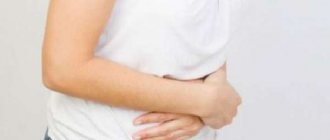
Prolonged constipation is one of the signs of chronic megacolon Photo: shutterstock.com
If the disruption of intestinal innervation is partial, then symptoms may appear later, after several months. Frequent constipation is common, even despite taking medications. Due to digestive disorders, anemia and malnutrition (physical underdevelopment) develop. The child is often sick and has a weakened immune system.
The acquired form of the pathology is diagnosed in older children and adults. Chronic megacolon manifests itself with the same symptoms, but they are less pronounced than with a congenital disease. The process of nutrient absorption is disrupted to a lesser extent, so the patient’s general condition is usually not impaired.
Constipation with secondary megacolon lasts more than a week. Sometimes it is possible to palpate compressed stool in the lower abdomen. In some patients, fecal incontinence occurs due to reflex atony of the anal sphincter.
If the disease develops acutely, for example, against the background of infectious colitis or obstruction, a clinical picture of intestinal obstruction develops. Sharp abdominal pain, cessation of gas discharge, vomiting, nausea, and increased body temperature occur. In this case, you should immediately seek medical help.
Diagnostics
If stool retention occurs in a newborn, then Hirschsprung's disease is first ruled out. In the secondary form of the disease, it is necessary to identify the etiological factor leading to intestinal dilatation. It is important to differentiate between congenital and acquired forms of megacolon. The treatment approach and prognosis depend on this.
Diagnostics includes the following instrumental research methods:
- irrigoscopy (x-ray of the intestines with the introduction of contrast);
- computed tomography;
- colonoscopy;
- anorectal manometry.
To confirm Hirschsprung's disease, aspiration biopsy of intestinal tissue is necessary. The absence of nerve ganglia on histological examination confirms the diagnosis.
In case of toxic megacolon, a bacterial examination of the stool is additionally performed to identify the causative agent of the disease. In this case, it is not recommended to do a colonoscopy due to the high risk of intestinal perforation.
Diagnostic methods
Diagnosis of Megacolon disease begins with a preliminary examination and medical history. The doctor pays attention to the size of the abdomen and its shape. The doctor palpates the intestinal loops, and also asks the patient about the presence of a predisposition to pathology and the transfer of certain diseases. The patient is given a referral to:
- radiography;
- intestinal colonoscopy;
- endoscopy;
- laboratory research;
- coprogram;
- biopsy;
- anorectal manometry;
- intestinal irrigoscopy.
During the initial examination of the patient, the doctor performs palpation of the abdomen.
The diagnosis can be established only after a comprehensive examination. Diagnostics allows you to identify infectious pathologies, visually examine the organ, take material for a biopsy, and detect narrowed segments of the intestine.
Pathology can only be recognized at an early stage using radiography. The diagnosis cannot be made knowing only the presenting symptoms.
To confirm the diagnosis, the patient is given a referral for examination of the large and small intestine. Additionally, the doctor may recommend an ultrasound. If you suspect the presence of pathology, you need to contact a gastroenterologist and surgeon. Independent selection of medications is strictly prohibited.
The video explains in detail how a colonoscopy is performed:
Treatment
Acquired megacolon is treated conservatively or surgically. As a rule, they begin with therapeutic measures aimed at eliminating constipation and normalizing intestinal activity:
- laxatives;
- cleansing enemas;
- prokinetics (Itomed).
A high fiber diet is prescribed. The menu must include the following products:
- vegetables and fruits;
- nuts;
- seeds;
- whole grain cereals;
- dried fruits;
- legumes;
- sea kale.
Vegetable oils (olive, flaxseed) and avocado improve intestinal function. You can add wheat bran or psyllium (psyllium husk) to your dishes.
For toxic megacolon due to enterocolitis, gastroprotectors (Rebagit), probiotics (Bifiform), and steroids or antibiotics, if indicated, are prescribed. At the same time, correction of water and electrolyte balance and detoxification therapy are carried out.
If there is no effect from conservative treatment, surgery is recommended. It is also indicated for congenital megacolon (Hirschsprung's disease). In the toxic form, the indication for surgery is the lack of effect of drug therapy within 24-48 hours.
The choice of technique is based on the degree of intestinal dysfunction. The following types of operations are possible:
- Ileostomy. Indicated for slow intestinal motility. Usually performed on middle-aged and older patients.
- Ileorectostomy. It is carried out with the defecation mechanism preserved. Recommended mainly for older people.
The following operations are recommended for children with Hirschsprung's disease:
- Colon decompression with colostomy;
- Resection of a section of intestine without nerve ganglia and formation of an anastomosis.
Megacolon in children
If previously it was believed that the incidence of the disease was 1: 20,000 - 1:30,000 newborns, then in the second half of the 20th century. it is defined as 1:10000 -1:15000, which is apparently due to earlier and more accurate diagnosis. Among the patients approx. 90% are boys.
Etiology and pathogenesis
Yu. F. Isakov, A. I. Lenyushkin proposed to distinguish between several etiopathogenetic forms of Megacolon in children. 1. Diseases of a functional nature: a) forms that have in their etiology damage to the intramural ganglia - congenital Hirschsprung's disease and acquired forms of M. in connection with hypovitaminosis B1, infectious diseases, Chagas disease; b) forms not associated with damage to the intramural ganglia or associated with their secondary damage - congenital idiopathic Megacolon, acquired forms due to psychogenic constipation, endocrine disorders, the influence of medications. 2. Diseases accompanied by mechanical disturbances of passage through the intestines: congenital malformations of the anal canal and acquired diseases due to its cicatricial stenosis after burns, inflammatory processes.
The greatest wedge of importance is M. due to congenital aganglionosis of the colon, that is, Hirschsprung's disease itself. The practical significance of this form is very great and the term “Hirschsprung’s disease” is often used as a synonym for M.
For a long time, the cause of this disease was considered to be congenital idiopathic expansion of the colon, accompanied by a violation of its motor and secretory activity. Hence the first name of the disease - “megacolon”. Morphological studies have shown that the expansion of the intestine is secondary.
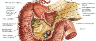
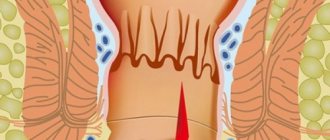
Rice. 1. Schemes of the main anatomical forms of Hirschsprung’s disease: a - rectal (1 - section of the aganglionic zone of the rectum, 2 - dilated sigmoid colon); b — rectosigmoidal (the arrow indicates the area of the aganglionic zone of the rectum and sigmoid colon); c — segmental (1 — with two segments and a section of normal intestine between them, 2 — with one segment); d — subtotal (1 — with damage to the rectum, sigmoid and transverse colon, 2 — with spread to the right half of the colon); e — total (the arrow indicates the aganglionic zone of the entire colon).
The study of the colon in congenital aganglionosis made it possible to identify 5 main anatomical forms of this disease (Fig. 1). The above grouping does not exhaust all anatomical variants; there are many transitional forms between individual forms. However, most often the lesion involves the rectosigmoid colon (70%), intermediate flexure and rectal ampulla (20%). As a result of impaired intestinal motility, feces stagnate above the site of the lesion, causing expansion of the overlying sections of the colon. The walls of this part of the intestine are hypertrophied as a result of increased peristalsis of the proximal parts of the colon in order to move the contents through the aperistaltic aganglionic site. The diameter of the dilated intestine can reach large sizes. The aganglionic segment, on the contrary, appears underdeveloped. Whitehouse, Kernohan, Swenson (FR Whitehouse, JW Kernohan, O. Swenson), 10. F. Isakov showed that the main element in the pathogenesis of Hirschsprung's disease is changes in the histostructure of the intramural nervous apparatus in a certain segment of the colon. Morphological studies revealed significant changes in the nodes of the musculo-intestinal (Auerbach) plexus and lesions of the submucosal (Meissner) plexus of “normal”-looking distal sections of the colon. Research allows us to characterize this form of M. as congenital aganglionosis of a section of the large intestine, in which peristalsis cannot occur in those areas of the intestine where the myenteric plexus is absent. Severe, even death, changes in the muscular aganglionic zone (with the formation of an irreducible aganglionic mass in the place of the former muscular membrane) aggravate disorders of intestinal motility and make them permanent. Morphologically the zone is aganglionic, clinically it is aperistaltic.
The term “idiopathic megacolon” can rather be called a collective one: it includes all unclear cases that do not fit into the framework of Hirschsprung’s disease. This pathology is characterized by hl. arr. expansion of the distal parts of the colon, including the rectum, sigmoid, less often descending and transverse colon. In this group, with a generally similar clinical and radiological picture, two options can be distinguished: a) the cause of M. is spasm or stenosis at the level of the internal sphincter of the anus, which is detected during digital rectal examination or sphincterometry; b) there are no pronounced functions or mechanical obstacles, and the cause of intestinal gigantism remains unclear. “Idiopathic megacolon” cannot be considered without taking into account data on the immaturity of the innervation apparatus of the colon. The immature structures of this apparatus can be affected by unfavorable factors in the postnatal period. The immaturity of the intramural ganglia at the time of birth is most pronounced in the caudal part of the colon, so this section most often suffers, for example, from vitamin deficiency, exposure to intestinal and viral toxins, as well as for other reasons. First there are functional, and then morphological changes. In advanced cases, more proximal parts of the colon undergo secondary changes.
Clinical picture and course
Rice.
2. Appearance of a child with Hirschsprung's disease (abdominal circumference is sharply increased). The leading symptom of Hirschsprung's disease is the absence of independent bowel movements (constipation). Dysfunction of the digestive tract is manifested by constipation from the day of birth or the first months of life, lack of independent stool, a gradual increase in abdominal circumference (Fig. 2) and the development of chronic fecal intoxication. The initial manifestations of constipation, their subsequent nature and persistence are largely determined by the length of the aganglionic segment, the nature of the child's feeding, and the compensatory capabilities of the intestine.
General disorders become especially important in children and are expressed by general developmental delays, exhaustion and anemia. Symptoms are most pronounced in children older than one year and less distinct in newborns and infants. The adaptation of the child’s body can be so complete that the disease is manifested only by constipation and the absence of independent stool.
There are three forms of wedge, the course of Hirschsprung's disease.
1. Light form. In the first days, or less often weeks, of life, patients differ little from healthy children. Sometimes their meconium passage is delayed, which may be accompanied by slight bloating and vomiting, but the overall picture is not alarming, especially since after a light enema or insertion of a gas tube, spontaneous bowel movements are observed. However, with the introduction of complementary foods, the condition of children worsens and stool occurs more often only after a cleansing enema. With insufficient care, as a result of prolonged coprostasis, fecal stones are formed (see). The general condition gradually worsens, which is associated with chronic fecal intoxication.
2. The moderate form is often transitional between severe and mild. The general condition is slowly but constantly deteriorating. Constipation becomes more and more persistent. Conservative measures provide a temporary effect. To empty the intestines, they are increasingly resorting to siphon enemas (see).
3. A severe form of congenital aganglionosis manifests itself from the first days of a child’s life. The phenomena of complete low intestinal obstruction are rapidly increasing (see Intestinal obstruction). Meconium is absent or very scanty, gases do not pass away. Abdominal bloating progressively increases, intestinal peristalsis becomes visible, and profuse vomiting appears. The general serious condition is rapidly increasing.
Diagnosis
The diagnosis is based on the study of anamnesis, wedge, symptomatology and X-ray data of the colon. With a survey fluoroscopy and radiography of the abdominal cavity in an upright position of the patient, a large accumulation of gases in the colon is detected. An increase in the volume of the colon and its swelling leads to a high position of the diaphragm, shortening of the pulmonary fields, and a horizontal position of the heart in the chest cavity.
The leading method of X-ray diagnostics is irrigoscopy (see), which, in case of suspicion of M., should be performed using a barium suspension prepared in an isotonic solution of sodium chloride. In case of Hirschsprung's disease, during irrigoscopy, a narrowing of a limited area of the rectum or sigmoid colon is detected, corresponding to the aganglionic zone, above which the intestine is sharply dilated. The aganglionic zone peristalts weakly or does not peristalt at all, its contours are rigid, smooth or jagged. The transition from the narrowed section to the widened part is funnel-shaped. When the aganglionic zone is located in the rectum, the dilated sigmoid colon hangs over the narrowed area, which makes it difficult to identify, so it is necessary to take pictures in different positions of the patient. The contours of the dilated part of the intestine are smooth, devoid of haustration. The folds of the mucous membrane are longitudinal, wide, and often not visible due to the accumulation of feces. Often, when studying the relief of the mucous membrane, its changes are found, characteristic of the inflammatory process and ulceration.
The X-ray picture of congenital gigantism of the colon due to anorectal malformations and idiopathic M. is characterized by expansion and elongation of the entire colon or part of it, usually the sigmoid. The dilation begins immediately above the anal canal in the form of a cone. Haustration is well expressed throughout the colon. Multiple central filling defects caused by coprolites are often detected. Intestinal peristalsis is preserved. Emptying of the colon from the contrast mass is slow. The relief of the mucous membrane is not clearly defined and not throughout the entire intestine. In case of secondary M. due to rectal stenosis, the expansion of the colon predominates over the lengthening and begins proximally from the pathologically altered section of the intestine. The transition to the expanded part of the intestine is sharp. There is no funnel shape characteristic of Hirschsprung's disease. Haustration in the expanded part of the intestine is smoothed. The relief pattern of the mucous membrane is unclear. In specialized institutions, endoscopic research methods (colonoscopy, sigmoidoscopy using fiber endoscopes) are widely used in the diagnosis of M.
Differential diagnosis
carried out with megadolichocolon, idiopathic megacolon, secondary megacolon due to congenital and acquired mechanical obstructions in the distal colon and various forms of constipation in children resulting from age-related dyskinesia. Differential diagnosis of these types of Megacolon is based on the symptoms described by rentgenol. Identifying the underlying pathological process in the large intestine helps to distinguish secondary M. due to developmental anomalies and acquired diseases from its other types. In patients with periodic constipation resulting from colonic dyskinesia, irrigoscopy may reveal areas of intestinal spasm resembling the aganglionic zone of narrowing in Hirschsprung's disease or organic narrowing of the intestine. In such cases, the absence of a characteristic increase in the overlying part of the colon, its study under conditions of artificial pharmacology, hypotension and dynamic observation facilitate the diagnosis of the function, the origin of the narrowing. In doubtful cases, a rectal biopsy is performed; in cases of Hirschsprung's disease, the absence of nerve cells in the test material is revealed.
Treatment
Treatment depends on the form of the disease. Patients with a mild form are subject to conservative treatment (enemas, Vaseline oil, suppositories, therapeutic exercises, diet, restorative treatment). For patients with moderate disease, conservative treatment is also indicated; if there is no effect, colostomy is recommended. The most favorable age for surgery is 2-3 years. Patients with severe forms are not subject to conservative treatment. When a diagnosis or suspicion of M. is made, an urgent colostomy is indicated, radical surgery after 1 year. Radical surgery is performed after 1.5-2 months. after the creation of anus praeternaturalis (see). The most widely used operations are the Swenson-Hiatt, Duhamel, and Soave method.
The classic version of the Swenson operation (O. Swenson, 1948) consists of removing the narrowed (aganglionic) zone of the colon with the imposition of a direct intraperitoneal anastomosis or an oblique one according to Isakov (Fig. 3, a).
Hiatt (R. Hiatt, 1951) introduced significant detail into Swenson's technique. First, laparotomy is performed (see), then mobilization of the resected section of the colon in the distal direction, not reaching 2-2.5 cm from the anus, and extraperitoneal resection of the aganglionic and dilated area. A fenestrated clamp is inserted through the anus stretched with the fingers, the Crimea is grasped by the wall of the mobilized sigmoid colon and turned outward. In this case, two cylinders are formed: external (rectum, turned outward by the mucous membrane) and internal (reduced sigmoid colon). Then the anterior semicircle of the rectum is dissected through all layers, departing 1.5-2 cm from the mucocutaneous junction. Through the resulting hole, the sigmoid colon is pulled up with tweezers. Interrupted nylon sutures are placed between the seromuscular membrane of the sigmoid colon and the muscular layer of the rectum. Then the posterior semicircle of the rectum is dissected and the first row of anastomotic sutures is completed. Next, the entire sigmoid colon is cut off in accordance with the previously planned level of resection and a second row of anastomosis is applied with catgut sutures (through all layers of both intestines). The anastomosis is inserted into the anus.
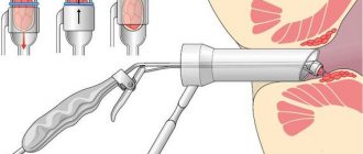
Rice. 3. Schematic representation of the main methods of surgery for Hirschsprung’s disease: a - Swenson’s method (extraperitoneal resection of the affected area of the intestine with the application of an oblique anastomosis according to Isakov, the arrow indicates the anastomosis between the rectum and the reduced colon); b — Duhamel’s method modified by Bairov (1 — opened lumen of the rectum, 2 — retracted colon, 3 — line of anastomosis between the colon and rectal ampulla); c — Soave method (1 — wall of the seromuscular cylinder of the rectum, 2 — retracted colon, 3 — anastomotic line).
During Duhamel's operation (B. Duhamel, 1960), the mobilized sigmoid colon is tied at the point of its transition into the rectum with two silk ligatures. To the left of the rectum, after dissection of the parietal peritoneum, a tunnel is created between the sacrum and the posterior surface of the rectum. The rectum is ligated distal to the ligated ligatures, transected and sutured tightly. Then, along the lower semicircle of the anus, retreating 0.5 cm in depth from the outer edge, the mucous membrane is peeled off by 1.5 cm and at this level all layers of the rectum are dissected (without damaging the internal sphincter) until it connects with the lumen of the previously formed tunnel. Using a forceps passed through the resulting tunnel, the mobilized sigmoid colon is captured and brought down to the perineum. The aganglionic zone to be removed and the extended part of the colon are resected at the level of the anus. The posterior semicircle of the reduced sigmoid colon is sutured along the edge of the wound at the anus, and the anterior semicircle is sutured with rare sutures to the posterior wall of the mobilized part of the rectum. At the suggestion of G. A. Bairov, a crushing clamp is applied to the formed “spur”, facilitating the independent formation of an anastomosis. The part of the intestine between the jaws of the clamp becomes necrotic and the clamp itself falls off (Fig. 3, b).
In modern clinics in our country and abroad, the operation proposed by Italy has become widespread. surgeon Soave (F. Soave, 1963). With this method of operation, a circular incision is made in the seromuscular layer of the sigmoid colon at a level of 6-12 cm from the transitional fold of the peritoneum, after which the muscular cylinder of the distal intestine is carefully isolated (without damaging the mucous membrane!) to the level of the internal sphincter of the anus. The large intestine is brought down through the muscular cylinder of the rectum and cut off, leaving a freely hanging section 5-7 cm long. The excess part of the intestine is cut off in the second stage 15-20 days after the formation of a sutureless anastomosis (Fig. 3, c).
The main advantage of this method is that the intestine is brought down through the natural anal canal, eliminating damage to the anatomical structures around the rectum. The nature of possible complications depends on the surgical method. The most common complications are peritonitis, cicatricial stenosis of the anastomosis, fecal and urinary incontinence. Postoperative management of patients is the same as for intestinal resection (see Intestine, operations).
Forecast
With early diagnosis and timely radical surgery, the prognosis is usually favorable.
Prognosis and prevention
With timely treatment, the prognosis for megacolon is favorable. After radical surgery for Hirschsprung's disease, improvement is observed in 90% of patients. If left untreated, life-threatening conditions may occur: infectious-toxic shock, water-electrolyte imbalance, sepsis.
Prevention of megacolon is aimed at preventing the etiological factor. To prevent constipation, you need to eat more foods with fiber (vegetables, fruits, whole grain bread, nuts). At the same time, exclude from the diet foods that slow down intestinal motility: sweet pastries, potatoes, rice, chocolate. To prevent congenital megacolon, pregnant women should adhere to a healthy lifestyle, give up bad habits and unnecessary medications.