Pancreas
The pancreas is a mixed secretion gland (see Fig. 2). Some of its cells synthesize pancreatic juice, which enters the lumen of the duodenum, and some synthesize insulin and glucagon.
Rice. 2
Through the ducts of the pancreas, inactive forms of enzymes enter the duodenum and are activated there (see Fig. 3).
Rice. 3
Trypsinogen is converted into trypsin, which breaks down proteins into amino acids. Lipase acts only in the presence of bile. Nucleases break down nucleic acids into nucleotides.
Library
S.T.
Metelsky Doctor of Biological Sciences, Chief Researcher of the State Research Institute of General Pathology and Pathophysiology of the Russian Academy of Medical Sciences; contact information for correspondence; Moscow, 125315, Baltiyskaya 8. Purpose of the lecture . Consider the physiological mechanisms of absorption in the gastrointestinal tract (GIT). Basic provisions . In the literature, these issues are covered from three sides: 1) topography of absorption of substances in various parts of the gastrointestinal tract - stomach, duodenum, jejunum, ileum and colon; 2) the main functions of enterocytes; 3) the main mechanisms of absorption in the intestine. 7 main mechanisms of absorption of substances in the intestine are considered. Conclusion. Of the entire gastrointestinal tract, the jejunum and ileum are characterized by the widest spectrum of absorption of various compounds.
Understanding the physiological mechanisms of absorption in the small intestine is of great importance in practical gastroenterology. Keywords:
Absorption, ions, sodium, nutrients, gastrointestinal tract, simple diffusion, facilitated diffusion, osmosis, filtration, pericellular transport, active transport, coupled transport, secondary energized transport, endocytosis, transcytosis, P-glycoprotein.
Basic mechanisms of absorption
The wall of the small intestine, where the most intensive absorption of essential nutrients, or nutrients, occurs, consists of the mucosa (villi and intestinal glands), submucosa (where the blood and lymphatic vessels are located), the muscular layer (where the nerve fibers are located) and the serosa. The mucous membrane is formed by villi, covered with single-layer epithelium interspersed with goblet cells; Lymphatic vessels, a capillary network, and nerve fibers pass inside the villi. A characteristic feature of the transport of substances in the epithelium of the small intestine is that it occurs through a monolayer of cells. The absorption surface of such a monolayer is significantly increased due to microvilli. Enterocytes of the small intestine, where the absorption of nutrients (nutrients) mainly occurs, are asymmetrical, or polarized: the apical and basement membranes differ from each other in permeability, a set of enzymes, the magnitude of the electrical potential difference and perform unequal transport functions. Ions enter cells using ion channels or special molecular machines - pumps. Energy for the entry of ions into the cell is usually provided through the plasma membrane by an electrochemical sodium gradient generated and maintained by the functioning of the Na+, K+-ATPase pump. This pump is localized on the basolateral membrane facing the blood (Fig. 1). The energy that can be obtained from the electrochemical potential of Na+ (ion concentration difference + electrical potential difference across the membrane) and which is released when incoming sodium crosses the plasma membrane can be used by other transport systems. Consequently, the Na+, K+-ATPase pump performs two important functions: it pumps Na+ out of cells and generates an electrochemical gradient that provides energy to the mechanisms of solute entry. The term “absorption” refers to a set of processes that ensure the transfer of substances from the intestinal lumen through the epithelial layer into the blood and lymph; secretion is a movement in the opposite direction.
Absorption in various parts of the gastrointestinal tract
20% of alcohol consumed is absorbed in the stomach, as well as short-chain fatty acids. In the duodenum - vitamins A and B1, iron, calcium, glycerol, fatty acids, monoglycerides, amino acids, mono- and disaccharides. In the jejunum - glucose, galactose, amino acids and dipeptides, glycerol and fatty acids, mono- and diglycerides, copper, zinc, potassium, calcium, magnesium, phosphorus, iodine, iron, fat-soluble vitamins D, E and K, a significant part of the vitamin complex B, vitamin C and alcohol residues. The ileum contains disaccharides, sodium, potassium, chloride, calcium, magnesium, phosphorus, iodine, vitamins C, D, E, K, B1, B2, B6, B12 and most of the water. In the large intestine - sodium, potassium, water, gases, some fatty acids formed during the metabolism of plant fibers and undigested starch, vitamins synthesized by bacteria - biotin (vitamin H) and vitamin K.
Main functions of enterocytes
The main functions of enterocytes include the following. Absorption of ions, including sodium, calcium, magnesium and iron, by the mechanism of their active transport. Water absorption (transcellular or pericellular) occurs due to the osmotic gradient formed and maintained by ion pumps, in particular Na+, K+-ATPase. Absorption of sugars. Enzymes (polysaccharidases and disaccharidases) located in the glycocalyx break down large sugar molecules into smaller ones, which are then absorbed. Glucose is transported across the apical membrane of the enterocyte using the Na+-dependent glucose transporter. Glucose moves through the cytosol (cytoplasm) and leaves the enterocyte through the basolateral membrane (into the capillary system) using the GLUT-2 transporter. Galactose is transported using the same transport system. Fructose crosses the apical membrane of the enterocyte using the GLUT-5 transporter. Absorption of peptides and amino acids. In the glycocalyx, peptidase enzymes break down proteins into amino acids and small peptides. Enteropeptidases activate the conversion of pancreatic trypsinogen to trypsin, which in turn activates other pancreatic zymogens. Lipid absorption. Lipids - triglycerides and phospholipids - are broken down and passively diffuse into enterocytes, and free and esterified sterols are absorbed in mixed micelles (see below). Small lipid molecules are transported into the intestinal capillaries through tight junctions. Sterols that enter the enterocyte, including cholesterol, are esterified by the enzyme acyl-CoA: cholesterol acyltransferase (ACAT), together with resynthesized triglycerides, phospholipids and apolipoproteins, is included in chylomicrons, which are secreted into the lymph and then into the bloodstream. Resorption of unconjugated bile salts. Bile that enters the intestinal lumen and is not used in the process of lipid emulsification is reabsorbed in the ileum. The process is known as enterohepatic circulation. Absorption of vitamins. For the absorption of vitamins, as a rule, the absorption mechanisms of other substances are used. A special mechanism exists for the absorption of vitamin B12 (see below). Secretion of immunoglobulins. IgA from mucosal plasma cells is absorbed through the basolateral surface through the mechanism of receptor-mediated endocytosis and released into the intestinal lumen as a receptor-IgA complex. The presence of a receptor gives the molecule additional stability.
Basic mechanisms of absorption of compounds in the intestine
In Fig. 2 presents the main mechanisms of absorption of substances. Let us consider these mechanisms in more detail. Presystemic metabolism, or metabolism (effect) of the first passage of the intestinal wall. A phenomenon in which the concentration of a substance decreases sharply before entering the bloodstream. However, if the administered substance is a substrate of P-glycoprotein (see below), its molecules can be repeatedly transferred into and out of enterocytes, as a result of which the likelihood of metabolism of this compound in enterocytes increases. P-glycoprotein is highly expressed in normal cells lining the intestine, renal proximal tubules, blood-brain barrier capillaries, and liver cells. P-glycoprotein transporters are members of the largest and most ancient transporter superfamily, present in organisms from prokaryotes to humans. These are transmembrane proteins whose function is to transport a wide range of
substances through extra- and intracellular membranes, including metabolic products, lipids and drugs. Such proteins are classified as ATP-binding cassette transporters (ABC transporters) based on their sequence and the arrangement of the ATP-binding domain. ABC transporters influence the drug resistance of tumors, cystic fibrosis, bacterial resistance to many drugs, and some other phenomena. Passive transport of substances through the epithelial layer. Passive transport of substances through a monolayer of enterocytes occurs without the expenditure of free energy and can be carried out either transcellular or pericellular. This type of transport includes simple diffusion (Fig. 3), osmosis (Fig. 4) and filtration (Fig. 5). The driving force for the diffusion of solute molecules is its concentration gradient. The dependence of the rate of diffusion of a substance on its concentration is linear. Diffusion is the least specific and, apparently, the slowest transport process. With osmosis, which is a type of diffusion transfer, movement occurs in accordance with the concentration gradient of free (not associated with the substance) solvent (water) molecules.
The filtration process involves the transfer of a solution through a porous membrane. The passive transfer of substances through membranes also includes facilitated diffusion - the transfer of substances using transporters, i.e., special channels or pores (Fig. 6). Loose diffusion is substrate specific. The dependence of the rate of the process at sufficiently high concentrations of the transported substance reaches saturation, since the transfer of the next molecule is inhibited by waiting for the transporter to be free from transfer of the previous one. Pericellular transport is the transport of connections between cells through the area of tight junctions (Fig. 7); it does not require energy expenditure. The structure and permeability of tight junctions of the small intestine are currently being actively studied and debated. For example, it is known that claudin-2 is responsible for the selectivity of tight junctions for sodium. Another possibility is that intercellular transfer occurs due to some defect in the epithelial layer. Such movement can occur along intercellular areas in those places where desquamation of individual cells occurs. This path may be a gateway for the penetration of foreign macromolecules directly into the blood or tissue fluids. Endocytosis, exocytosis, receptor-mediated transport (Fig. and transcytosis. Endocytosis is the vesicular uptake of fluid, macromolecules or small particles into a cell. There are three mechanisms of endocytosis: pinocytosis (from the Greek words “drink” and “cell”), phagocytosis (from the Greek words “eat” and “cell”) and receptor-mediated endocytosis or clathrin-dependent endocytosis. Violations of this mechanism lead to the development of certain diseases. Many intestinal toxins, in particular cholera, enter enterocytes precisely through this mechanism. During pinocytosis, the flexible plasma membrane forms an invagination ( invagination) in the form of a pit. Such a pit is filled with liquid from the external environment. Then it is detached from the membrane and, in the form of a vesicle, moves into the cytoplasm, where its membrane walls are digested and the contents are released. Thanks to this process, cells can absorb both large molecules and various ions that are unable to cross the membrane on their own.Pinocytosis is often observed in cells whose function is related to absorption. This is an extremely intensive process: in some cells, 100% of the plasma membrane is absorbed and restored in just an hour. During phagocytosis (a phenomenon discovered by the Russian scientist I.I. Mechnikov in 1882), the outgrowths of the cytoplasm capture droplets of liquid containing any dense (living or nonliving) particles (up to 0.5 microns), and draw them into the thickness of the cytoplasm, where hydrolyzing enzymes digest the absorbed material, breaking it down into fragments that can be absorbed by the cell. Phagocytosis occurs via a clathrin-independent actin-dependent mechanism; This is the main mechanism of defense of the host body against microorganisms. Phagocytosis of damaged or aged cells is necessary for tissue renewal and wound healing. In receptor-mediated endocytosis (see Fig., specific surface receptors are used for the transfer of molecules. This mechanism has the following properties - specificity, the ability to concentrate the ligand on the cell surface, refractoriness. If a specific receptor, after binding the ligand and its absorption, does not return to the membrane, the cell becomes refractory to this ligand. Using the endocytotic vesicular mechanism, both high molecular weight compounds such as vitamin B12, ferritin and hemoglobin, and low molecular weight ones - calcium, iron, etc. are absorbed. The role of endocytosis is especially great in the early postnatal period. In an adult, the pinocytotic type of absorption is of significant importance in provision of the body with nutrients apparently does not.Transcytosis is a mechanism by which molecules that enter the cell from outside can be delivered to various compartments within the cell or even move from one layer of cells to another.
One well-studied example of transcytosis is the penetration of some maternal immunoglobulins through the intestinal epithelial cells of the newborn. Maternal antibodies enter the baby's body with milk. Antibodies bound to the appropriate receptors are sorted into early endosomes of digestive tract cells, then, with the help of other vesicles, pass through the epithelial cell and fuse with the plasma membrane at the basolateral surface. Here the ligands are released from the receptors. The immunoglobulins then collect in the lymphatic vessels and enter the newborn's bloodstream. Consideration of absorption mechanisms from the point of view of individual groups of substances and compounds will be presented in one of the following issues of the journal. The work was supported by the Russian Foundation for Basic Research grant 09-04-01698 References:
1. Metelsky S.T. Transport processes and membrane digestion in the mucous membrane of the small intestine. Electrophysiological model. – M.: Anacharsis, 2007. – 272 p. 2. General course of human and animal physiology. - Book 2. Physiology of visceral systems / Ed. HELL. Nozdracheva. – M.: Higher School, 1991. – P. 356–404. 3. Membrane digestion. New facts and concepts / Ed. AM Ugolev. – M.: MIR Publishers, 1989. – 288 p. 4. Tansey T., Christie DA, Tansey EM Intestinal absorption. – London: Wellcome Trust, 2000. – 81 p
article taken from the website of the Russian Journal of Gastroenterology, Hepatology, Coloproctology
The article is posted at:
https://www.gastro-j.ru/article/33-fiziologicheskie-mehanizmyi-vsasyivaniya-v-nbsp-kishechnike/show/full/
Parietal digestion
There is also parietal digestion, discovered by Russian academician A.M. Coal (see Fig. 6).
Rice. 6
The intestinal mucosa forms a relief: villi, crypts, folds, which increase the surface area of the intestine (see Fig. 7).
Villi are protrusions of the intestinal epithelium that are capable of contracting, thereby accompanying the absorption of chyme.
Crypts are invaginations of epithelial plates.
Rice. 7
The mucous membrane of the small intestine is formed by enterocytes. The surface of these cells, facing the lumen, is covered with microvilli, which increases the surface area by 40 times.
Rice. 8. Microvilli
Intermediate hydrolysis products enter the brush border zone formed by microvilli, where further hydrolysis and absorption occur.
The main enzymes of parietal digestion are amylase, lipase, protease.
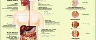
What happens to the nutrients?
After entering the blood, beneficial substances and microelements begin to be carried into the cells of the tissues that make up our body. There they take part in metabolism, or metabolism. Metabolic processes are incredibly important for maintaining our vital functions, since it is thanks to them that the necessary proteins, amino acids and other building blocks are formed from which the human body is built. Building materials go a long way from the place where nutrients are absorbed in humans, to every organ, every cell of our body.
A well-coordinated and harmonious metabolism lays a solid foundation for building a healthy body. A person whose metabolism is in order has good health, a lot of energy and a good mood. If this process is disrupted, then problems will not be long in coming. This can lead to disruption of the endocrine system, gout, excess cholesterol, impaired mental development and many other bad things.
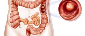
Large intestine and its role in nutrition
From the small intestine, undigested and unabsorbed food masses pass into the large intestine (Fig. 52). It is about 2 m long and includes three sections: blind, colon
and
rectum
.
Between the small and large intestines there is a special valve that allows food to pass through in portions and only in one direction. The glands of the walls of the large intestine (Fig. 53) do not produce enzymes, but secrete mucus necessary for the formation of feces - feces .
During meals, which most people eat 3-4 times a day, the walls of the colon contract vigorously, pushing the contents into the rectum. This occurs at the moment when a new portion of food enters the stomach and it is necessary to free up space in the large intestine. This is why, immediately after a heavy meal, a dull rumbling may be heard in the stomach.
The stool that enters the rectum consists of 70% water, and the rest is the remains of undigested food (mainly fiber) and bacteria. Their movement through the large intestine takes approximately 12 hours. During this time, partial absorption of water and substances dissolved in it occurs.
The contents of the large intestine contain a huge number of bacteria that cause the breakdown of fiber and undigested protein molecules. Bacterial flora
, living in the intestines, secretes a number of vitamins vital for humans: K, E, group B. When protein products are broken down, a whole set of toxic substances is formed: phenols, indoles, skatoles. All these substances are neutralized as blood passes through the liver. The processes of fermentation and putrefaction occurring in the large intestine must be strictly balanced, otherwise diseases of the gastrointestinal tract may develop.
Thus, undigested food remains form feces, 1/3 of which consists of microorganisms. Feces are removed through the rectum. Emptying the rectum - defecation
- is a complex reflex act under the control of the brain.
Formation and composition of feces
Water is absorbed in the large intestines, and the liquid food gruel coming from the small intestines becomes denser. The amount of absorbed water can be judged from the following data: from 4000 g of food gruel, 3850-3800 g are absorbed back and 150-200 g of formed feces remain. The formation of feces is facilitated by lumps of intestinal mucus, which glue together undigested food particles.
Stool contains undigested food particles, mucus, dead intestinal cells, decayed bile pigments that give stool a dark color, and large amounts of bacteria; the latter make up 30-55% of feces.
Barrier role of the liver
Nutrients absorbed through the intestinal walls through the bloodstream first enter the liver. In liver cells, substances harmful to health that accidentally or intentionally enter the intestines are destroyed. At the same time, the blood passing through the capillaries of the liver contains almost no chemical compounds toxic to humans. This function of the liver is called barrier function
.
For example, liver cells are capable of destroying poisons such as strychnine and nicotine, as well as alcohol. However, many substances harm the liver, causing its cells to die. The liver is one of the few human organs capable of self-healing (regeneration), so for some time it can tolerate tobacco and alcohol abuse, but up to a certain limit, followed by the destruction of its cells - cirrhosis of the liver
and death.
The liver is also a storehouse of glucose, the most important source of energy for the entire body, and especially the brain. In the liver, part of the glucose is converted into a complex carbohydrate - glycogen. Glucose is stored in the form of glycogen until its level in the blood plasma decreases. If this happens, glycogen is converted back into glucose and enters the bloodstream for delivery to all tissues, and most importantly, to the brain.
Suction theories
It was assumed that absorption is due to diffusion, osmosis and filtration, that is, it is exclusively a physicochemical process (Dubois-Reymond, 1908). However, absorption cannot be the result of filtration alone, since the blood pressure in the capillaries is 30-40 mm Hg. Art., and in the lumen of the small intestine - much less, about 5 mm Hg. Art., and with intestinal contraction it increases to 10 mm Hg. Art., but absorption increases with its increase in the intestine. Diffusion and osmosis also play a role in absorption, but they cannot explain it, since hypotonic solutions and water are absorbed contrary to osmosis and diffusion, i.e., against the diffusion gradient.
A study of absorption in an isolated segment of a dog's intestine when its own blood was introduced into this segment showed that, despite the fact that on both sides of the intestinal wall there was the same liquid - the dog's blood - this blood was absorbed after some time. Absorption temporarily stops when drugs act on the intestine and completely stops after the intestine dies. This proves that absorption cannot be only a physicochemical process, but is a physiological process characteristic of intestinal epithelial cells under normal conditions of their life.
This is also proven by the fact that absorption increases oxygen consumption in the intestinal epithelium, its membrane potential increases and morphological changes occur in it.
Absorption is regulated by the nervous system and can be changed by a conditioned reflex. The nervous system also influences absorption through the vasomotor nerves and the nerves that regulate intestinal movements.
The vagus nerves increase absorption, and the sympathetic, splanchnic nerves sharply reduce it. Some hormones (pituitary gland, thyroid gland, pancreas) enhance the absorption of carbohydrates (R. O. Faitelberg, 1947). Bile accelerates the absorption of fats not only in the intestines, but also in the stomach.