Gastric intubation, technique
Gastric intubation, purpose : obtaining gastric juice to assess the secretory function of the stomach. Indications for gastric intubation . Stomach diseases. Contraindications. Stomach bleeding; varicose veins of the esophagus; acute inflammatory diseases of the esophagus and stomach; hypertonic disease; angina pectoris; difficulty breathing through the nose. Equipment . Sterile thin gastric tube; syringe with a capacity of 20 ml; towel; clean kidney-shaped coxa; seven large-capacity test tubes or clean, dry jars with directions on each; trial breakfast (200 ml of 7% decoction of dry cabbage, meat broth or 5% alcohol); glass of boiled water.
Indications for insertion of a gastric tube
A nasogastric tube is used in the presence of the following diseases and disorders (the placement algorithm in these cases is the same):
- Injuries in the mouth, pharynx, abdomen.
- Acute pancreatitis.
- Coma state.
- Postoperative period for surgical interventions in the abdominal and thoracic cavities, gastric resection, suturing of a perforated ulcer.
- Post-stroke condition causing difficulty swallowing.
- Narrowing of the esophagus.
- For the treatment of intestinal obstruction.
- Exhaustion of patients.
This is interesting: What is blood clotting time according to Sukharev and deciphering the analysis
What is a feeding tube?
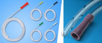
As already mentioned, the phrase “nutritional tube” means a special device that is inserted into the human body through the nasal passage, nasopharynx and esophagus directly into the stomach; such a tube is also called a nasogastric tube.
The design of this device is simple; it consists of a long hollow tube, rounded at one end, thereby preventing damage to internal organs and tissues. This tube has a small diameter and is made of completely hypoallergenic materials, which eliminates any threat to the patient’s health. In addition, the material from which the probes are made is very elastic, and when it comes into contact with the humid and warm environment of the human body, it becomes even more flexible.
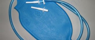
On the outside of the probe, the tube is equipped with a special funnel-shaped hole, through which liquids are introduced (a Janet syringe and specially prepared food are used).
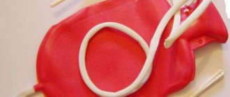
Syringe Janet
This hole is covered with a special lid, which prevents even the smallest foreign particles or objects from getting inside.
It is also important to understand that depending on the age of the person, the specifics of his problem and physiological factors, the feeding tube may differ slightly; the length of the tube varies, as well as its diameter. This allows the device to be used even for infants, and not only for adult patients.
How to prepare for probing
Before examining the stomach, you should try to normalize your mental state and talk with your doctor about the purpose of the sounding. The calmer the patient is, the easier and more comfortable the procedure will be. Overexcitation can negatively affect the composition of gastric juice and strengthen the reflex that causes the urge to vomit.
On the eve of probing, you should not eat - at least 12 hours must pass from dinner or breakfast to the start of the examination. To obtain an objective picture of the composition of gastric juice, it is advisable not to drink or smoke anything.
With these simple measures, preparation for diagnosing the stomach is completely exhausted. There is no need to carry out special drug preparation in advance. On the contrary, if the patient must follow the doctor's orders and take pills, he must inform the gastroenterologist performing the intubation. It is advisable to avoid the use of drugs that directly affect the functioning of the stomach: atropine, caffeine, antacids.
Preparation before the examination
The patient lies on the couch on his side, a saliva collection tray is placed next to him, and the front of the chest is covered with a napkin. This precaution is necessary so that salivation (salivation) does not interfere with normal breathing, since saliva is spat out, but not swallowed. First you need to remove the dentures, a special ring is installed in the mouth in order to eliminate the possibility of clenching the jaws. To reduce sensitivity, an aerosol with an analgesic effect is sprayed into the oral cavity.
Caring for the decompression tube
A gastric decompression tube is installed for a short period of time (a few days at most). The goal is to aspirate gastric contents to relieve the underlying parts of the digestive tract
a (for obstructive and paralytic intestinal obstruction, pyloric stenosis, after operations on the abdominal organs).
Aspiration is carried out several times a day with a syringe or suction. To prevent the probe from becoming clogged, it is periodically purged with air and changed position (twisted, pulled).
A two-channel probe is often used for continuous aspiration (air flows through one of the channels).
It must be remembered that in this case the patient loses fluid and electrolytes, so the corresponding losses must be replenished by intravenous administration of blood electrolytes under laboratory control.
After aspiration, the probe is washed with saline.
The amount of aspirate is measured and recorded (subtracting the volume of lavage fluid).
You should think about removing the probe if:
- Aspirate per day does not exceed 250 ml.
- Gases are released.
- Normal bowel sounds are heard.
Indications for the procedure
Examination of the stomach using this method is carried out for various indications:
- diagnosis of gastrointestinal diseases;
- the need to feed or administer medications directly into the stomach (premature infants, patients with pathologies and injuries of the esophagus, pharynx, oral cavity, as well as unconscious persons);
- washing in case of intoxication of the body with chemicals or low-quality products.
Diseases for the diagnosis of which gastric intubation is prescribed:
- ulcerative lesions;
- gastritis with high and low acidity;
- reflux esophagitis.
The examination helps to identify features of the course of the disease, changes in the tissues of the stomach, the appearance of neoplasms of any etiology, and features of the structure of the mucous membrane. In case of toxic damage, probing with a special type of probe will allow you to quickly remove poisons from the body and prevent their harmful effects on the organs and systems that support human life.
Probe installation process
The process of installing a nasogastric tube consists of a series of clear and practiced steps. The main requirement for its correct installation is that the patient is conscious; the whole process must first be explained to him.
The fact is that in an unconscious state there is an increased risk that the tube will end up in the respiratory tract instead of the esophagus. To prevent this from happening, the doctor must insert two fingers into the patient’s throat, facilitating the correct passage of the probe tube. If a person is conscious, at the moment the body passes through the device, he must make a swallowing movement.
Installation is not a very complicated process, but in the case of installing a nasogastric tube at home, it is better if a specialist does this. In general, the process takes place in several stages.
Preparation
It consists of preparing everything you need (a probe of a certain length and diameter, a Janet syringe with a volume of 150 to 200 milliliters, several clamps, a marker, anesthetic, glycerin or lidocaine). It is also necessary to explain the upcoming procedure to the person if he is conscious.
Installation
Before starting installation, it is recommended to place the device used in the refrigerator in order to stiffen the tube, which will facilitate its easier passage. In addition, the cold body of the tube will reduce the gag reflex.
It is also necessary to first disinfect your hands, and the patient, even if it concerns a bedridden patient, should be placed in a sitting or half-sitting position.
The further procedure is as follows:
- Check the patency of the nostril for insertion. To do this, each nostril is pinched in turn and breathing movements are performed; in some cases, you have to clear your nose;
- Several marks are made on the probe. First, the distance from the earlobe to the mouth, then from the oral cavity to the xiphoid process of the sternum. The first segment indicates reaching the larynx, the second shows the length at which the tube must be placed inside;
- To reduce the gag reflex and eliminate unpleasant sensations, the nasal cavity and pharynx are treated with lidocaine;
- The end of the probe, which will be placed into the human body, is lubricated with the same lidocaine or glycerin, which ensures its easy and unhindered advancement;
- Through the nasal passage, the tube is brought to the larynx (mark 1), after which the person must make swallowing movements, promoting its further advancement;
- As soon as the advancement of the device reaches the second mark, the probe is in the stomach, further movement stops;
- Now you need to check the correct position of the tube. To do this, take a syringe and spray up to 30 milliliters of warm boiled water through the upper funnel. If, when listening to the abdominal cavity, some kind of “gurgling” is heard, everything is done correctly;
- The funnel at the outer end of the probe must be covered with a cap, and the end itself must be secured by fastening it with a pin to the collar or gluing it with a plaster.
Installing a feeding device is not so difficult, however, you need to act clearly, confidently and correctly. If you are not confident in your abilities, it is better to seek help from a specialist. Detailed instructions with a video explanation of the device installation process can be found in the section
Evaluation of the study results
Fractions of gastric juice obtained as a result of intubation are sent for laboratory testing to diagnose possible gastric pathologies. The following indicators are taken into account:
Analysis of this data will allow the attending physician to draw up a complete picture of the disease. If the laboratory test results for gastric secretions differ from those shown in the table, then you need to undergo treatment from a gastroenterologist.
Normal parameters of gastric juice in a healthy person
Nutrition process
Nutrition for bedridden patients with a tube is also carried out according to a certain scheme, consisting of several points:
- The patient should take a semi-sitting position;
- The outer end of the probe falls below the level of the neck and is pinched;
- A syringe with a nutrient mixture heated to 38-39 degrees is attached to the funnel;
- The funnel with the syringe is raised to a distance of over 50 centimeters above the stomach and the clamp is removed;
- Food is introduced slowly, with virtually no pressure (150 ml in about 5-6 minutes);
In order to have a complete understanding of the condition of the mucous membrane of one of the most difficult organs of the digestive system to diagnose, an informative examination such as gastric probing is prescribed.
This examination is carried out on an outpatient basis by a gastroenterologist. A probe is inserted through the mouth or nose into the esophagus and advanced into the stomach. Using a specially developed algorithm, the doctor performs diagnostic manipulations to study indicators such as:
- composition of gastric juice taken in several stages;
- secretory function of the mucous membrane;
- acidity level;
- the presence or absence of food residues in the mucous membrane;
- the volume of fluid pumped out of the stomach.
The examination causes virtually no discomfort, despite the fact that it is carried out without anesthesia or pain relief. The position of the tube in the patient's stomach is monitored using an X-ray machine.
The probe can be inserted through the nose.
The use of modern materials makes it possible to conduct research using a very thin probe with a diameter of 0.4-0.5 cm. The minimum possible dimensions will help avoid gagging and do not create unpleasant sensations.
Modern medical technology has further improved and simplified gastric probing. An exceptionally thin probe can now be equipped with a miniature video camera that transmits an image of the internal contents of the organ and its structural features to a computer screen. Based on the data obtained, the doctor will be able to confirm or refute the alleged diagnosis, differentiate it from similar pathologies, and promptly detect the onset of cancer damage to the mucous membrane and muscular wall of the organ.