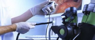
What the doctor sees on an ultrasound of the liver Ultrasound allows you to evaluate the size of the liver, shape, contours and tissue structure.
Ultrasound is the most popular diagnostic method when the patient complains of discomfort in the right side, nausea, weakness and general loss of strength.
Articles on the topic
- “How to fight weight?” – master class from Lady Gaga
- Ursodeoxycholic acid is included in the GFAC recommendations
- Russian surgeons performed a unique liver operation
- Zero without a stick. What are the safe doses of alcohol?
- Omega-3 fatty acids: how they affect the liver
Author of the article:
Karina Tveretskaya
- Site Editor
- Work experience - 11 years
Views: 15809
Reading time: 3 min.
If you come to the doctor with pain in the right or left side, or in the abdomen strictly in the center, most likely, after an examination, he will refer you for an ultrasound examination. Ultrasound is one of the most informative methods for diagnosing abdominal organs. It gives the doctor a fairly large amount of information, especially when it comes to the liver. What the gastroenterologist learns about you during an ultrasound scan – we’ll talk about this further.
Let's examine the liver. Who needs a fibroscan? Elastometry is an ultrasound examination that allows you to identify the presence and degree of fibrosis (damage to liver tissue). How does this diagnostic method differ from ultrasound? And can it replace a biopsy? We answer in the article.
Anatomical features of the liver
The liver is one of the largest organs in the human body. In newborn babies, it occupies almost half of the abdominal cavity. In adults, its weight reaches 1.8 kg, and the average dimensions are 18/13 cm (length/width). The liver is located in the right hypochondrium under the diaphragm. It has a triangular shape, consists of two lobes separated by a ligament. Moreover, the right lobe is approximately 6 times larger than the left.
The organ is surrounded on all sides by peritoneum, except for the hilum and posterior wall. Consists of soft pinkish-brown tissues covered with a fibrous membrane.
Functions of the organ:
- detoxification of the body: liver cells cleanse the blood of toxic substances, control its quality,
- participates in the synthesis of bile necessary for digestion,
- Metabolism: responsible for the absorption and transformation of fats, proteins and carbohydrates, which pass along with the blood through the collar vein.
Indications and contraindications for the procedure
An ultrasound of the liver is necessary for the following symptoms and conditions:
- abdominal injuries,
- pain in the right hypochondrium,
- feeling of heaviness after eating,
- jaundiced tint of the skin, mucous membranes and sclera of the eyes,
- discoloration of stool and bright urine color,
- long-term use of strong drugs,
- radiation and chemotherapy.
The procedure allows you to clarify the presence, location and size of the affected lesions or abscess, which are identified through tests and other studies. The size of the organ, the structure of its tissues, and the presence of neoplasms are also determined.
The advantage of ultrasound is that it does not involve invasive intervention, does not have a negative effect on the body, and is performed on an outpatient basis. Therefore, there are no absolute contraindications, but the study may be postponed due to the use of certain medications that affect the clinical picture, as well as when foci of inflammation appear on the skin in the projection of the organ.
Before the procedure, you should not drink alcohol or eat food that causes bloating, as this may result in an incorrect diagnosis.
Carrying out the procedure
The study should only be carried out by an experienced specialist who is able to correctly interpret the information received. During the study, the size of the organ, its shape and the condition of the segments (which are clearly visualized by ultrasound) are assessed.
The liver segments look like this:
- I (caudate lobe);
- II and III – lateral (lying further from the median plane) left segment;
- IV – left medial (lying closer to the median plane) segment;
- V and VIII (right anterior segment);
- VI and VII (right posterior segment).
Indications and contraindications
This procedure is prescribed in the following cases (indications for use):
- if, as a result of a biochemical blood test, changes in liver test parameters were diagnosed;
in the presence of injuries in the abdominal area;- in case of changes in the color of the skin or sclera of the eyes (yellowing);
- if you suspect the development of an abscess;
- in the presence of helminthic infestation;
- in the case when palpation reveals changes in liver volume (its increase);
- if you suspect the presence of any neoplasms (benign tumors or cancer);
- in the presence of gynecological diseases;
- to monitor the treatment of certain liver diseases (during therapy);
- during dispensary observation of some patients with chronic diseases;
- if viral hepatitis is suspected.
Contraindications for use are:
- the presence of purulent skin damage;
- burns in the area where the study is planned;
- patient's refusal to undergo the procedure.
Preparation
As a rule, the procedure does not require special preparation and can be performed at any time (any time of day), but to obtain more reliable information, the patient should still adhere to some recommendations. Nutrition plays an important role in this case. A few days before the procedure, you should avoid certain foods.
Before an ultrasound, it is forbidden to eat the following foods:
- bread (rye flour);
- baked goods (yeast);
- legumes;
- fruits and vegetables (raw);
- sweets;
- bakery products;
- porridges prepared with milk and added sugar;
- certain dairy products;
- carbonated drinks;
- alcohol;
- juices (mostly fruit).
Meals should be fractional (4-6 times a day), and the recommended volume of fluid consumed is up to 1.5 liters per day. You should also know about other equally important conditions for preparing for the procedure:
- there is a need to use certain medications several days before an ultrasound scan, to improve digestion or to prevent flatulence (activated carbon was previously widely used, but today there are a lot of more effective and modern medications that help in this situation, for example, Espumisan , Duphalac, Linex);
if the drugs do not help (do not eliminate bloating, constipation and other problems in the gastrointestinal tract) or if the patient does not have time to prepare for the procedure, he is recommended to cleanse the intestines with an enema;- the procedure should be carried out on an empty stomach, so it is advisable to refuse dinner and breakfast;
- You should not stop taking previously prescribed medications before an ultrasound scan.
You should also know that ultrasound is not prescribed earlier than 2 days after contrast radiography or endoscopy of the stomach, as well as within 2 days or a week after laparoscopy.
Parameters and stages
The importance of the procedure lies in examining parts of the organ and measuring the following parameters:
- anteroposterior size of the liver lobes;
- biliary tract;
- portal vein (its diameter).
Ultrasound of the liver
Ultrasound is carried out in several stages:
- The examination begins with a transverse scan of the upper abdominal cavity. This helps to obtain more accurate information about the anatomy of the liver.
- The most important stage is oblique scanning, which is carried out along the edge of an arch-shaped osteochondral formation (costal arch). During the research process, it is possible to study the surfaces, the structure of the parenchyma of the liver lobes, make precise measurements, examine the networks and bile ducts.
Performance during pregnancy
Ultrasound during this period is an important and extremely necessary method that helps to identify (exclude) various types of fetal malformations. But there are also certain time limits (sometimes in quantity) that must be taken into account when prescribing such a procedure.
Ultrasound during pregnancy
In the early stages, ultrasound should be performed only according to strict indications. These are the following:
- detection of discrepancy between the size of the uterus and fetal parameters at the established time;
- presence of bloody discharge;
- the occurrence of pain in the lower abdomen (on any side) in women;
- to exclude (confirm) frozen pregnancy;
- with IVF (done to confirm the normal development of the embryo);
- with serious damage to the liver and other gastrointestinal organs.
When should an ultrasound be done during pregnancy (if its course is normal):
at 12 weeks - helps to exclude congenital anomalies, clarify the gestational age, and exclude developmental defects;- at 20-21 weeks - at this period the structure of all fetal organs is examined, the presence of malformations of the central nervous system and cardiovascular system is excluded (confirmed);
- at 32 weeks - during this ultrasound, it is important for specialists to determine the position of the fetus, exclude developmental delays and the presence of previously undetected defects.
At later stages, ultrasound replaces Doppler examination of the fetus. Using Doppler, blood flow, the state of the fetal heart and the functioning of the placenta are studied. This method also helps to diagnose hypoxia or umbilical cord entanglement (which one is specified: 1,2,3-fold).
Ultrasound of the liver: preparation for the procedure
Preparation for an ultrasound of the liver involves following a diet before the examination so that gases do not accumulate in the abdomen at the time of the hardware procedure.
Should I come for the examination on an empty stomach or not? For the most accurate diagnosis, you should not eat before the ultrasound. In this case, 3 days before the procedure, you need to exclude dishes with legumes, cabbage, rich in fiber, white bread and fruits.
If necessary, a specialist may prescribe a cleansing enema or taking sorbents (Smecta, activated carbon), anti-bloating drugs (Espumizan) and enzymes (Creon or Pancreatin).
The specialist tells the patient how to prepare for the study individually at the appointment.
In emergency situations, ultrasound is performed without preparation.
Features in children
The characteristics of the liver parenchyma (structure, echogenicity, sound conductivity and contours) are unchanged regardless of the age of the subject. There are differences in size and some features of preparation for manipulation for younger children.
Preparation
Before the ultrasound, the essence of the planned operation should be explained in a manner understandable to the child. If, due to his age, he cannot understand what is happening, it is necessary to provide him with maximum comfort: the presence of his mother, normal temperature conditions and a dry diaper.
To make the procedure enjoyable for the child, you can promise him a reward upon completion, or take his favorite toy with you. On the day of the examination, it is better to wear clothes that easily allow access to the abdomen - shirts, sweaters with buttons or a zipper.
Pre-study diet requirements for older children are identical to those for adults.
If an infant has been sent for an ultrasound, vegetable and fruit purees and juices should be excluded for 2-3 days. He should be fed only breast milk or formula.
If there is pathology of the gastrointestinal tract, for a better overview, the doctor prescribes enzyme preparations and sorbents.
Methodology and results of the study
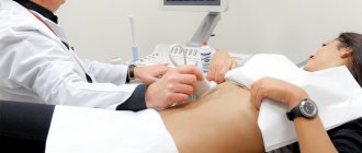
The patient is placed on his back, and a special conductive gel is applied to the area where the organ is projected. Using an ultrasound sensor, the doctor receives a picture on the screen, from which he draws up a description and conclusion. If necessary, a specialist can examine the entire abdominal cavity. Depending on the complexity of the case, the procedure takes from 15 to 30 minutes.
What will a liver ultrasound show? The following are the characteristics that the doctor determines during the study:
- Organ dimensions. Normally, they should be within the following limits:
- oblique diameter - up to 15 cm,
- length/thickness of the right lobe - 11-15/11-13 cm,
- length/thickness of the left lobe - 10/5-7 cm.
- Uniformity of the structure to exclude nodules, compactions and determine what they are, recognize calcifications - all these anomalies have increased echogenicity. If neoplasms are detected, then their density or liquid content is determined.
- Contour boundaries: the edge should be smooth, with clear outlines.
- Condition of the vessels (the diameter of the collar vein in healthy people is up to 1.8 cm, the hepatic artery - up to 1.3 cm, the hepatic vein - up to 1 cm).
Children may have different results: the size of the organ depends on age.
Ultrasound can also detect abnormal development of an organ, including congenital hypoplasia in a child and other pathologies:
- Agenesis of the right or left lobe of the liver is the absence of one of them, and more often it affects the left lobe.
- Rigel's lobe is a modified form of an organ (for example, lingual).
- Accessory lobes are additional formations above the diaphragm or in the hernial sac, which are connected to the main ones through a fibrous cord.
- Cystic or polycystic - multiple neoplasms affect the liver wall during intrauterine development. They may not show any symptoms for many years.
One of the parameters that is assessed by ultrasound is the granularity of the structure. Hepatocytes have a flattened shape, resembling grains. At the same time, the tissue structure itself is porous - normally it is soft and fine-grained.
Since diseases develop gradually, changes in tissues also occur in stages: first, the grain size is assessed as medium, then - coarse.
The latter occurs with hepatitis and may be associated with severe obesity or diabetes.
Diffuse changes
These are abnormal conditions of liver tissue that develop in advanced and severe diseases. The parenchyma becomes thinner and may become deformed, which leads to a change in its integrity and functioning. This condition is manifested by yellowness of the skin and sclera, the appearance of plaque on the tongue, and aching pain in the abdomen on the right side.
Reasons for changes in parenchyma:
- infectious and non-infectious hepatitis,
- cirrhosis, in which tissue scarring occurs,
- liver obesity,
- sudden change in body weight,
- long course of antibiotics.
Liver cysts
These benign neoplasms can appear in any segments and ligaments of the organ. More often found in women. Their size can reach up to 25 cm, which causes pain, nausea and asymmetry of the abdominal cavity. The development of pathology is provoked by cholelithiasis, polycystic ovary syndrome and cirrhosis of the liver. Today, this disease is diagnosed in less than 1% of the population.
There are also congenital cysts that are formed due to disturbances in the development of the bile ducts and their blockage. Such cysts have their own capsule. On ultrasound, cavities with fluid are visible as dark spots - anechoic zones, including multiple forms.
Echinococcal changes
Associated with parasitic invasion of the liver, in particular, tapeworms and echinococci. They usually occur without symptoms, and most often echinococcosis is detected by ultrasound.
The disease can take one of two forms, sometimes mixed variants are found:
- alveolar - in the form of tumors,
- hydative - in the form of a cyst.
What does an ultrasound of the liver show in this case? Round areas of abnormal tissue with liquid contents. To clarify the diagnosis, an immunological blood test is performed. Echinococcosis is dangerous due to the further spread of parasites from the liver throughout the body.
Traumatic changes
They may appear after strong blows, falls, rib fractures due to a central or subcapsular rupture, as well as after an abscess. Typically, traumatic lesions are accompanied by hematomas, which on ultrasound are displayed as roundness without clear boundaries with a heterogeneous structure.
Tumors
Ultrasound can detect malignant and benign neoplasms.
Benign:
- liver adenoma - a tumor with clear boundaries,
- vascular hemangioma - an abnormal formation has a heterogeneous structure and unformed contours,
- lipoma - a tumor of fat cells in the capsule, which, when examined, resembles a hemangioma or cancer metastases,
- cystadenoma is a multilocular cystic tumor with a rich blood supply that can develop from the tissues of the liver and external bile ducts. Unlike other benign tumors, it is characterized by frequent relapses and the possibility of degeneration into oncology.
Using ultrasound, you can identify carcinoma, hepatoblastoma, angiosarcoma - dangerous malignant formations with a compacted structure.
What may cause concern from an oncological point of view:
- the liver is enlarged,
- there are changes in blood vessels,
- seals are determined in the area of the collar vein,
- the lower edge of the organ has a rounded shape,
- attenuation of ultrasonic waves, blurry picture on the device.
Why is ultrasound necessary?
The liver has two types of surface: visceral and diaphragmatic. The visceral surface is distinguished by a complex relief with grooves and depressions from neighboring organs, and the diaphragmatic surface is smooth and convex in shape.
The liver also has sagittal grooves. In one of them lies the gallbladder and the inferior vein, and in the second there is the round hepatic ligament. The furrows are connected to each other by gates. The liver has an unusual vascular system, which includes two venous and one arterial parts.
This organ is the only one in the entire human body that has two afferent vessels at once. The common hepatic artery provides access to arterial blood, and the portal vein provides access to blood from the spleen and gastrointestinal tract. If the outflow from them is disrupted, an internal examination is required, i.e. Ultrasound.
An ultrasound examination of the liver is performed if the following signs are present:
- abdominal pain;
- hepatomegaly;
- "acute" abdomen syndrome;
- yellowness of the skin and sclera;
- the risk of developing cancer, tumors or cysts on internal organs;
- bleeding in the gastrointestinal tract.
The simplicity of ultrasound allows it to be performed on both adults and children.
Possible causes of liver enlargement
A pathological change in the size of the liver indicates a dysfunction of the organ. This is a symptom, not a disease.
Not only tumor formations, but also other factors can provoke liver enlargement:
- viral and bacterial infections,
- hepatitis,
- fibrosis and cirrhosis,
- parasitic infestations,
- gallstones,
- alcoholism,
- lipid metabolism disorder,
- heart failure.
A significant enlargement of the liver is accompanied by jaundice, a feeling of heaviness in the side, discoloration of stool, and emotional instability. A change in size may indicate the development of serious diseases: cancer, cirrhosis, liver failure.
The causes and treatment of the disease that caused the enlargement of the organ are determined by the doctor based on hardware diagnostics and laboratory tests.
Normal liver size according to ultrasound
Liver sizes in women, men, children
- Liver sizes according to ultrasound in men
The table shows the average liver size in healthy adult men.
| Share | Index | Dimensions, cm |
| Whole organ | Height | 20 – 22,5 |
| Thickness | 10 – 12 | |
| Length | 15 – 18 | |
| KVR | 15 | |
| Right | Height | 9,5 – 12,5 |
| Thickness | 11,5 – 13 | |
| Length | 12 – 15 | |
| KVR | Up to 15 | |
| Left | Height | To 10 |
| Thickness | Up to 7 | |
| Length | Until 3 | |
| KVR | Not defined |
- Liver sizes according to ultrasound in women
Typically, women have smaller livers than men. This is due to the fact that women do not eat large amounts of unhealthy foods, unlike men.
| Share | Index | Dimensions, cm |
| Whole organ | Height | 18,5 – 22,5 |
| Thickness | 9 – 12 | |
| Length | 14 – 18 | |
| KVR | 15 | |
| Right | Height | 8,5 – 12,5 |
| Thickness | 11 – 13 | |
| Length | 11 – 15 | |
| KVR | Up to 15 | |
| Left | Height | To 10 |
| Thickness | Up to 7 | |
| Length | Until 3 | |
| KVR | Not defined |
- Liver sizes according to ultrasound in children
In children, the size of the liver may vary depending on age. Moreover, the size of the organ is not affected by gender until the age of 18. The table shows the size of the right and left lobes of the liver in children from the upper edge to the lower:
| Share | Age | Dimensions |
| Right | From 1 year to 14 years | Starting from 6 centimeters, annual increase in size by 6 millimeters |
| 15 years | 10 centimeters | |
| 18 years | 12 centimeters | |
| Left | From 1 year to 14 years | Starting from 4 centimeters, annual increase in size by 2 millimeters |
| 15 years | 5.5 centimeters | |
| 18 years | 5 centimeters |
Ultrasound with elastography
With a standard ultrasound examination, the initial stages of hepatitis, fibrosis and cirrhosis are visible with identical signs. To make a correct diagnosis and assess the degree of development of fibrosis, an elastographic technique is used.
It involves the use of a low-frequency ultrasonic sensor. Its radiation reaches the necessary tissues through the intercostal spaces and is converted into electromagnetic waves. By their speed of distribution one can judge the elasticity of tissues. The doctor simultaneously sees 2 images: an ultrasound and a color picture indicating the density of the structure.
Elastometry does more than just make an accurate diagnosis. It helps avoid liver biopsy, an invasive procedure with a risk of side effects and high cost.