Indications for the study
Ultrasound is a diagnostic method that can detect serious liver diseases.
Doctors recommend that even healthy patients undergo this examination at least once a year. As you get older, the frequency of procedures should increase. Often, specialists send patients for an ultrasound of the liver when:
- yellowness of the skin, mucous membranes and sclera of the eyes;
- darkening of urine;
- discoloration of feces;
- the appearance of discomfort in the right hypochondrium after eating;
- abdominal injuries;
- undergoing chemotherapy or radiation treatment;
- long-term drug therapy;
- painful sensations in the right hypochondrium;
- the presence of inflammation determined by palpation.
What is the human liver
This is an exocrine gland. It is located in the abdominal cavity and occupies 2.5% of the total body weight. The surface of the organ is a convex shape. The inner part, which is located below, has a concave appearance. The convex side of the gland is attached to the diaphragm due to the presence of the falciform ligament. Inside it are ligaments and veins.
How much does a human liver weigh?
- In the male body - on average about 1.5 kg,
- In women, the weight of the liver is 1.2 kg.
The indicated standard sizes are typical for a healthy person. Its neighboring organs are the stomach and intestines.
Therapy
Treatment of changes in the liver parenchyma consists of identifying the cause of their occurrence and further elimination. If the patient has viral infections, he is prescribed antiviral drugs, and for autoimmune diseases - immunosuppressive drugs.
The patient is strictly prohibited from drinking alcoholic beverages. Basic therapy is combined with the intake of vitamins, amino acids and phospholipids. Another component of treatment is a diet that excludes the consumption of fatty, fried, salty and spicy foods.
Related publications:
- Can the human liver feel pain?
- Hepatocyte is a cell of the liver parenchyma. Structure and functions of hepatocytes
- Where is the gallbladder located in humans? Anatomy and structure of the gallbladder
- What function does the liver perform in the human body?
Preparing for an ultrasound
To get more accurate ultrasound results, you should follow some rules. Don't forget the following recommendations:
- 3 days before the study, stop eating fatty and fried foods;
- stop smoking and drinking alcoholic beverages;
- the day before the procedure, you should avoid foods that contribute to the appearance of gases in the intestines;
- before the procedure, cleanse the body with tablets or an enema;
- 8 hours before the procedure you should stop eating.
Other diagnostic methods
Modern medicine includes many ways to detect diseases in the liver, thanks to which the most accurate diagnosis can be made.
In order to obtain adequate research results, it is not enough to perform only ultrasound diagnostics and echography on the patient. The examination should be comprehensive and include:
general tests: blood, urine, feces; biochemical blood test; ELISA, PCR, which detect antibodies capable of resisting viruses, their DNA or RNA and thereby confirm the role in the development of the disease. Tomography (CT); liver biopsy with histological examination of a fragment of the affected area , is done if cirrhosis is suspected (formation of fibrous nodes in the liver).
Normal liver sizes for women and men
| Thickness | 11.2 -12.6 cm. |
| Length | 11-15 cm. |
| Oblique vertical dimension | 15 cm. |
| Thickness | Up to 7 cm. |
| Height | 14-18 cm. |
| Length | 14-18cm. |
| Sagittal size | 9-12 cm. |
| Cross section | 22.5 cm. |
A gland can be considered the correct size if it does not exceed the given parameters.
The vessels located inside the organ must also comply with established standards.
| Artery diameter | Central vein diameter | Diameter of the inferior vena cava | Portal vein |
| 4-6 mm | No more than 10 mm | 10-14 mm | No more than 25 mm |
The size of the organ can be influenced by lifestyle, physical condition and other factors. However, there is an average norm, relative to which small deviations in men are acceptable:
- the entire organ - its height does not exceed 18 cm and cannot be less than 15 cm, the length ranges from 20 to 22.5 cm, while the thickness of the gland is from 10 to 12 cm. The oblique incision is 15 cm;
- the right lobe is 9.5-12.5 cm in length, and no more than 15 cm in height with a minimum of 12 cm. The thickness of the right lobe is 11.5-13 cm with a CVR of up to 15 cm;
- the left lobe is 10 cm in length, no more, and up to 3 cm in height, while the thickness can be 7 cm.
For women, the rates will differ, although only slightly. Sizes depend not only on a person’s build, but also on the fact that women eat and drink much less harmful foods. Normal liver sizes according to ultrasound in women will be as follows:
- the total length is at least 18.5 cm, but the maximum, as in men, is up to 22.5 cm. The thickness of the organ reaches 12 cm. Height is up to 18 cm in the largest area. KVR – 15;
- the length of the right lobe reaches 12.5 cm with a minimum of 8.5 cm. The thickness of the right lobe can be at least 11, but not more than 13 cm, and the height can be from 11 to 15;
- The length of the left lobe does not exceed 10 cm, and the height is 3 cm, while the thickness of the left lobe reaches 7 cm.
In both cases - for men and women, the size of the liver according to CVR for the left lobe is not determined.
Along with the size of the liver, ultrasound must determine the parameters of the liver parenchyma, which include:
- General echogenicity is an indicator of the ability of the gland to transmit ultrasound waves. A healthy liver with normal dimensions transmits impulses evenly. For comparison, they can compare the echogenicity with the pancreas - it should be the same or slightly higher.
- Grain. Normally, this parameter looks like a fine-grained image on an ultrasound machine, and structural abnormalities are not noticeable. The diseased organ may be covered with large grains with various inclusions and formations.
- Volumetric neoplasms. A healthy person does not have white, black or gray tumors.
Another important indicator that is studied on ultrasound along with the size of the liver is the condition of the blood vessels.
When measuring the size of the liver using ultrasound, blood vessels are studied in all respects. Several important veins run through the gland:
- Portal vein. If a person breathes calmly and is not nervous, then the transverse incision is approximately 13 mm. Blood normally flows into the liver; when breathing is held, the blood flow speed remains within 24 cm per second.
- Hepatic artery. Normally in humans it branches into 2 branches, their total diameter is 6 mm. The speed of blood flow in this zone reaches 75-70 cm per second.
- Hepatic veins. The structure of these elements of the circulatory system can vary greatly. In some patients they consist of large vessels, in others - of small ones. To average the indicators, it is customary to measure the transverse size - from 5 to 10 mm.
- Caval inferior vein. The diameter of the structural element is 2.2 mm, and deviations should be no more than 0.2 mm. Any parameters exceeding this value or falling short of normal indicate possible vascular pathologies.
Sometimes duplex scanning is required - during it, structures are displaced during blood flow.
Changes in the liver parenchyma, treatment and prevention
If the examination reveals a disease that has caused changes in the liver tissue, then it is necessary to undergo treatment as soon as possible aimed at removing toxic substances from the liver and restoring liver cells. It is necessary to take medications that improve the functioning of the digestive system (enzymes to restore intestinal flora), anti-inflammatory drugs. An important role is played by the intake of choleretic agents and antispasmodics, which can facilitate the passage of bile and pancreatic stones.
It is necessary to cleanse the liver of toxins, including using folk remedies - using decoctions of medicinal herbs or infusions of medicinal plants. In some cases, it is necessary to take painkillers and vitamins (especially group B).
Before you start taking medications, you need to quit smoking, completely forget about alcoholic beverages, and also be sure to follow a diet. Food should be low in calories, without pepper, spices and salt. You can only eat boiled or steamed dishes and low-fat dairy products. Sweet and sour foods should also be excluded from the diet. Fruits are very healthy, but only ripe and not sour. We need to fight obesity. Diet and the fight against bad habits play a therapeutic as well as a preventive role in improving the health of the liver parenchyma.
Is frequent ultrasound dangerous?
Often patients refuse ultrasound due to the effects of ultrasound. With frequent testing, serious negative reactions can occur in the body, which cause a degenerative process. In fact, this is not true.
For the body to receive a serious dose of radiation, it needs to receive about 10 watts per square centimeter. During an ultrasound examination, each centimeter receives about 0.05 watts.
The duration of the ultrasound examination procedure is no more than 20 minutes. During this period, the body cannot receive a sufficient amount of radiation, which could cause serious harm to the body. Even if you carry out the procedures every day, nothing bad will happen.
It is extremely dangerous to refuse ultrasound: this method allows you to diagnose many pathological conditions in the early stages. Among them are polyps, stones, and malignant neoplasms.
The size of the liver is normal according to Kurlov
The measurement of an organ according to Kurlov is based on the value between the lower and upper extreme points of the gland. Oblique dimensions are taken into account; they are measured from a point on the left costal arch to the limit of the gland at the top.
The upper and lower borders of hepatic dullness are 9-11 cm,
The midline at the level of the upper border of hepatic dullness and the border of the xiphoid process and the navel is 8-9 cm,
The midline at the level of the upper border of hepatic dullness and the lower sharp edge of the organ is 7-8 cm.
Changes in indicators
According to anatomical standards, the size of the liver in a healthy person should fit into the following indicators. Right lobar part: thickness (fullness), oblique vertical size, length is from 110 to 130 mm, not more than 150 mm, from 110 to 150 mm, respectively. The angle of the right lobe is ideally 75 degrees.
Left lobar part: height or craniocaudal size, thickness or anteroposterior size are - no more than 100 mm, about 70 mm, respectively. The angle of the left lobe is 45 degrees. Entire gland: length, transverse parameter size, thickness (liver size in the sagittal plane) correspond - from 140 to 180 mm, from 190 to 230 mm, from 100 to 120 mm.
The size of the hepatic portal vein normally does not exceed 13 mm, the vena cava - 15 mm, the splenic vein - 10 mm, the artery - 7 mm. The artery should normally be 5–7 mm. The size of the common bile duct is half that of the vena cava. A healthy organ is represented by a homogeneous structure consisting of small grains. The liver should be without protrusion or depression, with clearly defined boundaries.
The liver image is deciphered by the attending physician who prescribed ultrasound diagnostics.
An indicator of echogenicity is the speed and degree of reflection and absorption of an ultrasonic wave by cells. Ultrasound shows how much liver tissue is capable of transmitting ultrasound. Waves are absorbed worse and reflected faster when the amount of fluid in the tissues is low. The predominance of liquid, on the contrary, promotes wave absorption. Overestimated or underestimated echogenicity data indicate malfunctions of the gland and the presence of pathologies.
For women during the perinatal period, liver ultrasound is performed according to indications, and the results should not differ from anatomical norms.
If one or another normal indicator is changed towards a decrease or increase, we can talk about the presence of abnormal processes and serious diseases.
The size of the gallbladder is normal in adults on ultrasound
When there are no clear foci of inflammation, and changes in the active cells of the liver epithelium (parenchyma) are distributed evenly throughout the gland, each individual section of the organ reflects the waves differently, that is, the echogenicity of the liver changes. In the medical research protocols, this situation is hidden under the label “diffuse changes.”
When the liver grows and increases in volume (hepatomegaly), steatosis (fatty hepatosis) or cardiogenic ischemic hepatitis (the disease is directly related to the work of the heart and blood vessels) is suspected. If ultrasound conductivity decreases, this is a sign of water retention and swelling. The medical term used is hypoechogenicity.
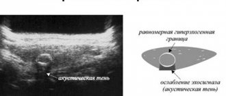
Hyperechogenicity or an increased rate of wave conductivity is characteristic of changes in fat composition, the presence of oncological processes, polycystic disease and hepatitis (including alcohol etiology), cirrhosis. The presence of local dark spots of various shapes and sizes in the image indicates hollow formations in the liver (cysts). These pathologies can be congenital or acquired.
The latter are divided into two types:
- parasitic (echinococcal), provoked by helminthic infestation
- traumatic, those that the organ received as a result of mechanical damage.
Consolidation of the gland structure against the background of increased echogenicity may be an indicator of the formation of a benign angioma or fatty tumor (lipoma). Also, benign neoplasms are determined by the curvature of the liver vessels and its shape.
Schematic representation and ultrasound image of a liver affected by cirrhosis: the affected organ increases in size
It is very important to regularly conduct liver examinations: with the help of ultrasound you can diagnose serious diseases in the initial stages. To prevent serious complications, you should undergo this procedure every year.
During an ultrasound examination, the doctor examines the condition of the liver in detail: determines the number of segments, lobes, size of the organ, and exact location. Ultrasound can detect incipient cirrhosis and hepatitis. A healthy adult liver has the following dimensions on ultrasound:
- organ length – usually ranges from 12 to 18 centimeters;
- cross-sectional length – normally ranges from 20 to 23 centimeters;
- the diameter of the portal vein is no more than 1.5 centimeters;
- the diameter of the liver artery does not exceed 6 millimeters;
- liver width is usually 23-28 centimeters;
- the size of the left lobe is about 6-8 centimeters;
- the size of the right lobe is no more than 12.5 centimeters.
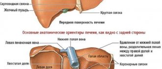
The gland is located in the upper right part of the abdominal cavity. The exception is people with transposition of internal organs. The diaphragmatic surface of the liver has a convex shape; it is adjacent to the lower surface of the diaphragm. The falciform ligament approaches this surface, dividing the liver into lobes (right and left).
Posterior to the liver, it connects to the coronary ligament. The second surface of the gland is visceral. It has several grooves. The first groove is located at the level of the falciform ligament, the second forms the bed of the gallbladder and the groove of the inferior vena cava. Both of these grooves are connected to each other, forming the porta hepatis.
The visceral surface of the right lobe of the liver is represented by the quadrate and caudate lobes. The first is located anterior to the gate of the gland, the second - posterior. The visceral surface of the organ is in contact with the esophagus, stomach, duodenum, kidney and adrenal gland, and colon. The outside of the gland is covered with visceral peritoneum, under which there is Glisson's capsule. Experts are of the opinion that the liver is divided into three lobes and eight segments.
Right lobe
The lower edge of this lobe, provided there is a healthy liver, is at the level of the right costal arch. It protrudes slightly along the midclavicular (10–20 mm) and midline (up to 60 mm). An increase in these indicators, provided the size of the gland is normal, is possible in patients with bronchial asthma, bronchitis with an asthmatic component, and inflammation of the pleura. The liver “rises” against the background of abdominal bloating after resection of the right lung. The angle of the bottom edge should not exceed 75o.
Measurements of the right lobe are taken along the midclavicular line. Normal for adults (centimeters):
- KVR (oblique vertical dimension) – up to 15;
- height – 8.5–12.5;
- length – 11–15.
The size of the right lobe of the gland in a child depends on age. From the top to the bottom edge of an infant, the figure is 6 cm, at 15 years old it increases to 10 cm, after another 3 years - to 12 cm.
Left lobe
The size of this part of the liver from the lower edge to the diaphragmatic surface should not exceed 10 cm. Normal thickness values are within 5–6 cm - this is one of the most important parameters for diagnosing hepatomegaly in the early stages. The angle of the lower edge is 30–45°. The normal size of the left lobe of the liver in children (from the upper to the lower edge) also depends on the age category: in an infant - up to 4 cm, at 15 years old - up to 5.5 cm, at 18 years old - up to 6 cm.
Caudate lobe
Carrying out measurements of this portion of the organ allows us to obtain additional data for the differential diagnosis of a number of diseases. Its thickness normally in adults should not cross the threshold of 3–3.5 cm. Length – up to 7 cm, dimensions of the papillary process – 1–3.8 cm. The ratio of the caudate lobe to the right lobe is less than 0.55.
Other structures
The size of the common hepatic artery (CHA) is assessed by scanning in the epigastric region. The sensor is moved from the xiphoid process to the junction of the hepatic and splenic arteries. The PCA has a tubular structure and is characterized by a lack of echogenicity. Its diameter is 0.5 cm, the volumetric blood flow rate is 6.9 ml/min/kg (calculated depending on the patient’s body weight).
Coarse liver structure
The proper hepatic artery lies in front of the portal vein. Her echographic picture corresponds to the indicators of OPA. When switching to B-mode, you can examine the place of vessel bifurcation and its lobar branches. Smaller structures are not visualized. Diameter – about 0.44 cm.
The portal vein is located in the region of the gate of the gland. It also has a tubular structure with no echogenicity, but its walls are hyperechoic, which makes it easy to examine the condition of the vessel during ultrasound examination. In rare cases, a specialist may detect low echogenicity signals in the lumen of the portal vein, which is mistakenly perceived as the presence of blood clots.
It is important to remember that as you inhale, the diameter of the vessel increases, and as you exhale, it decreases. To view the portal vein, the probe is placed perpendicular to the right costal arch and moved from the area of the xiphoid process to visualize the gate of the gland. Diameter on inhalation – 0.12–0.14 cm, on exhalation – 0.08–0.12 cm. Volumetric blood flow rate – 13.5 ml/min/kg.
The superior mesenteric vein is visualized by moving the ultrasound probe along the longitudinal axis from the splenic artery. Its diameter is 0.55–0.95 cm, the volumetric blood flow rate is 640–980 ml/min. The assessment of the bile ducts begins with the lobar ducts. They have walls with high echogenicity and a small diameter of 0.1–0.3 cm.
Features and types of changes in the liver parenchyma
Liver tissue can change in its composition, shape, and density. Depending on the nature and extent of these changes, they can be divided into:
Focal; Local; Diffuse.
Using ultrasound, an echoscopy of the liver is performed, which produces a picture from which a diagnosis of the disease can be made. Focal changes indicate the presence of a single damaged or compacted lesion. With local changes, the pathology affects individual areas of the parenchyma.
Diffuse changes affect the entire organ. They can be caused not only by disease of the liver itself, but also by cardiac pathology or disruption of the pancreas. Of the two lobes of the liver, the right one, which is larger in size, experiences a greater load. Therefore, diffuse changes in the right lobe are detected more often.
Pronounced signs of diffuse changes do not always indicate serious illnesses. Moderate changes may occur with certain foods. A slight enlargement of the liver in a small child can also be explained by the characteristics of children's physiology. And only if diffuse changes in the liver parenchyma progress with age, then such a pathology should cause concern.
Our readers recommend
Our regular reader recommended an effective method! New discovery! Novosibirsk scientists have identified the best remedy for cleansing the liver. 5 years of research!!! Self-treatment at home! After carefully reviewing it, we decided to offer it to your attention.
EFFECTIVE METHOD
Even if the detected diffuse changes do not affect the person’s well-being, it is necessary to undergo an examination, blood tests, and a test for markers, since this anomaly may be a symptom of latent viral hepatitis or malignant tumors.
Features of ultrasound of children
Newborns are characterized by an enlarged liver. She is not yet able to perform all functions. When the child is about 1.5 years old, the organ begins to acquire the correct shape and size. Also, by this age its segments are formed.
To determine the size of the liver in children under three years of age, the palpation method (feeling the organ) is used. In children under 3 years of age, the lower edge protrudes two centimeters to the right of the lower rib.
After reaching this age, the organ becomes smaller in size and no longer extends beyond the ribs. The Kurlov method of determining size is mainly used when the child turns seven years old.
| Child's age, years | 1-2 | 3-4 | 5-6 | 7-8 | 9-10 | 11-12 | 13-18 |
| Right lobe, mm | 60 | 72 | 84 | 96 | 100 | ||
| Left lobe, mm | 33 | 37 | 41 | 45 | 47 | 49 | 50 |
In children under eight years of age, connective and superficial tissues are not developed. The structure of the organ takes on a similar appearance to the liver of an adult after the child reaches this age.
All of the listed parameters are general indicators, that is, the tables show normal liver sizes for healthy adults and children. When determining the size of the liver, it is necessary to take into account the individual characteristics of each person. When discomfort appears, you should contact an appropriate specialist.
To find out what condition the organ is in, a blood test and ultrasound are performed. When using a biochemical blood test, you can see if there are any deviations:
- albumin;
- bilirubin;
- cholesterol;
- triglycerides;
- lipoproteins;
- glucose;
- squirrel.
The use of ultrasound can record indicators of all deviations from the norm.
The norms for ultrasound results depend on the age of the child. This parameter is also influenced by its growth.
To determine normal indicators, doctors use a special mathematical formula that can be used to calculate each parameter. Modern doctors use special centile tables. They can indicate age and height, after which the characteristics of the liver are shown.
Typically, in newborn children, the size of the liver is no more than 5 centimeters; by the age of 2 it grows to 6.5, and after another 3 years – to 8. Only at the age of 18 is a full-fledged healthy liver formed. A healthy organ must meet the following characteristics:
- the contours are clear, even, divided into separate segments;
- the structure is homogeneous, without neoplasms;
- the superior vena cava is characterized by its lack of echogenicity;
- The liver parenchyma is echogenic.
Features of pathological formations of liver tissue
Hypodense (or hypodense) formation in the liver can be detected by detailed examination on computed tomography or magnetic resonance imaging. The fundamental parameter for deciphering a tomogram is density. X-ray radiation, which is used in computed tomography, when passing through the body encounters resistance on its way - the altered structure of the liver - and becomes weaker at the exit. Each zone has its own attenuation coefficient, which is expressed in Hounsfield units.
Normal parenchyma density ranges from +50 to +70 units, while pathological density can be represented by:
- fatty nodes (-100...-10 units) for liposarcoma, lipoma, fibrolipoma, adenoma, angiolipoma;
- cysts (0…+20 units) regular, parasitic, metastatic;
- bilomas (+4…+10) - limited accumulations of bile due to damage to the bile ducts after surgery or injury;
- soft tissue or fluid lesions (+20...+40 units), in which their composition must be taken into account, since the differential range of diseases is wider.
In relation to the liver parenchyma, the detected formation can be either higher in density or lower. The most difficult structures to diagnose are those consisting of soft tissues: nodular hyperplasia, hematoma, biloma. It is possible to more accurately recognize dense calcification structures: areas of calcification formed in the place of dead or irreversibly changed cells.
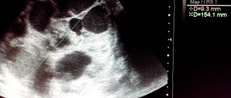
Computed tomography provides a detailed image showing hypodense formations in the liver.
Reasons for deviations
The functions performed by the liver include:
- removal of toxins;
- participation in the synthesis of hormones;
- preventing the appearance of excess hormones;
- normalization of carbohydrate metabolism;
- proper distribution of glucose and glycogen throughout the body;
- participation in the process of blood formation.
Obesity (metabolic disorder). When a person is overweight, a change occurs in the structure of the gland tissue.
Alcoholism. If a person drinks alcohol in large quantities, the cells of the organ cease to function fully, as fat accumulates in them, and the normal size of the liver changes to an enlarged one.
Viral liver diseases. Viruses negatively affect organ tissue. They cause inflammation and can also disrupt the structure of hepatocytes.
Liver damage from toxic substances. The gland can be affected by medications, antibiotics, household chemical products, radiation, and harmful substances.
Changes in the normal state of the gland can occur due to hereditary pathology, as well as subsequently impaired myocardial functionality, blood disease and liver disease. When deviations occur and the normal size of the liver is deformed, the following diseases may occur:
- hepatitis;
- cirrhosis;
- cystic formation;
- metastases;
- tumors.
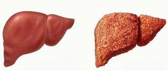
With the development of fatty hepatosis, the structure and homogeneity of the liver changes.
Patients are not concerned about the first symptoms of glandular diseases. Serious consequences can occur if treatment is not started on time or the patient self-medicates.
First of all, it should be said about possible anomalies of the gland. Women, men and children may experience:
- absence of all lobes of the liver (incompatible with life) or one of them (hypertrophy of the existing part is observed);
- localization elsewhere in the body, for example, on the left under the ribs. This condition is combined with transposition of all abdominal organs;
- rotation – a change in the location of the gland along one of the axes;
- hypertrophy of one of the lobes of the organ without the development of a pathological condition;
- the appearance of additional lobes;
- the appearance of additional furrows.
Steatohepatosis
This is a disease characterized by fatty degeneration of liver tissue. Occurs when there is a violation of lipid metabolism, abuse of fatty and fried foods, against the background of defects in the structure of hepatocytes. What changes the doctor will see during diagnosis depends on the severity of the disease. In the diffuse form, the pathological process covers almost the entire tissue of the gland, sometimes unchanged areas remain (more often found in the gates of the liver, the first, fourth and fifth segments).
The focal and local forms are characterized by separate areas of degenerated cells. The shape of the gland usually remains unchanged, the size increases, and the contours are smooth. A specialist may detect a decrease in the sound conductivity of the organ. The vessels become less pronounced on the echographic picture, which is associated with significant degenerative changes.
Hepatitis
The disease is characterized by inflammation of the liver and can occur in acute and chronic forms. Hepatitis is a group of pathological processes that occur against the background of damage to gland tissue by viruses, long-term treatment with drugs, and exposure to toxic and poisonous substances. In acute cases, the liver retains clear contours and its shape, the capsule is visualized more clearly than in a healthy person. The structure of the gland loses its homogeneity, areas of varying echogenicity appear, and the walls of large vessels are more clearly visible.
In the chronic course of the disease, the size of the organ increases due to all departments. The contours remain smooth and are clearly visualized, although the liver capsule is not as visible as normal. The lower edge of the gland is rounded and its angle increases. The echogenicity of tissues increases, which can be uniform or uneven.
Cirrhosis
At the initial stage, ultrasound practically does not detect changes. Some patients may experience an increase in the size of the liver - first the right lobe, then the left and only then the caudate. The thickness of the latter reaches more than 4 cm, which means compensatory hypertrophy. The formation of scar retractions leads to a change in the contours of the affected organ, they become lumpy and uneven. The lower edges of both lobes increase their angle.
Changes in the structure of the liver are also observed. Foci of varying echogenicity appear. The sound conductivity of the organ decreases. If the patient suffers from jaundice of the skin, sclera and mucous membranes, the specialist should check the condition of the intrahepatic and extrahepatic bile ducts. With the development of portal hypertension, the diameters of blood vessels change (in the table).
| Vessel | Changes |
| Portal vein (extrahepatic part) | More than 1.4 cm |
| Splenic vein | More than 0.9 cm |
Tumors
Hemangiomas (vascular neoplasms) are the most common among other liver tumors. The capillary form of hemangioma is represented by small cavity structures that look on the monitor as a homogeneous conglomerate. The cavernous form is represented by large cavities, which are visualized as areas with absence or decreased echogenicity. Lipomas look like round or oval formations with clear, even contours.
Glandular adenoma can occur in any part of the liver, but more often in the right lobe. If the tumor is located very close to the capsule, the surface of the latter becomes deformed. The adenoma has an irregular round shape and clear contours. Tumors of a malignant nature are manifested by a high density of contents, changes in the vascular pattern, the appearance of additional vessels feeding the tumor, and lack of clear contours. As cancer grows, the size of the liver in children and adults increases noticeably.
Interpretation of liver ultrasound is most often done by gastroenterologists and hepatologists - doctors who specialize in the health and pathologies of the gastrointestinal tract in general and the liver in particular. After receiving the results form, doctors determine the need for additional research methods.
Most often, it is the viral form of hepatitis that leads to an increase in the size of the organ.
If the liver is enlarged, we can talk about serious diseases that require additional research. Most often, the organ changes in structure due to viral hepatitis. These are microorganisms that specifically affect hepatocytes, which leads to their destruction. In place of the damaged cells, connective tissue develops, which during ultrasound examination appears as an enlarged liver.
Other reasons include:
- chronic alcoholism;
- hydatid cyst;
- ascites;
- liver failure;
- Gilbert's syndrome;
- pathologies of the cardiovascular system;
- blood diseases;
- malignant and benign neoplasms.
The development of hypertension can be judged by an increase in the diameter of the portal vein of the organ.
Increased size of the portal vein is observed with venous hypertension. The right lobe also changes parameters, which is reflected in the ultrasound results. The maximum value often reaches 20-25 mm. The portal vein can withstand high pressure, therefore, if treatment is prescribed in time, normal values can be restored and serious consequences can be prevented.
At the birth of a child, parenchymal organs are measured to identify congenital pathologies. Incorrect values in an infant indicate disturbances in the blood supply, intrauterine formation of the structures of the hepatobiliary system and spleen. Such abnormalities require immediate treatment through surgery. Interpretation of research results must necessarily include laboratory diagnostics.
Ultrasound examination with high reliability determines a number of diseases of the hepatobiliary system, the main ones are:
- wounds, mechanical injuries, ruptures;
- purulent inflammatory lesions (abscesses), hepatitis of any etiology, degenerative changes caused by metabolic disorders in gland cells (fatty and cholestatic hepatosis) - inflammatory diseases;
- cysts, tumors, lipomas, angiomas (neoplasms of various nature);
- vascular pathologies;
- infectious inflammation of the bile ducts (cholangitis);
- helminth infections;
- fatal liver disease (cirrhosis);
- proliferation of connective tissue with scar changes (fibrosis).
For a more detailed examination, the patient may additionally be recommended other hardware techniques: duodenal intubation (diagnosis with parenteral administration of a stimulus), X-ray or X-ray cholangiography, MRI cholangiography.
The liver is the only organ that has the property of self-regeneration, but with a disregard for health, its capabilities are limited. Diseases of the hepatobiliary system are always severe pathologies that require a forced change in lifestyle. The gland should not be subjected to excessive stress in the form of alcohol, unhealthy foods, or fatty foods.
Determination of diffuse changes using echoscopy
The ultrasound method is based on the ability of body tissues to reflect ultrasound. For various painful conditions of the liver, examination helps detect the following abnormalities:
When examined using ultrasound, echo signs of the liver are studied
Parenchyma compaction; Heterogeneity of structure; Increase in size.
When examined using ultrasound, echo signs and characteristic indicators are studied:
The size of the right and left lobes; Clarity of outlines; Structure of parenchymal tissue; Correctness of the pattern of liver vessels; Echogenicity is the ability to reflect ultrasound.
There is a norm for echogenicity of the parenchyma for a healthy liver. If the echo pattern is increased or decreased, suspicions of a specific diagnosis should be confirmed by laboratory tests.
The device displays data on the intensity of the sound reflection process. The echogenicity of healthy tissue is taken as normal. The sound reflection of damaged tissue differs depending on the changes that have occurred. Thus, excess fat in parenchymal tissue reduces this characteristic, and compaction of the parenchyma means that it is increased.
Healthy liver tissue contains a lot of water. With fatty degeneration, diffuse changes in the liver parenchyma occur, which is expressed in a decrease in water content due to the accumulation of fat. At the same time, the speed of sound reflection increases, and the echogenicity index decreases. This picture is observed in liver hepatosis.
Yellowness of the skin
In infectious liver diseases (viral hepatitis) and inflammatory processes, the density of the parenchyma increases due to tissue swelling. The speed of sound reflection becomes lower than normal, and increased echogenicity is observed. The human condition is characterized by the following symptoms:
Jaundice; Digestive disorder; Hepatomegaly (enlarged liver); Pain in right side; Vomit.
Similar signs can be observed with liver injuries, diabetes mellitus, and cirrhosis. In addition, an increase in echo characteristics can occur with obesity, as well as with drug intoxication.
Decoding the results
During ultrasound diagnostics, the doctor conducts a detailed examination of the liver. To diagnose pathological changes, a specialist must record the results on a special form. The protocol specifies the exact dimensions of the internal organ, the degree of echogenicity, and additional parameters.
Only with a comprehensive study of the liver will it be possible to identify serious deviations in the functioning of this internal organ. If there are indications, the patient is sent for further diagnostics
When evaluating ultrasound results, the following diseases can be identified:
- Hepatitis, heart failure, fatty hepatosis manifest themselves in the form of an increase in the size of the internal organ.
- The early stage of liver cirrhosis looks like a stagnant process inside the organ.
- Benign and cancerous formations, helminths, metastases, the appearance of local islets with increased echogenicity.
- Moderate and severe cirrhosis manifests itself as heterogeneous echogenicity.
- Cirrhosis and metastases look like a lumpy surface of the liver.
- Pathologies of the cardiovascular system - the liver acquires a rounded edge.
- Bile duct cysts are an expansion of the organ.
- GSD is the presence of stones or sand in the organ cavity.
- Atrophy or dyskinesia - the gallbladder decreases in size.
While the child is in the womb, this organ performs a hematopoietic function, which determines its increased parameters.
Sizes for different age categories have their own indicators. This is due to the fact that during intrauterine development the liver performed the functions of the hematopoietic system, so its size, in relation to other organs, is quite large. It can be palpated because it is located below the costal arch, which is not typical for adults.
| Age | Length (mm) | Width (mm) | Thickness (mm) |
| Newborn | 42 | 22 | 18 |
| 5 months | 55 | 31 | 19 |
| 1 year | 70 | 37 | 26 |
| 4 years | 79 | 43 | 28 |
| 10 years | 98 | 51 | 33 |
| 15 years | 107 | 53 | 35 |
You need to understand that in newborns, normal indicators depend on weight and height. In premature babies, these values may differ, but when the baby reaches the required parameters, the indicators should normalize. For a one-month-old baby, the dimensions are the same as at birth, which is explained by a gradual change in its functions. The norm in children is measured in millimeters, since deviations may indicate organ pathologies or developmental abnormalities.
The nature of diffuse changes in the parenchyma
In case of disease of the liver, as well as other organs directly related to it (gall, bladder, intestines, pancreas), degeneration processes of various types can occur in the parenchymal tissue:
Fibrous changes are associated with the proliferation of scar tissue. This pathology can manifest itself due to alcohol or toxic poisoning, or infection with parasites; Hypertrophic - thickening of the parenchyma; Sclerotic - with damage to the liver vessels; Dystrophy - when the liver tissue is gradually replaced by a fatty layer (with hepatosis); Changes associated with tissue swelling due to traumatic or inflammatory edema.