Causes of the disease
In most cases, hyperplasia appears because the patient has not completed treatment for any disease, such as stomach ulcers, gastritis or other inflammations. This leads to active cell division, which contributes to the formation of polyps. The bacterium Helicobacter pillory can also provoke these changes. Sometimes pathology appears due to various infectious diseases. But these are not the only reasons for the appearance of hyperpasia; there are others:
- disruption of the patient’s hormonal levels, for example, excess estrogen;
- heredity, so if a woman has adenomatous polyposis, her daughter or granddaughter can inherit it; with this disease, polyps also form in the human stomach;
- the patient has been taking certain medications for a long time, which damage the walls of the stomach;
- carcinogens have entered the body, which also contribute to the growth of the gastric epithelium.
Etiology
Gastric hyperplasia develops due to unfinished treatment of diseases of the gastrointestinal tract. As a result, active cell growth begins and polyps appear.
The main causes of hyperplasia:
- changes in hormonal balance, especially when the amount of estrogen is increased;
- genetic predisposition, in particular adenomatous polyposis (characteristic of polyps in the stomach) - if the pathology is diagnosed in a woman, the disease can be inherited by a daughter or granddaughter;
- long-term use of medications that can affect changes in the structure of the gastric mucosa;
- unfavorable environment - a pathological increase in the number of cells may begin.
The cause is the bacterium Helicobacter pylori and other infectious diseases.
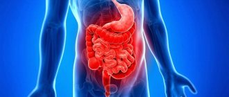
What is intestinal hyperplasia
This pathology means excessive production of intestinal lymphoid tissue cells, which leads to its growth in the mucosal and submucosal layers of the organ. At the same time , the mass of the intestine increases, its functioning is disrupted .
The disease is diagnosed in people of both sexes of any age. The occurrence of intestinal hyperplasia is not associated with eating certain foods and does not depend on the area of residence.
There are many reasons for the development of pathology. They look like this:
- Various disorders of the secretory processes of the intestinal mucosa.
- Hormonal disorders of the body.
- Damage to organ tissues by autoimmune, carcinogenic cells.
- Violation of the nervous regulation of the gastrointestinal tract.
- Long-term chronic stressful situations.
- Colonization of the intestines by pathogenic bacteria.
- Disorders of the immune system.
- Disorder of gastrointestinal motility.
The clinical picture of the disease largely depends on which part of the intestine is affected. The general condition of the body may suffer, the patient becomes weak, and body temperature rises periodically. There are also often complaints of spastic abdominal pain.
Patients may be bothered by long-term diarrhea (stool often contains bloody and mucous impurities), flatulence. In the case of a protracted course of the pathology, a decrease in the patient’s body weight is often diagnosed.
How to treat inflammation of the colon?
Find out how rectal pain is treated.
Classification
In medicine, experts distinguish between many different types of hyperplasia:
- Focal developing hyperplasia of the gastric mucosa. The first stage of development of the anomaly, when certain polyps begin to appear. Focal diagnosed hyperplasia of the stomach covers only some areas (“foci”), which is why it got its name. The lesions look like growths of various shapes and sizes, painted in a different color, so they are clearly visible during examination. Formations arise at the site of previously received damage or erosion.
- Follicular identified gastric hyperplasia. This type of pathology is often diagnosed. Lymphatic cells begin to grow. The reasons for the development of the anomaly are different: the influence of carcinogens, hormonal imbalance, the presence of the bacterium Helicobacter pylori, stressful situations and much more. A distinctive feature of this type of disease is the formation of follicles in the stomach.
- Hyperplasia of the integumentary pitted epithelium of the stomach. A dangerous type of pathology that can contribute to the appearance of a malignant tumor in the intestine. The structure of the epithelium changes under the influence of unfavorable factors: cells grow and become larger.
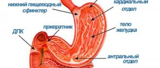
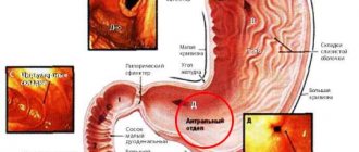
- Hyperplasia involving the antrum of the stomach. The antrum is the last part of the organ before it exits into the intestine. In this place, with the development of hyperplasia, multiple small growths, pits and ridges begin to form.
- Foveal hyperplasia of the gastric mucosa. This type of pathology is characterized by an increase in the length of the folds of the mucous membrane and an increase in their curvature. Pathology occurs due to prolonged inflammation or self-administration of anti-inflammatory drugs.
There are also other types of pathology: glandular, polypoid, lymphoid.
Hyperplasia of the antrum of the stomach
Prevention of follicular gastritis
To avoid suffering from the disease again, you need to do the following:
- Do not give up the diet at the first improvements. Ideally, it should be followed for at least a year.
- Drastically switch to a healthy diet, forever give up junk food, smoking and alcohol.
- Schedule a visit to your gastroenterologist every six months.
Lymphoid follicles in the stomach are a rare but completely curable form of gastritis. As with other types, the patient must undergo a course of drug treatment, follow a special diet, and when curing, do not forget about prevention.
Symptoms
If the patient has an early stage of the disease, it will be very difficult to diagnose it based on symptoms, since the person does not experience any discomfort when epithelial tissue grows. Even the appearance of hyperplastic polyps, if they are small, is not felt by the patient; only large polyps can impede the passage of food and cause severe bleeding or provoke pain.
However, as the pathology progresses, the functioning of the stomach is disrupted, which causes digestive problems. This leads to the patient experiencing a number of symptoms that may indicate the appearance of hyperplasia:
- pain, it can be either temporary or constant, makes itself felt after eating or when the patient has been fasting for a long time;
- suffers from heartburn;
- the stomach is bloated, there is constipation;
- belching occurs with a long sour taste;
- in later stages, the patient may complain of nausea and vomiting;
- he loses his appetite;
- The patient complains of weakness, body aches, and suffers from dizziness.
If these and other symptoms appear, you should consult a doctor and undergo a full examination.
Treatment of diseases of the ileum
Crohn's disease and other dangerous illnesses can be fatal. Lymphoid hyperplasia appears against the background of an immunodeficiency state. Proliferative changes are observed on the intestinal walls. They can be transitory and disappear just as easily as they appear. This is how the body reacts to external stimuli. Diagnosis of diseases allows us to find the causes of a person’s illness. If the function of the ileum is impaired, a comprehensive examination will help determine this. The doctor examines the patient and asks what he is complaining about. An accurate diagnosis can be made using the results of stool, urine, blood tests and fiber endoscopy.
Delayed presentation may require surgery. In the first stages of disease development, drug treatment is used. The doctor also prescribes a strict diet. The patient should eat only foods that are quickly and easily absorbed by the body.
You need to include a lot of vitamins in your menu. The patient is prohibited from drinking alcohol and smoking. Addiction to bad habits negatively affects the health of the intestines. Heavy foods are excluded from the diet. The patient is contraindicated in consuming large amounts of salt and fat. He is not recommended to eat bitter, spicy and fried foods. You will need to give up mushrooms and meat and fish, which take a long time to digest.
The diet may include porridge, omelet, tea and compote. An excellent drink for the immune system is rosehip decoction. Sea buckthorn oil will help a lot. 50 ml should be drunk on an empty stomach every day. You can have breakfast only 2 hours after eating. A healing drink can be prepared from flax seeds. A teaspoon of seed is poured into a glass of boiling water. The infusion is left for 30 minutes.
It needs to be stirred from time to time. The drink is drunk on an empty stomach 1 hour before meals. Compliance with the diet and strict doctor’s instructions will help put the patient on his feet. The main thing is to be treated until complete recovery. It is necessary to include in the daily menu products that do not cause discomfort and allergies. If you monitor all the changes in your body, then everything can be corrected. After completing the course of treatment, you can return to normal life.
The ileum continues the jejunum and does not have a clearly defined border with it, but has thicker walls and a larger diameter.
The length of the organ is 1.5-2.6 meters, the internal diameter is approximately 2.5 cm.
The ileum is separated from the cecum by the ileocecal valve, which acts as a gate and allows the bolus of food to pass from the ileum into the cecum in one direction. It protects the small intestine from bacteria from the large intestine. Disturbances in the functioning of the ileocecal valve lead to the backflow of contents with colonic bacteria.
The area in front of the ileocecal valve is called the terminal section. Fatty acids are absorbed in it. The ileum loops are located in the right iliac fossa. A small part is located in the pelvic cavity. The organ is covered with peritoneum and is fixed to the posterior abdominal wall using a fold (mesentery).
The mucous membrane of the walls of the ileum is covered with villi with columnar epithelium. In the center of the villus there is a lymphatic sinus. Through it, fat breakdown products enter the bloodstream. Monosaccharides enter through the capillaries.
The muscularis propria consists of two fibrous layers, between which lies tissue with blood vessels and muscle plexuses.
Due to contractions of the muscular membrane, the chyme (gruel) is mixed and pushed through.
Types of gastric hyperplasia
The classification of gastric hyperplasia is determined by the nature of the tissue confirmation and the type of cells that have undergone proliferation.
Focal gastric hyperplasia
Focal hyperplasia of the stomach is an early form of a polyp, manifests itself in the form of a benign tumor in one of the sectors of the stomach, in the so-called “focus”, hence the name. It can have a different size, usually resembles a small growth, with a modified structure, this can be seen especially well during a study with contrast, when paint gets on the foci of hyperplasia, they immediately change color and stand out against the background of normal tissues. The outgrowths may look like a tubercle or have a stalk; they can be single or multiple. They are also called wart hyperplasia.
Most often they are transformed from erosion of the mucous membrane. Detected by endoscopic examination.
Which guts does it hit?
This pathological process can be diagnosed along the entire length of the gastrointestinal tract. But the most common location of hyperplasia is the small intestine .
This is explained by the fact that this department is constantly in contact with pathogenic microflora, viral and autoimmune agents.
It is important to note that the final section of the small intestine is rich in lymphoid tissue, which functions as the body’s immune defense, so it is most susceptible to hyperplasia. This is often observed with viral infections and helminthic infestations.
This applies to the crypts of the large intestine. These formations also perform the function of immunoprotection and contain hormonal cells. For this reason, they often increase in size. Therefore, focal hyperplasia of the crypts of the colon mucosa is quite often diagnosed in gastroenterology.
It is important that various helminths also often affect this part of the digestive tract. This pathology of the mucous membrane is a reaction of the intestine to invasion.
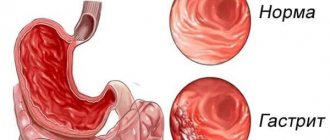
Lymphoid gastritis
This is a rare and insidious disease: the symptoms are mild, and serious pathological changes occur in the stomach that do not allow it to function fully. The exact cause of the development of this particular pathology is unknown. One thing is clear - lymphoid gastritis occurs against a background of chronic gastritis.
A rare form of gastritis has distinctive features in the form of accumulations of lymphocytes that unite into follicles in the area of irritation and degeneration of the epithelium. Lymphatic vesicles formed in large quantities, necessary for the restoration of areas affected by Helicobacter, are often confused with hypertrophied gastritis or tissue atrophy - this is the difficulty of diagnosis.
Another name for the disease - follicular gastritis - was given to it due to the appearance of formations - follicles, which can grow to significant sizes and form a dense layer, thereby complicating not only the digestion process, but also the diagnosis of the pathology itself.
A rare form of gastritis has distinctive features in the form of an accumulation of lymphocytes uniting into follicles in the area of irritation and degeneration of the epithelium
The development of a rare form of the disease is facilitated by inflammation of the mucous membrane, which was ignored for a long period of time, and there was no necessary treatment and prevention.
Diagnosis of gastritis is often difficult due to the lack of specific manifestations of lymphoid pathology, and at the initial stage – very disturbing symptoms. Follicular gastritis is easily confused with the hypertrophic or atrophic form. Advanced lymphoid gastritis can lead to a disease of the lymphatic tissues of the stomach, which at the initial stage of examination is sometimes confused with cancer, but with timely treatment, the chances of recovery are much greater.
The duration and effectiveness of treatment for gastritis depends on how long the sick person decided to seek help from a doctor, on a full examination, and on following all prescribed therapeutic recommendations. Traditional medicine reference books have a huge number of recipes to help overcome this unpleasant disease. Whether it is worth resorting to unconventional methods is something everyone has the right to decide for themselves, but this issue must be agreed upon with the attending physician.
Follicular gastritis, which appears against a background of chronic gastritis, requires the use of various medications that relieve inflammation of the gastric mucosa, eliminate pain, have an enveloping effect, the ability to reduce and regulate the production of hydrochloric acid, and help break down food with the help of enzymes. If Helicobacter is detected in the gastric environment, a course of antibiotics is prescribed to suppress pathogenic microorganisms.
Chronic lymphoid gastritis
An illness of this form is a fairly rare pathology that occurs against the background of chronic gastritis, with the causes of the disease still unknown to specialists. Its main symptom is the accumulation at the site of damage in the epithelium of the digestive organ of a large number of lymphocytes, which look like follicles.
Main symptoms of the disease
It is difficult to identify any specific signs that characterize this particular form of inflammation of the gastric mucosa - for the most part they are common to all types of the disease. Symptoms of lymphoid gastritis may include the following sensations and processes:
- frequent and prolonged heartburn;
- unpleasant taste in the mouth;
- sour belching;
- morning stomach pain, usually occurring 1-3 hours after breakfast;
- loss of appetite - to the point that the patient refuses to eat;
- both frequent constipation and diarrhea;
- heaviness in the stomach, bloating;
- nausea, often ending in vomiting.
With advanced forms, the following may also be observed:
- conspicuous thinness;
- weakness;
- pale and dry skin;
- dense white coating on the tongue;
- jams, cracks in the corners of the lips.
Diagnostics
Due to its asymptomatic onset, the disease is difficult to diagnose in time; its presence is often discovered by chance during a routine examination. Therefore, it is recommended to undergo them once every six months, especially if a person is aware of his predisposition and risks of developing hyperplasia.
It is necessary to contact a gastroenterologist, and, if necessary, an oncologist.
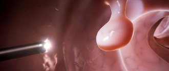
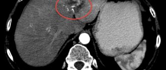
The main diagnostic method is fibrogastroduodenoscopy
An examination in a doctor’s office begins with collecting an anamnesis (the course of the disease according to the patient, a story about his usual lifestyle and family). FGDS (fibrogastroduodenoscopy) is the main diagnostic method. Allows you to examine the stomach from the inside and evaluate the lesions, their scale, nature and specificity. It is during this procedure that focal foveal hyperplasia of the stomach becomes noticeable.
Sometimes FGDS is supplemented by a biopsy (sampling of foreign tissue), which, through histological laboratory examination, helps determine the presence of bacteria and the nature of the neoplasm (benign, malignant).
An X-ray with contrast is indicative - the patient drinks barium, after which an examination is carried out. Allows you to determine the size of polyps, their shape and contours. Since the root cause may be another disorder in the functioning of the body, to complete the picture, they take a blood test (general and chemical), feces and urine, and sometimes gastric juice. They also help identify Helicobacter, which can be diagnosed by the presence of antibodies in the blood, antigens in the stool, the bacterium itself in a biopsy, or a positive urea breath test. In addition, to establish the root cause, an ultrasound of internal organs (pancreas, liver) can be performed.
Follicular hyperplasia of the stomach develops and is asymptomatic, except for a general deterioration in health. It can only be detected during a special examination!
Early diagnosis is the basis of treatment
All diagnostic measures are carried out to establish the characteristics of the disease; it is impossible to diagnose the disease without the use of medical equipment. Treatment of lymphofollicular hyperplasia begins with diagnosis and examination of the patient. For this they widely use:
The FGDS procedure will help determine the presence of pathology.
- Radiography, which helps determine the contours, shape and size of polyps on the walls.
- Endoscopy. Carry out for histological analysis of polyp tissue.
- Fibrogastroduodenoscopy. Used for visual inspection of the gastrointestinal tract. The procedure is appropriate for making a diagnosis and determining the nature of the formation: polyp or tumor.
Diagnosis of gastric hyperplasia
Since it is almost impossible to diagnose gastric hyperplasia without special tests and examinations, doctors use a number of specific studies:
X-ray - shows the presence of polyps in the stomach, you can see its contours, shape, whether it has a leg, what contours are smooth or intermittent. In addition to polyps, you can see a tumor, or rather only its outline.
A more accurate study is fibrogastroduodenoscopy - using a special apparatus, an examination of the internal walls of the stomach is carried out and all neoplasms can be specifically examined and a polyp can be distinguished from a tumor and from other growths.
A biopsy is done after the above tests, since this study is aimed at establishing the malignancy of the tumor and its morphological composition.
Diagnosis of the disease
An inflammatory process of the stomach such as lymphoid gastritis is a difficult disease to diagnose. To ensure that the diagnosis is correct, gastroenterologists prescribe the following procedures to the patient:
- mandatory endoscopic examination - insertion of a probe with a miniature video camera into the stomach;
- biopsy - collection and subsequent cytological, histological, morphological analysis of tissue samples from the gastric mucosa;
- blood, urine, stool tests;
- collection of anamnesis - information for medical history;
- echocardiogram of the abdominal organs.
Based on the research results, assessment of the patient’s symptoms and complaints, he is given a specific diagnosis.
Treatment
A gastroenterologist treats gastric hyperplasia; if necessary, he can refer you to an oncologist or a surgeon, but surgical intervention is required in rare cases; conservative treatment is more often prescribed.
Helicobacter pylori eradication
If a medical examination reveals the presence of Helicobacter pylori bacteria in the stomach, therapy will include their eradication - destruction.
Treatment is similar to that of type B (type 2) gastritis. In order to destroy the bacteria, it is necessary to do a culture and antibiotic sensitivity test. After this, a course of antibacterial drugs is prescribed for 7-14 days. The list of medicines includes:
- Metronidazole;
- Tetracycline;
- Clarithromycin;
- Amoxicillin.
Proton pump inhibitors are prescribed along with antimicrobial drugs. Helicobacter gastritis is almost always accompanied by an increase in stomach acidity. The fact is that acid production is a natural measure to protect the organ from pathogenic bacteria. However, Helicobacter pylori is resistant to hydrochloric acid, so the acid attacks the walls of the stomach, causing inflammation, which can lead to hyperplasia.
Proton pump inhibitors are drugs:
- Omez;
- Laxoprazole;
- Esomeprazole.
The doctor also prescribes medications to protect the gastric mucosa from the effects of hydrochloric acid - antacids:
- Almagel;
- Gastal;
- Maalox.
It is important to emphasize that any prescriptions can only be made by the attending physician.
Forecast
The prognosis depends on the grade of the tumor; The 5-year survival rate for patients with early-stage indolent maltoma is 50%. In later stages, the prognosis is poor; The five-year survival rate is 25%.
Early treatment can significantly prolong the life of patients with lymphofollicular hyperplasia.
Patients are prohibited from self-medicating, since improper therapy can aggravate the disease. Traditional methods are allowed to be used only after consultation with a doctor, since some remedies can cause serious side effects and interact with chemotherapy drugs.