1
11950
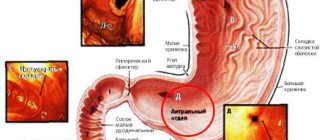
Focal formations of the liver are a serious disease of this internal organ. It is characterized by the formation of one or more cavities in the liver, inside of which there is biological fluid. This is a separate group of pathologies that can be both benign and malignant.
This condition requires mandatory consultation with your doctor. You shouldn’t let everything take its course, because even the simplest condition can take on a negative course.
The lesions may contain pus, blood, and other fluids. In this case, emergency surgery is performed.
What it is?
Focal formations of the liver are understood as pathological neoplasms in the structure of the organ. We can talk about pathological foci of two types:
- Cystic cavities. These are sac-like formations filled with cellular fluid or purulent exudate (if we are talking about abscesses).
- Neoplasms. Tumors of benign and malignant nature.
Focal formations can be either single or multiple. In general, we are talking about a huge number of possible diseases, united by a single term.
Clinical case
Today we have covered a fairly voluminous topic, and finally I would like to talk about one of the most interesting cases related to the identification of focal liver formations.
So, a 34-year-old man came to see me, who for several months had been bothered by discomfort in the right hypochondrium, periodic flatulence, and nausea. Sometimes he noticed moderate pain in the epigastrium. The patient also complained of weakness and told me that his body temperature was often elevated – within the range of 37.2–37.7 °C. Such symptoms are usually called nonspecific - they are characteristic of many pathologies. But the low-grade fever alerted me; In addition, upon palpation of the abdomen, the presence of a tumor-like formation was determined, and upon questioning, I found out that the man was training dogs. There was a suspicion of echinococcosis, an infection that leads to the formation of parasitic cysts in the liver. After the examination, the diagnosis was confirmed, and since it was possible to “intercept” the disease in an uncomplicated form, the patient successfully underwent treatment.
What threatens those people who are sick but don’t know about it? Echinococcosis can be asymptomatic for several years, but allergies, intoxication inevitably occur, and in the later stages - complications in the form of compression of the bile ducts and blood vessels, ascites, perforation and suppuration of the parasitic cyst, up to sepsis. Therefore, using the example described today, I want to show the need to take focal liver lesions seriously and emphasize the importance of timely seeking medical help.
Reasons for formation
There are many possible reasons for the formation of nodes of various etiologies in the structure of the liver. Among them:
- Burdened heredity. It goes without saying that tumors and cystic cavities are not directly inherited. We are talking only about the inheritance of metabolic characteristics, as well as the functioning of the gastrointestinal tract. If someone in the ascending line has been diagnosed with a liver tumor, the risk of developing the same lesion is approximately 5%. If more than one relative suffered, the probability increases to 10-15%. However, these are not absolute numbers. It all depends on the lifestyle and habits of the individual patient.
- History of liver disease. Inflammatory lesions of the liver, such as hepatitis of a medicinal or infectious nature, or cirrhosis, can cause the formation of pathological foci in the organ.
- Presence of gallbladder diseases. Inflammatory diseases of the gallbladder (cholecystitis, biliary dyskinesia) contribute to the development of secondary liver damage.
- Infectious diseases. We are talking about unsanitized foci of chronic inflammation. Paradoxically, even carious teeth can cause the growth of pathological formations in the organ.
- Echinococcal cysts in the liver
Parasitic liver lesions. We can talk about damage by echinococci and liver flukes. The routes of infection are very different. Most often, infection occurs through the nutritional (food) route. Through the bloodstream, the described helminths enter the internal organs. The liver is the first to suffer. Having settled in the organ, pathological organisms begin active activity. The body reacts to “invaders” by responding with inflammation. A huge number of white blood cells and other protective cells attack the “intruders” without much success. As a result, the patient’s body delimits pathological microorganisms with a fibrous capsule (encapsulates them). Such capsules are visible on ultrasound.
- Pathologies of intrauterine development. Usually we are talking about inflammation of the bile ducts while still in the womb. In such cases, cysts form.
- Unfavorable environmental conditions in the place of residence. This factor negatively affects health in general and, in particular, the condition of the liver.
- Excess fatty foods in the diet.
These are the most common reasons why focal formations form in the liver.
Lesions of infectious origin
We have already said that when certain pathologies of infectious origin occur in the body (abscess, hepatitis, candidiasis, tuberculosis, etc.), various types of focal lesions of the parenchyma can occur in the liver structures.
These are, however, lesions that have every chance of being completely eliminated with adequate treatment.
Among focal liver lesions of an infectious nature are usually called:
- Conditions of hepatosis (primarily of fatty origin) - when focal deposition of fatty areas occurs in the cytoplasm of liver cells.
The problem arises when, against the background of a primary infectious disease, a lipid metabolism disorder occurs;
- hepatitis of various forms and origins;
- cirrhosis, in which healthy hepatocytes are replaced by connective tissues, with the appearance of regenerative nodes.
In any case, our readers should understand that it is important to detect any focal formation or liver damage in a timely manner and undergo the most thorough check.
Timely detection of any liver lesions (benign or malignant) can be considered the main factor in the effectiveness of subsequent treatment procedures.
Classification
All organ neoplasms can be divided into benign and malignant.
Benign
Among benign neoplasms in the liver, the following are distinguished:
- Cystadenomas. They are cyst-like structures. Heterogeneous in structure. They consist of many small chambers filled with liquid. They produce special proteins - mucins.
- Liver cysts. Benign formations. A cyst is a sac-like structure filled with cellular fluid and, a little less frequently, another exudate (bile, purulent). They are characterized by a stable character, not prone to growth and malignancy (malignancy). Most often found in the right lobe of the liver.
- Hamartoma. They occur in children and adolescents and, extremely rarely, can be found in adult patients. They are tumor-like structures that arise as a result of spontaneous proliferation of liver tissue. Despite this, they are benign in nature and are not prone to malignancy. Hamartoma is a congenital malformation and is most often discovered by chance.
- Liver adenomas. They are extremely rare. Such tumors are formed from glandular cells of the organ. The main risk group is women aged 20 to 50 years. It is difficult to differentiate from other types of tumors, therefore, when detected, a tissue biopsy is required for subsequent histological examination.
- Liver hyperplasia. It is a violation of fragmentation (lobulation) of an organ. It is difficult to distinguish from malignant degeneration of hepatocytes, so a histological examination is required.
- Hemangiomas. Hemangioma is a benign tumor of an organ. Like other benign tumors, hemangioma is not prone to infiltrative growth and does not metastasize. Well vascularized (provided with blood) because it comes from vascular tissues. Despite its benign quality, it requires mandatory follow-up. It grows extremely slowly. These tumors account for up to 85% of all tumors found in the liver.
- Lipoma. Otherwise - fatty formation in the liver. It is benign in nature, but it is problematic to distinguish a lipoma from other types of tumors using ultrasound. A computed tomography scan is required.
- Parasitic lesions. Benign in nature, but difficult to treat.
- Abscess. It is similar in nature to a cyst, but abscesses are filled with purulent discharge. It is extremely dangerous because upon opening, pus escapes into the surrounding tissues of the abdominal cavity, causing peritonitis.
- Liver hematoma. Usually formed after injury.
Malignant
- Hemangioendothelioma (angiosarcoma). Originates from cells of lymph nodes and blood vessels. It has a relatively mild flow. By ultrasound, such focal liver lesions are defined as dense nodes in the liver tissue.
- Fibrolamellar carcinoma. Formed from liver tissue. It has a relatively slow flow.
- Hepatocellular liver cancer. Formed from hepatocyte cells. Characterized by rapid destruction of the organ. Provokes cirrhosis and impaired hepatic circulation.
- Hepatoblastoma. May differ in infiltrative growth. At the same time, it is an unpredictable tumor that can have a more benign course.
- Kaposi's sarcoma. An extremely aggressive tumor characterized by diffuse growth and rapid proliferation. Fortunately, it is rare.
- Cholangiocarcinoma. This space-occupying formation is characterized by infiltrative growth. It metastasizes early. Such a tumor has a tendency to block veins.
These are the most common focal changes in the liver.
Cystic formations
These neoplasms differ in origin, structure and size. They can be inflammatory, parasitic or congenital, and have a capsule and liquid content. Usually they are filled with a clear or yellowish liquid, but a brown or green tint may appear, which indicates an admixture of blood or bile.
Cysts can be located superficially or inside the gland, and can also reach 25 cm. If during diagnosis a cyst is found in each segment, it is customary to talk about polycystic disease.
Non-parasitic cysts
They are liquid formations with a capsule that are formed from the bile ducts. They are registered in 5% of the population, mainly in women. They can be single or multiple, affecting no more than 30% of the liver tissue.
In most cases, cysts are located in one lobe. With polycystic disease, more than 50% of the glandular tissue is affected, and the cysts are localized in both lobes without preserving the normal glandular tissue between them.
If we consider false cysts, they form in the post-traumatic period. The wall of the neoplasm is represented by fibrous tissue. In addition, such cysts can form after treatment of ulcers or excision of an hydatid cyst. Their contents are a light liquid, which can sometimes contain an admixture of bile.
Clinically, non-parasitic formations do not appear; only occasionally, with a significant increase in size, heaviness or pain in the right hypochondrium is observed. Discomfort can be associated with both stretching of the gland capsule and compression of surrounding organs.
Parasitic cysts
Thanks to modern ultrasound machines, a diagnostician can accurately determine the location of the tumor and the nature of its contents. Immunological methods are also used in diagnosis, for example, RIFA.
Alveococcosis develops as a result of infection with cestodes of echinococcus, which differs from the causative agent of echinococcosis in morphological and biological characteristics.
First, let's take a closer look at echinococcosis. It is considered a rather serious disease and develops as a result of infection of the body with echinococcus. The main problem of diagnosis is the long asymptomatic course, which is why a person consults a doctor at a late stage of the pathology. The volume of cyst contents can reach 5 liters.
Postoperative and post-traumatic cysts
Considering the liver abscess, it should be said about the infectious origin of the pathology. Pathogenic microorganisms penetrate the gland tissue with bile, lymph or blood flow. Often the lesions are localized in the right lobe, have a round shape and are accompanied by discomfort and pain in the right hypochondrium.
An infected cavity in the gland can form in the presence of intra-abdominal infection, after wounds, traumatic damage to the organ, or surgical interventions.
In addition to pain, the disease is manifested by fever, pronounced sweetness and profuse sweating. In terms of frequency of occurrence, the leading cause of abscess is infection of the bile ducts (cholecystitis, cholangitis). Inflammation can also occur after endoscopic manipulation or parasitic infection of the biliary tract.
In second place among the causes of abscesses is an intra-abdominal infection, which penetrates the liver through the portal vein. This is observed with diverticulitis (inflammation of the intestinal processes), a violation of the integrity of the intestine or its ulcerative lesion.
Hematomas form after injury or surgery, when blood accumulates in the cavity, which has entered the parenchyma from a damaged blood vessel.
During an ultrasound examination, the following may be detected:
- formation filled with liquid with clots, which indicates the initial stage of cyst formation;
- lesion with dense masses, partitions of varying thickness and dense walls (stage of progression);
- at the last stage, a false cyst with liquid contents or fibrous areas may be detected, which indicate resorption of the cyst.
Associated symptoms
There are usually no symptoms. Pathological manifestations occur with large formations and are caused by two mechanisms of action: compression of surrounding tissues and focal changes in the organ. Among the characteristic manifestations:
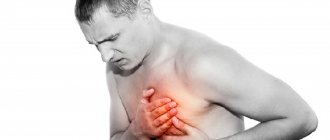
- Pain syndrome. The pain is localized in the right side (hypochondrium). By nature - aching, pulling, monotonous. Nonspecific sign. May be accompanied by hepatitis and cirrhosis.
- Heaviness in the right side, feeling of fullness.
- The phenomena of obstructive jaundice with yellowing of the skin and sclera of the eyes.
- Disturbances of the digestive processes with characteristic heartburn, stool disorders, flatulence.
With malignant neoplasms on the liver of large size and helminthic lesions, the following symptoms develop:
- pale skin;
- feeling of weakness, weakness;
- phenomena of general intoxication: headache, nausea, fever;
- persistent hyperthermia;
- sudden weight loss;
- lack of desire to eat.
This symptomatology is not specific. Only a doctor can put an end to the question of the reasons after conducting a thorough diagnosis.
Malignant tumors
During laparoscopic examination of the abdominal cavity, material is collected, which is subsequently sent for histology. In addition, laparoscopy makes it possible to examine surrounding organs, which is necessary to determine the extent of the malignant process.
The morphological structure of the tumor can only be determined using histological analysis.
It is not always possible to use the puncture technique under ultrasound guidance, since the material can be collected from the unaffected part of the organ. In most cases, the pathology is diagnosed at a late stage, when the tumor is considered inoperable and metastasis is observed.
It is not always possible to suspect a malignant lesion using ultrasound examination, since it may have the same echogenicity as normal gland tissues. Only computed tomography and magnetic resonance imaging make it possible to more accurately establish the localization of the lesion, assess its size, density, and relationship with surrounding tissues.
When using elastography, as well as elastometry, the information content of ultrasound increases significantly. An important part of the diagnosis is the assessment of blood flow in the tumor.
A malignant lesion can be of primary or secondary origin. In the first case, malignant transformation of cells occurs directly in the liver. As for the secondary process, the gland is affected by metastases from the main tumor, which may be located in another organ. Often the liver is affected secondarily.
Among the types of cancer it is worth highlighting:
- hepatocellular carcinoma, which is characterized by rapid progression and high mortality. The risk group is the male part of the population after 50 years of age;
- angiosarcoma, which is also characterized by high aggressiveness;
- hepatoblastoma - manifested by nodes without a capsule, yellowish tint. Pathology is diagnosed in infants.
Symptomatically, the malignant process manifests itself:
- severe malaise;
- icteric syndrome (yellowing of the skin, mucous membranes, darkening of urine and discoloration of feces);
- rapid loss of body weight;
- pain syndrome in the area of the right hypochondrium;
- dyspeptic disorders (nausea, vomiting and flatulence);
- lack of appetite.
On palpation, the gland is felt as a dense, lumpy, painful formation. Therapeutic tactics depend on the stage of the oncological process and the morphology of the tumor. If the formation is considered operable, it is removed.
Treatment of liver tumors is based on:
- type of disease;
- stages of the pathological process;
- functional state of the gland;
- general condition of the patient (presence of allergic reactions and concomitant pathologies);
- the risk of complications (this applies to cases where the formation affects large vessels, intestines and diaphragm).
A feature of the malignant process is the rapid growth of the tumor, metastasis, germination into surrounding organs, inhibition of organ functions and often an unfavorable outcome due to late diagnosis and aggressiveness of the tumor.
Diagnostics
The examination begins with a visit to a specialized specialist. Both benign and malignant liver tumors are treated by gastroenterologists and gastroenterological oncologists. At the initial appointment, the doctor asks questions to identify the nature of the complaints and create a primary picture of the disease. Next comes the turn of palpation. A physical examination allows the doctor to evaluate the structure of the liver and identify tumors. Then it is necessary to conduct a series of instrumental studies. Among them:
- Ultrasound diagnostics. It is the main way to identify any space-occupying formations in the liver tissue. Such a study is considered universal, but will not determine the type of focal liver damage. Necessary both for identifying the disease and for assessing the effectiveness of treatment.
- MRI/CT. It is considered the gold standard in diagnostics. The most informative is computed tomography with contrast. Makes it possible to obtain detailed images of the liver and volumetric processes.
- Biopsy. Necessary for histological examination of tissues. Allows you to determine the type of tumor.
In addition, they resort to blood tests for tumor markers.
For benign formations, dynamic observation is indicated. An ultrasound and/or CT scan is performed every 2-3 months.
Liver adenoma
Occurs in patients using steroid hormones and oral contraceptives.
Education is growing slowly
Types of tumor:
- hepatocellular – adenoma cells resemble hepatocytes;
- cystadenoma (from small bile ducts in which mucus accumulates and cystic formations form).
The tumor is a node with a diameter of 1 to 20 cm, delimited from the liver tissue and having a capsule. Does not grow into blood vessels.
The histological difference from hepatocellular cancer is the preservation of intercellular connective tissue structures. Ultrasound and computed tomography are used for diagnosis.
Adenoma on ultrasound
To determine treatment tactics, the size of the tumor is important.
For small focal formations (less than 5 cm), women are advised to stop hormonal contraception, as well as change their diet aimed at reducing body weight. In men, liver adenomas often become malignant, so surgical treatment is prescribed immediately, regardless of the size of the tumor.
Treatment
The basis of treatment for both benign and malignant lesions is surgery. For benign tumors and tumor-like structures, dynamic observation is indicated. If the tumor begins to grow, surgical resection is indicated. With malignant tumors the situation is somewhat more complicated. Immediate removal of the tumor is indicated, and in severe cases, resection of the entire liver with further organ transplantation. Subsequently, a course of chemotherapy and radiation therapy is carried out. Complete cure is also possible for malignant tumors. It is important to identify pathology at the initial stage. Only in this case will the treatment be effective.
Treatment and symptoms of liver tumor
Liver tumors
Have you been struggling with LIVER PAIN for many years without success?
Head of the Institute of Liver Diseases: “You will be amazed how easy it is to heal your liver just by taking it every day...
Read more "
Liver tumors, including in children, are dangerous neoplasms of the parenchyma, which from an early age disrupt the functions of the organ and, if left untreated, lead to the death of a large gland and the entire body. The diagnosis becomes more widespread every year and is often a consequence of progressive forms of hepatitis. Every year, malignant tumors in the liver are the main cause of death in 250,000 patients of different ages (5% in early childhood). As for benign tumors, a clinical patient with adequate treatment can be saved by surgically removing the cysts.
Diagnosed liver tumors are benign and malignant, with progressive liver cancer predominating in the primary and secondary forms. The primary diagnosis is caused by increased activity of degenerated hepatocytes of the parenchyma; secondary is a complication of a malignant process of another internal system. Already with primary cancer of a large gland, metastases appear, which become symptoms of the disease, provoke complaints from the patient, and force them to consult a doctor for advice. If it is hepatocellular carcinoma, it is preceded by cirrhosis; whereas cholangiocarcinoma and sarcoma appear against the background of one of the forms of hepatitis.
There is the following classification: cholangiocarcinoma is formed from the bile ducts, hepatocellular carcinoma is formed directly from the liver cells.
Treatment begins after diagnosis; in advanced cases, there is no stable therapeutic effect. Secondary formations in the liver accompany metastases that appear during malignant processes of the organic resource. The size of the lesions can exceed 10 cm; such a voluminous neoplasm disrupts the functions of the “filter”, and the body dies from intoxication products.
A benign liver tumor is a less dangerous diagnosis, since the structure of the neoplasm does not contain cancer cells or hepatocyte mutations. These are small formations filled with liquid, which, when growing, lead to dysfunction of the organic resource. The predominance of increased echogenicity, increased bilirubin, and enzyme imbalance are appropriate.
Causes of the disease
Malignant liver tumors, and in children too, have a ramified etiology. Among the pathogenic factors, the following should be highlighted:
- genetic predisposition;
- long-term drug therapy with potent drugs;
- one of the forms of hepatitis;
- complicated pregnancy;
- pathological childbirth;
- developmental defects;
- Wiedemann-Beckwith syndrome;
- adenoma;
- cirrhosis of the liver;
- hepatoblastoma;
- capillary hemangioma;
- progressive cholelithiasis;
- diabetes.
Patients who are close to such pathogenic factors represent a risk group, and the prognosis for the future is not the most comforting with the additional influence of the social factor. Whatever the reasons, a detailed diagnosis begins with collecting medical history data and identifying provoking factors for their prompt elimination.
Symptoms and stages of the disease
An oncologist can determine the stage of the disease after a lengthy diagnosis, visualize the size of the affected organ, and the area of metastases.
- At the first stage, there are no restrictions on the size of the malignant formation; there is a single formation in the liver. Neighboring organs, tissues, lymph nodes are not affected - a positive clinical outcome with a timely response and successful surgical intervention.
- At the second stage, the neoplasm grows into the thickness of the blood vessels, exceeds 5 cm in diameter, and spreads to the lymph nodes. The danger lies in the potential damage to neighboring organs.
- The third stage offers the following types, which make it possible to diagnose a specific disease with maximum accuracy: in stage IIIA there are several foci, extensive damage to the organ, predominantly the right lobe, with predominant growth into neighboring organs and lymph nodes;
- in the ShV stage, cancer cells of the parenchyma penetrate the portal and hepatic veins, while not affecting the lymph nodes, eliminating metastases to neighboring organs;
- in the ShS stage, metastasis of only the gallbladder is excluded, the lymph nodes retain normal functionality.
Focal formations in the liver in a progressive stage reduce the patient’s quality of life; moreover, the size of the liver increases, which is already felt upon palpation at home. A clinical patient can feel the first symptoms without additional hospitalization. This:
- pain under the ribs on the right side;
- signs of dyspepsia of varying intensity;
- sudden weight loss;
- lack of appetite;
- yellowing of the skin, sclera of the eyes;
- general weakness;
- fever;
- iron deficiency anemia relapse stage;
- Cushing's syndrome;
- ascites;
- nose bleed;
- decreased physical activity.
If there is no treatment, the malignant tumor grows in size, and urgent surgery is required to save the clinical patient. You need to pay attention to the first signs of the disease and seek diagnostics.
Diagnosis and treatment
Single or multiple parenchymal tumors, affected areas, metastases - all this will help identify ultrasound as the main method for diagnosing cancer. The organ on the screen has a modified structure and a loose surface; metastases and cysts are visualized; a hypodense or hyperechoic formation in the form of a black circle predominates. To determine whether the disease is cancerous or not, a biopsy is mandatory, as one of the effective invasive diagnostic methods.
To complete the clinical picture, laparoscopy is also indicated to determine the spread of the pathological process in the abdominal region and other organs and systems. Angiography shows that this is a malignant tumor or hemangioma, similar in symptoms. The diagnosis is complemented by the collection of anamnesis data, laboratory tests with an emphasis on biochemical blood tests, mts in the liver. Isoechogenic formation is determined exclusively by clinical methods.
Cysts and malignant neoplasms of the parenchyma are treated surgically; surgery is required. Complete removal of a tumor in the liver is possible only at an early stage of the disease; in advanced clinical situations, excision of the organ itself is not excluded. Extermination of the primary focus increases the risk of internal bleeding, which can result in death.
Intravenous chemotherapy is indicated alone or after surgery, but does not always provide a favorable prognosis. It is much more effective to administer specialized drugs directly into the hepatic artery. Damage to the right or left lobe allows the tumor to be removed; bilateral lesions and numerous neoplasms require different surgical solutions.
Here are several solutions of modern surgeons that give a chance to prolong the patient’s life:
- Resection with removal of only part of the organ. The operation has many contraindications, and the risk of repeated relapses is high. It is advisable to do it at an early stage, a long recovery period is indicated, disability with a non-working group.
- Transplantation of the affected organ is appropriate when the diameter of the tumor is less than 5 cm, the tumor is single or multiple in the amount of 2 - 3 units, each 2 - 3 cm in size. Among the main disadvantages is the difficulty of finding or waiting for a donor organ.
- Palliative procedures are an alternative if surgery is excluded.
- Radiofrequency destruction is performed for single tumors up to 3 cm in size under strict ultrasound and CT control.
- Percutaneous destruction with ethanol.
- Local ablation as an innovative method of treating cancer. This is an opportunity to remove cancer cells, prevent their spread, eliminate relapses and metastases, and partially preserve the affected organ.
- Embolization is the injection of trichloroacetic acid, which promotes the death of cancer cells. The procedure is appropriate in cases where the tumor is less than 3 cm in diameter. Metastases must be absent, otherwise the risk of relapse is high.
- Selective embolization is a blockage of an artery that is directly involved in feeding the neoplasm or cyst.
- Cryoablation is the destruction of the pathological process in the human filter by exposure to low temperatures.
- Radiofrequency ablation is exposure to radio waves of various frequencies.
Traditional treatment is less effective and is indicated as a complement to rehabilitation therapy. It does not provide positive dynamics, aggravates the clinical picture, increases the risk of relapses and complications. The disease is considered fatal and incurable; surgery can only extend the vitality of the body by an average of 5 to 7 years. If there is no treatment, the doctor diagnoses the impossibility of surgical intervention, the death of the clinical patient occurs within 3 to 4 months from complete intoxication of the body. If surgery is performed, several courses of chemotherapy are indicated to exclude repeated relapses and the spread of metastases to neighboring organs and systems.
After successful surgery, symptomatic therapy, lifestyle changes and a therapeutic diet are necessary. Complications include the possible rupture of a malignant neoplasm with general intoxication of the body, followed by death. If tumors in the liver progress, treatment should be timely.
Focal formations of a malignant nature
This category is divided into groups of diseases of primary and metastatic types. In the first case they call:
- fibrolamellar and hepatocellular carcinoma;
- Kaposi's sarcoma;
- peripheral cholangiocarcinoma;
- hepatoblastoma;
- hemangiosarcoma;
- epithelioid hemangioendothelioma.
Focal liver formations of a metastatic type appear if the patient has a tumor of the ovaries, breast, gastrointestinal tract or lungs. Hypervascular space-occupying formation in the liver can be triggered by the presence of an infectious disease, for example, hepatitis, tuberculosis, toxocariasis and others.
As for diffuse diseases, they can be as follows:
- Hepatosis. This type can be either a hypodense focus of formation or a malignant one. It occurs due to the fact that fat drops begin to accumulate in the cytoplasm of the organ cells. Hepatosis occurs when there is a violation of lipid metabolism, due to alcohol abuse, if a person is a lover of tasty, fatty and unhealthy food. It also occurs in people with diabetes who are on a hunger strike. This disease can occur in people taking hepatotoxic drugs. In this case, the mass formation of the right lobe of the liver or the left gives a diffuse increase in echo signals, respectively, and the organ itself increases in size.
- Diffuse echogenicity may increase in the case of alcoholic or chronic viral hepatitis.
- Liver cirrhosis is characterized by the replacement of organ tissue with neoplasms, and regeneration nodes may occur.
Whether it is a hypodense formation or some other lesion, as soon as the first signs appear, you must contact a doctor. In this case, the patient must sequentially undergo computed tomography, ultrasound, and blood tests, and tumor markers are used. If controversial issues arise, the doctor may prescribe an additional fine-needle biopsy of the organ, angiography or laparoscopy. It is easier to treat an identified hypodense formation at the initial stage than to later eliminate the disease, and even treat its consequences.
Return to contents
Lesion formation in children
In order to carefully examine the child, the first impulse is hepatomegaly. The reasons for going to the doctor may be the following:
- Metabolic disorders in the liver. In this case, an increased size of the organ will be observed, and the echogenicity will be higher than normal.
- For congestive heart failure. The level of echogenicity is higher than expected, with the veins of the liver and vena cava dilating.
- Genuine hepatitis. If you check a child who has just been born, you can see increased echogenicity of the liver.
- Hemangioendothelioma and hemangioma are quite common in children under one year of age, and such a formation can be detected by six months. If the symptoms indicate multiple small hemangiomas, then most likely there are formations in the spleen. Cavernous lesions appear as rough, irregular cystic growths. In this case, Doppler analysis is used, which helps to identify feeding and draining vessels and arteriovenous shunts.
- Neuroblastomas. This form occurs in half of the cases among newborns. Most of these tumors are detected during the period when metastases are in the process of separation. On echography, the liver is enlarged and there are metastases. But it is possible to identify the primary focus in the adrenal glands only in half of the cases.
As for diffuse changes, they appear as a result of systemic or pathological processes. Echography shows neoplasms in combination with jaundice and hepatomegaly. But if there is a place for cirrhosis of the liver, and the stage of the disease is already advanced, then the organ will not have an increased, but a decreased size. In such cases, hyperechogenicity is often observed, but if acute hepatitis or organ edema is not observed.
When diagnosing diffuse changes, the doctor determines their size, what their surface is: smooth or bumpy, as well as the edges, since they can be sharp or rounded. In parallel with this, the condition of the spleen, kidneys, lymph nodes, pancreas, and liver vessels is being examined.
Return to contents
Methods for treating focal liver lesions
More and more people prefer not surgical interventions, but more “gentle” ways to solve the problem. But, alas, they do not always help, so in the end you have to lie down on the operating table. The same applies to liver diseases.
As noted above, to identify the problem, it is necessary to undergo an ultrasound and computed tomography. Based on the examination results, the doctor draws attention to the size of the formation. They can be:
- small (diameter - up to 20 mm, volume up to 10 ml);
- medium (diameter - 20-40 mm, volume up to 80 ml);
- large (diameter - 40-80 mm, volume up to 600 ml);
- gigantic (diameter exceeds 80 mm and volume more than 600 ml).
Medium and large formations are in most cases eliminated using the puncture method. But as for small ulcers, this method is used here only with significant localization. Otherwise, antibacterial therapy gives successful results.
If formations of large and gigantic sizes are observed, then they are dealt with using the puncture-drainage method of treatment.
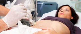
A stylet catheter is used, due to which it is possible to install a drainage of the required diameter so that pus does not enter the abdominal cavity. But depending on how deep or close to vital organs the lesion is located, the technique and equipment may change.
It is very important to pay attention to the problematic condition of the organ in a timely manner, so if you experience any pain or other sensations in the liver or abdominal cavity, you should immediately consult a doctor.
Rate this article:
Currently, there are more and more patients who are diagnosed with hepatic neoplasms (benign and malignant tumors, cysts). They are detected using modern diagnostic methods. CT, ultrasound and MRI are widely used to study internal organs.
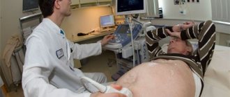
The safest and most accessible research method is an ultrasound examination of the liver. An ultrasound scan of the liver requires preparation. Diagnosis is carried out in the supine or left side position.
Liver ultrasound procedure
The purpose of liver ultrasound is to study its parts, anatomy, and search for pathological changes. Focal formation in the liver is a concept that combines diseases with pathological growth in the liver tissue.
Local benign neoplasms on ultrasound
1. Adenoma is a benign glandular tumor. According to ultrasound, adenomas look like simple formations with smooth contours. Regarding the degree of blood supply, they either do not contain vessels or contain extremely few.
2. Cysts (single, multiple) - formations that have a cavity, a capsule on the surface and liquid inside. Cysts are divided into congenital and acquired. Congenital ones contain bile. There are also simple and multiple cysts. Most often, cysts form in the right lobe. In ultrasound diagnostics, a cyst is an anechoic (liquid) local or diffuse formation with a capsule on the surface.
3. Hemangiomas (cavernous and capillary) are formed from pathologically overgrown vessels in the tissue - a benign vascular tumor. The ultrasound picture is represented by a formation with uneven contours and a heterogeneous structure.
4. Liver lipoma is a fatty tumor. It consists of fat cells (adipocytes) - 90%, 10% are pathologically dividing other cells. It is similar in structure to hemangioma and tumor metastases, so computed tomography with contrast is used to confirm the diagnosis.
5. Focal nodular hyperplasia is a benign neoplasm characterized by excessive diffuse cell growth and the absence of a capsule. On the ultrasound picture it appears as single lesions. They have a round shape, smooth contours. The diagnostic criterion is the presence of hepatic veins in the formation, which confirms the diagnosis.
Benign neoplasms
6. Biliary cystadenoma is a benign liver tumor that is extremely rare. It is a simple cyst with many chambers. The walls of the chambers produce mucin (a mucus-like substance consisting of protein and glucosamine). Characteristic ultrasound signs that distinguish them from simple cysts are a rich blood supply to the cyst walls and multiple papillary foci in them. Does not form metastases.
7. Hamartoma of mesenchymal origin. Characteristic signs are chaotically located vascular and cystic nodes and the connective tissue around them. Does not form metastases.
8. Bile duct hamartoma is a benign developmental defect. Detecting hamartoma using ultrasound is very difficult, since this disease is asymptomatic, and the hamartoma itself is small. It can be easily confused with metastases, so additional research methods are needed.
Distinctive features of all benign neoplasms:
- slowly increase in size;
- do not grow into surrounding tissues and organs;
- do not metastasize;
- respond well to treatment and do not recur;
- can turn into cancer.
It is clear from the above that benign tumors differ from malignant tumors in a favorable course. But there are pitfalls here. A benign formation tends to disrupt the function of an organ.
The following complications may also subsequently arise:
- bleeding into the abdominal cavity;
- organ rupture;
- hemorrhage into tissue.
In order to avoid the above complications, it is necessary to regularly conduct diagnostic studies (computed tomography, ultrasound and magnetic resonance imaging) once every 3 months.
Local malignant neoplasms on ultrasound
Malignant tumors are divided into primary and metastatic.
The primary ones include:
1. Fibrolamellar carcinoma.
- Ultrasound confirms the presence of tumors up to 3 cm in size. The lesions are usually dense.
- Ultrasound examination with contrast. Due to increased blood supply, ultrasound with contrast detects cancer.
- Ultrasound angiography. Contrast is injected using a catheter into the tumor artery and its accumulation is monitored. This is the most informative way to assess the blood supply to cancer.
2. Hepatocellular carcinoma (hepatocellular carcinoma). The ultrasound image reveals neoplasms up to 3 cm in size. The use of contrast agents increases the accuracy of the study. Ultrasound examination reveals changes in the portal vein, organ compaction and cirrhosis.
3. Kaposi's sarcoma is a rare disease. The clinical feature is rapid growth and rapid tissue infiltration. When the tumor disintegrates, bleeding occurs into the abdominal cavity. The tumor has an elastic structure and the shape of a cyst. An ultrasound examination will not be enough to make a diagnosis; laboratory testing and taking into account the medical history are necessary.
4. Peripheral cholangiocarcinoma. The ultrasound picture shows an increase in the lumen of the hepatic ducts. Damage to the portal vein and blockage of its lumen are also detected. Lesions of the hepatic artery are not recognized.
5. Hepatoblastoma. An ultrasound and CT scan should be performed to reveal a simple tumor. Its relationship with surrounding normal tissues is established using magnetic resonance imaging.
6. Hemangiosarcoma of the liver. The node has a heterogeneous structure on ultrasound.
7. Epithelioid hemangioendothelioma. The cancerous tumor is dense on ultrasound examination.
Metastatic tumors arise from tumors of the ovaries, breast in women, gastrointestinal tract and lungs in both sexes.
Distinctive features of all malignant neoplasms:
- rapid growth of tumors and progression of cancer;
- cancer metastases to organs and tissues;
- damage to the structure and function of the affected organs.
Focal lesions due to infections
- viral hepatitis in acute and chronic forms;
- tuberculosis;
- candidiasis;
- toxocariasis;
- echinococcosis;
- abscess.
Diffuse liver lesions on ultrasound
1. Lipid hepatosis - deposition of fat vacuoles in hepatocytes. Ultrasound reveals a diffuse increase in signal and compaction of the organ.
Fatty hepatosis has 3 degrees:
- 1st degree of fatty hepatosis - simple: the fat content in the liver tissue begins to exceed the norm;
- 2nd degree of fatty hepatosis - steatohepatitis: manifested by diffuse changes in the tissue;
- Stage 3 fatty hepatosis - fibrosis: a diffuse change occurs around the vessels, the organ becomes dense.
Separately distinguished:
- alcoholic hepatosis;
- non-alcoholic hepatosis;
- hepatosis of pregnant women;
- hepatosis in diabetes mellitus.
2. Cirrhosis is the replacement of normal tissue with connective tissue. A distinctive feature of cirrhosis on ultrasound is a compaction node in the tissue. Later, if the disease is not treated, it turns into cancer.
It should be remembered that if doubtful changes occur, additional studies are used in the form of computed tomography and magnetic resonance imaging. These methods make it possible to detail formations and detect cancer metastases. Timely detection of cancer, like any liver disease, is the key to successful and effective treatment.
Focal liver formation refers to a number of diseases that can occur in different ways, but have a common feature - the replacement of liver tissue with a cavity filled with fluid. Such a pathological change can be single or multiple, located inside the organ or on its membrane, have a capsule or not.
Focal formations in the liver parenchyma can be benign or malignant. It is important to establish in time the exact nature and stage of development of the pathology, for which you need to undergo an examination.
Infectious formations in the liver
It happens that, against the background of chronic infectious diseases, focal formation of the liver occurs in the human body. It is usually preceded by candidiasis, liver abscesses, echinococcosis, acute and chronic hepatitis, tuberculosis and toxocariasis, and they provoke large foci of pathology. As is known, a liver abscess is of a bacterial nature, and it is possible that the infection will penetrate the liver, causing the death of once healthy tissues of this organ. The first signs of the disease that should alert the patient are:
- acute abdominal pain;
- lack of appetite;
- vomiting and nausea;
- chills and fever.
The disease is dangerous due to its complications, therefore, effective treatment should be followed by antibacterial therapy and immediate surgical intervention. An amoebic liver abscess progresses when an amoebic infection is active in the body, which enters the body exclusively with contaminated water. The intestinal parasite from the intestines enters the circulatory system and soon reaches the liver. The main symptoms are fever, abdominal pain and jaundice. Treatment in most cases is surgical.
Hydatid cysts in the liver are caused by the activity of parasitic echinococcal worms Echinococcus granulosus and Echinococcus multilocularis. Route of infection: through contaminated food or through contact with animals and lack of sanitary standards. Echinococcus first affects the intestines, and then spreads throughout the body, affecting the brain, heart, lungs and liver. The main symptom of the disease is severe pain on the right side of the abdomen, which does not subside even after taking painkillers. Treatment involves chemotherapy and surgery. In order to choose the most effective therapy for such pathological conditions, it is important to correctly determine the diagnosis that accompanies a tumor in the liver and the cause of its occurrence in the human body.
IT IS IMPORTANT TO KNOW! Doctors are dumbfounded! IT EVEN TREATS HEPATITIS C! All you need is after breakfast... Read more–>
Malignant focal formations in the liver
It is possible that benign tumors, over time and under the influence of pathogenic factors, turn into malignant neoplasms, the exacerbation of which can result in the death of the patient. The liver is no exception, in the structure of which such dangerous cavities can also appear. Doctors distinguish metastatic and primary formations in the liver, where the former belong to an increased risk group. They progress in the presence of malignant tumors of the ovaries, gastrointestinal tract, breast and lungs. They are not amenable to conservative treatment, and eliminating them surgically is often pointless, since here it is first necessary to treat the underlying oncological disease with a surgical method. As for primary tumors in the liver, they are eloquent symptoms of the following diagnoses:
- Kaposi's sarcoma;
- hemangiosarcoma;
- fibrolamellar carcinoma;
- peripheral cholangiocarcinoma;
- hepatocellular carcinoma;
- epithelioid hemangioendothelioma;
- hepatoblastoma.
After a detailed diagnosis, the doctor determines which treatment method is most effective. The choice depends on the size of the formation in the liver, its behavior in the body (increases in size or not), and the individual characteristics of the body, in particular the presence of chronic diseases, are also important. The clinical outcome may be unpredictable, and the specialist also informs his patient about this.
Benign focal formations in the liver
So, if benign formations are present in the liver, then the following diagnoses for the body cannot be excluded:
Our readers recommend
Our regular reader recommended an effective method! New discovery! Novosibirsk scientists have identified the best remedy for cleansing the liver. 5 years of research!!! Self-treatment at home! After carefully reviewing it, we decided to offer it to your attention.
EFFECTIVE METHOD
- adenoma;
- hepatic polycystic disease;
- liver lipoma;
- biliary cystadenoma;
- liver hemangioma;
- focal nodular hyperplasia;
- bile duct hamartoma.
It is worth immediately noting that there is no threat to the patient’s life, however, neoplasms in the liver are prone to rapid growth, as a result of which they can rupture, worsen the functioning of the human filter, or put increased pressure on neighboring organs. Therefore, doctors strongly recommend undergoing a control ultrasound once every 2–3 months.
Neoplasm in the liver