You eat something tasty at lunch, and the pieces crushed in the oral cavity travel through a hollow tube, the esophagus, into a kind of bag - our stomach. To get an approximate idea of where the stomach is located and its size, mentally draw a vertical line through the navel and place your left palm on the left ribs so that your fingertips point toward your right shoulder and the line runs through the middle of the second phalanx. The shape of the stomach resembles a bean, the lower curvature is large, the upper curvature is small. The shape may vary slightly from person to person depending on their age and body type. The size of the stomach after a heavy lunch can increase very significantly. The wall of the stomach mainly consists of muscles and is therefore very elastic - the maximum capacity of this hollow organ is 12 or even 16 glasses.
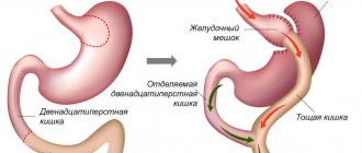
Schematic representation of the stomach (parts of the stomach are named as is customary in anatomy): 1 - cardiac part; 2 - fundus of the stomach; 3 - body of the stomach; 4 - gatekeeper cave; 5 - gatekeeper channel.
The stomach consists of several parts - the cardiac region, the fundus of the stomach, the body of the stomach and the pyloric region. Since each section has its own purpose, removing part of the stomach can have different effects on its overall function and have different consequences. In any case, the diet after removal of part of the stomach should be adjusted by specialists.
What is the stomach
The stomach is an unpaired hollow organ that is located in the upper left region of the abdominal cavity. It is one important part of the human digestive system. The size of the organ changes with age, and its different shapes are also possible. Traditionally, the main function of the organ was considered to be the production of enzymes and hydrochloric acid, which contribute to the processing of food mass. However, in the last century, several more important roles of the stomach were discovered, which significantly changed the approach to the treatment of many diseases.
The wall of the stomach consists of three layers - mucous membrane, smooth muscle fibers and adventitia, which is almost completely covered by peritoneum. Active peristaltic movements occur regularly (4-5 times per minute) after eating food, which contribute to its grinding and movement to the intestines.
What is the purpose of gastric histology?
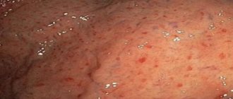
One of the organs of the digestive tract in which neoplasms often develop is the stomach. Histology should be performed if there is any change in the mucous membrane. The following diseases are considered indications for this study:
- Atrophic gastritis. This pathology is characterized by depletion of the cellular composition of the mucous membrane, inflammatory phenomena, and decreased secretion of hydrochloric acid.
- Rare forms of gastritis. These include lymphocytic, eosinophilic and granulomatous inflammation.
- Chronic peptic ulcer of the stomach and duodenum.
- Development of “small signs” according to Savitsky. These include general weakness, decreased appetite and performance, weight loss, and a feeling of abdominal discomfort.
- Detection of stomach polyps and other benign neoplasms.
- A sudden change in the clinical picture of a long-standing peptic ulcer. These include a decrease in the intensity of pain and the development of an aversion to meat food.
The listed pathologies refer to precancerous diseases. This does not mean that the patient has a malignant tumor and its location is the stomach. Histology helps determine exactly what changes are observed in the tissues of the organ. To prevent the development of malignant degeneration, it is worth conducting research as early as possible and taking action.
Structure of the stomach
The stomach itself begins at the level of the entry of the esophagus (XI thoracic vertebra). Anatomically, the following parts of the human stomach are distinguished:
- cardia or cardiac section;
- vault or bottom;
- body;
- pyloric or pyloric region.
There is also another division of the organ into the following parts:
- front wall;
- back wall;
- small curvature;
- greater curvature.
A diagram of the human stomach can be seen in the following figure.
If we consider the wall of the organ, we can distinguish three layers:
- The mucous membrane consists of a single-layer columnar epithelium. It also contains numerous cells that produce hydrochloric acid, mucus and bicarbonates. It also contains small endocrine glands that regulate the functioning of the digestive tract.
- Muscularis - consists of three layers of smooth muscle fibers that run in different directions. They contract under the influence of both local and central nervous regulation. The muscles are very elastic, which allows the organ to expand significantly after eating.
- The outer shell consists of a layer of epithelial cells (mesothelium) and connective tissue. Externally it is covered with peritoneum, which contains part of the arteries that supply blood to the organ.
Organ size in adults and children
In a newborn
The capacity of the ventricle of a newly born baby is small, slightly larger than a thimble. A child can eat no more than 5 or 7 ml of food. The organ in a newborn does not stretch, its walls remain hard, so babies are characterized by belching, which relieves them of excess food. Very quickly, approximately on day 3, the capacity of the ventricle increases immediately to 22-25 ml. Therefore, feeding is recommended frequently, but in small portions. Gradually, the baby’s ventricle becomes elastic and acquires the ability to stretch. A week after birth, the size of the ventricle increases slightly, and by the end of the 2nd week of life it becomes the size of a small chicken egg.
Return to contents
In a baby
The thoracic period is calculated from the first month of life to one year. The child begins to grow and develop rapidly; internal organs also confidently adapt to the external conditions that affect them. If a newborn has a normal stomach volume of 30 ml, then by the 3rd month it is 100 ml (that’s half a glass of liquid). Up to a year, the growth rate of the organ's capacity decreases slightly; by the end of the 1st year of life, the baby's ventricle can accommodate a little more than a glass of liquid food (250 ml).
Return to contents
From 2 years and older
In children 4-5 years old, the capacity of the stomach increases to 450 or 500 ml.
In a 2-year-old child, the maximum stomach size is 350-400 ml of food, including solids. In older children (4-5 years old), the capacity of the empty organ increases to 450 or 500 ml. From the age of 8, the baby approaches the normal volume, which is equal to half a liter. At this age, we can begin to say that the size of the stomach is already close to the mature level. Due to the ongoing development of the gastrointestinal tract, children at this age are recommended to eat soft foods, as well as hard foods cut into small pieces.
Return to contents
In an adult
The volume of a mature person's stomach is 500 ml. This indicator gives an empty organ. It is highly elastic and can stretch up to 4 liters, depending on how much is eaten. The filled organ has a length of about 26 cm along, and approximately 14 cm across. The distance between the lesser and greater curvature is 10-12 cm. If the stomach is not full, these figures are much smaller. The length of the stomach is 20 cm, with its back and front walls almost touching, and the distance between the curvature is 8 cm. These are standard indicators, but, naturally, there are variations. The initial size of the organ is the same for both overweight and thin people. Excess weight depends only on your eating habits and the amount of food you eat at a time.
Return to contents
Functional Features
The main function of the organ is to participate in digestion processes. This is accomplished through the following processes:
- mechanical processing of food mass with regular peristaltic movements of the wall;
- production of enzymes (pepsin, chymolysin and lipase) for the breakdown of proteins and fats;
- chemical treatment of food with hydrochloric acid, which increases the activity of enzymatic breakdown and also activates the production of enzymes in the duodenum.
In addition, the following functions were discovered:
- Protective - hydrochloric acid destroys a significant number of pathogenic microorganisms that enter the stomach along with food. The production of immunoglobulins type E and interferons by the mucous membrane also occurs.
- Some amount of water, salts, and glucose are absorbed.
- In the body and bottom of the organ, internal Castle factor is produced - an enzyme that promotes the absorption of vitamin B12. Its deficiency (which usually occurs after organ resection) leads to the development of megaloblastic anemia.
- Excretion of harmful substances (protein metabolism products, creatinine, urea). In a healthy person it is at a low level, but increases significantly with impaired renal function.
It is necessary to examine somewhat separately the endocrine function - the production of biologically active substances that regulate the functioning of the digestive system. In the wall of the stomach there are glands that secrete:
- gastrin – activates the production of pepsin, hydrochloric acid, increases acidity in the stomach;
- somatostatin – growth hormone;
- histamine – stimulates the secretion of gastric juice, is one of the most important protective mediators of the mucous membrane;
- substance P – increases motor activity and peristalsis of the stomach and postbulbar intestine;
- serotonin – regulates the motility of the digestive system;
- enteroglucagon – activates glycogenolysis processes in the liver.
The vicious circle of overeating
Constant overeating leads to an enlarged stomach and food addiction.
Dense and plentiful food that overwhelms the stomach leads to difficulty breathing, the enlarged stomach puts pressure on the lungs, on the heart, resulting in rapid heartbeat, shortness of breath, and fatigue. From below, the stomach compresses the liver, a stabbing pain appears in the side, puts pressure on the hematopoietic organ - the spleen, the blood begins to move more slowly through the veins, and the person becomes sleepy.
If a person does not work, then it is quite natural for him to try to take a nap after a hearty lunch. The energy received along with fats and carbohydrates is not consumed, but is stored in fat. As a result of regular overeating, the muscle tissue of the stomach stretches and stops contracting, the stomach enlarges and requires more and more food, the person gains weight and becomes fat.
He finds himself in a vicious circle: the more he eats, the more his stomach stretches, the more food he requires. The more he eats, the less he moves, and the more fat he gets, the more he wants to eat. And sleep. People who have retired or office workers who lead a sedentary lifestyle usually fall into this vicious circle. Sometimes stress leads to this condition. A person must break this vicious circle himself.
Where to begin? You need to start by taking a beaker or a 200 gram glass. Count how much you eat at one time. This can be done simply by pouring water into the container with which you eat. The water will show you how much food your plate can hold. Have your measurements shown that you eat much more than what is written above? And now it seems to you that by consuming only the specified amount of food, you will die of hunger?
The volume of the stomach also depends on the age of the person.
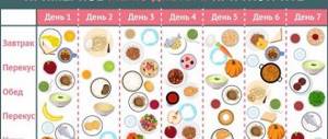
Start reducing your portions gradually. 30-50 grams daily. Keep a diary and write down for yourself how much and what you ate. Secondly, stop sleeping after lunch. Do you want to sleep? Look around, there’s probably something waiting in the house, waiting for you to clean, repair, and put it in order. Are you a wonderful host and everything is fine with you? Amazing. Go out and take a walk. A half-hour walk will discourage the desire to sleep.
Read: Where is the stomach: location, structure and functions in the body
If you are not working, try to find something to do: working on a plot of land, carpentry, cars - this is for men. Keep yourself busy with something - it will help you use your energy productively and take your mind off the refrigerator. And it may even bring in some money, at least for gifts for grandchildren. Practice shows that increasing physical activity and working in the house even for 2 hours allows you to burn up to 5 kg. in Week.
Electrical appliances have greatly simplified our lives. We stopped doing laundry, washing dishes, and cleaning the floor. All this is done by smart machines. On the one hand, this is good. But it's good for a really busy, working woman. People began to move less. And man was created for movement, for creation. And it’s good when he has a favorite activity for which smart machines free him. But often this does not happen, and food becomes a hobby.
Read along with this article:
- Why does the size of an adult’s stomach vary from person to person?
- What are proteins and amino acids, and what are the dangers of excess proteins?
- What foods contain collagen? How to eat for health benefits?
- Foods containing large amounts of protein: benefits for the body
- What causes stomach cancer and how to treat it
- The work of the pancreas, its special role and significance for everything...
- Anatomy: where is the human stomach located, what functions does it perform?
- Products that reduce acidity and other ways to combat...
- Where is the stomach: location, structure and functions in the body
Sections of the stomach
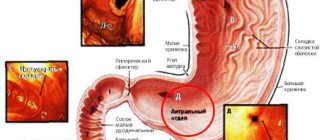
The sections of the stomach differ from each other, both in the structure of the mucous membrane and in acidity, which is measured during FGDS. The cardiac section is separated from the esophagus by a special muscular sphincter, which prevents gastric contents from entering the upper parts of the digestive system.
The largest section of an organ is the body. It is here that most enzymatic reactions take place, hydrochloric acid is secreted and food mass is crushed. The fundus of the stomach is a section of the organ that is topographically located above the cardia, but in its function is almost identical to the body. Gases often accumulate here, which can be detected by x-ray examination.
In the prepyloric part of the stomach, the lumen of the organ narrows. Here the acidity also begins to decrease. Often, endoscopic examination reveals ulcers of the mucous membrane or gastritis.
In the pyloric section, the lumen of the organ narrows significantly. The distal part of the organ is called the antrum (or cave). It is limited by the pylorus - a powerful sphincter that regulates the entry of food into the duodenum.
Gatekeeper part
The pyloric or pyloric part of the stomach is called pars pylorica in Latin. This is the final part of the stomach, it is limited by the pyloric opening, which opens into the duodenum. The pyloric part of the stomach can be divided into the pyloric cave - a place adjacent to the body of the stomach, a canal that is equal in diameter to the duodenum and the pylorus itself - the sphincter that separates the stomach from the intestines, which is facilitated by a special valve. The pyloric sphincter is of great importance: it regulates the flow of food from the stomach into the intestines and prevents it from going back. It should be noted that the environment in the duodenum is radically different from the environment in the stomach - firstly, it is alkaline, not acidic, and secondly, there are aggressive enzymes against which the gastric mucosa is defenseless. If the sphincter does not close completely and food still goes back into the stomach, this can lead to pain and belching.
Activities of the digestive system
The digestive system is the basis for the normal development and existence of the human body. It is through it that all nutrients (carbohydrates, proteins, fats, vitamins) that are necessary for the development of tissues and organs enter the body.
When these molecules are broken down in cells, energy is released, which is used to support all biochemical processes and perform the physical work of the muscles. Also, water and many different salts dissolved in it are absorbed in the stomach and intestines. At the same time, parts of the digestive system are also capable of releasing unnecessary or harmful compounds from the body.
How to shrink your stomach and lose weight
To reduce the size of your stomach, you can reduce the amount of food you eat.
In order for all the above components to enter the stomach, the diet must be varied. The daily menu may include various cereals, soups, pasta, borscht, stewed vegetables, fresh vegetable salads, meat, fish, etc.
Read: Where is the human stomach: projections, anatomical landmarks
The question is - how much? Each person decides for himself individually. Someone eats a lot, does not gain weight, and does not suffer from it. Some people eat little and don’t lose weight. Much depends on individual vital activity, on the energy consumption of a particular organism.
But let's return to the stomach. How much can it hold? As practice shows, up to 3 liters. But is it necessary to overload it like that? Let's calculate how much the average Russian eats for lunch. A plate of borscht or soup – 300 grams. Another 250 grams will be the second, 100 grams - bread. 650-700 grams is half the stomach full. And that's enough. This is the norm that doctors constantly repeat. Traditional healers echo them, persuading us not to overeat. Compote, jelly or cocoa, that is, the third, you can drink about two hours later, as an afternoon snack.
Most nutritionists believe that breakfast should be hearty. What does tight mean? Should I eat more? Organize your breakfast according to the principle: less is more. In other words, make your breakfast energy-rich. Let the food be smaller in volume so as not to waste extra energy carrying a heaviness in the stomach. But energetic. A difficult working day lies ahead. You need concentration and physical strength. A buttered bun with a banana on top wouldn't hurt here; or a cake, a piece of chocolate or a chocolate candy.
It's great if you have walnuts in your diet. This is a charge for the whole day. Do you like salads? Please. But only from sweet fruits and sour cream. Save your diet cabbage salads for the evening. So in the morning you will eat no more than 350 grams (counting tea or coffee), and recharge your energy. By the way, hot drinks in the morning help the body warm up and invigorate.
Snacks are not forbidden, but this does not mean that you have to run to the refrigerator every half hour. Have one snack between breakfast and lunch. And one - between lunch and dinner.
This can be a saucer of vinaigrette or a nutritious salad, but not more than 100 grams. Between lunch and dinner, the snack should be light in calories. Fruits, a glass of juice, vegetable salad. And for dinner, it is advisable to reduce proteins, fats and carbohydrates to a minimum. Let it be salads and fruit juices with light bran bread. And also no more than 300 grams. Help your body get ready for the coming sleep.
Read: How to reduce the size of your stomach in different ways
Blood supply
The blood supply to the stomach is provided by branches of the abdominal aorta - the proper hepatic, left gastric and splenic arteries.
The left gastric artery reaches the lesser curvature of the stomach from the cardiac side. It supplies blood to the lower part of the esophagus, the cardia, and also partially the body and fundus.
The right gastric artery departs from the proper hepatic artery. It passes between the leaves of the hepatoduodenal ligament and gives off branches to the body of the stomach and the pylorus. Short gastric arteries (2-6 pieces) supply blood to the bottom of the organ. They arise from the splenic artery.
In the greater curvature, the gastroepiploic arteries pass through the thickness of the peritoneum, which arise from the splenic or gastroduodenal. They form anastomoses with other branches of vessels in the body and pylorus.
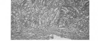
Sampling of stomach tissue for histology: methodology
To perform a histological examination of stomach tissue, it is necessary to perform a biopsy of the organ. In most cases, it is performed using endoscopy. The apparatus for performing FEGDS is placed into the lumen of the stomach and several pieces of organ tissue are cut off. It is advisable to take biopsies from several distant sites. In some cases, tissue for histological examination is taken during surgery. After this, thin sections from the biopsy are taken in the laboratory and examined under a microscope.
Stomach glands
The mucous membrane of the organ contains more than 14-15 million different glands. They are usually divided into the following groups:
- Cardiac - located in a similar department. They produce mucoid secretions, bicarbonates, and chlorides. This secretion reduces stomach acidity and also protects the mucous membrane.
- Fundic or own glands are located in the body and fundus. They are the most numerous. Contains several groups of cells:
- chief cells - produce pepsinogen (an inactive form of the enzyme);
- parietal – hydrochloric acid;
- cervical mucous membranes – young, functionally immature cells;
- accessory mucous cells - produce mucus and bicarbonates;
- endocrine cells - secrete biologically active molecules (somatostatin, motilin, gastrin, substance P, enteroglucagon.
3 . Pyloric glands are located in the same section of the stomach. Their secretion reduces the acidity of the food mass before it enters the duodenum. Produce bicarbonates, mucus and local hormones.
Histological characteristics[ | ]
Histologically, only 3 sections of the stomach are distinguished, since the mucous membrane of the fundus and body of the stomach have a similar structure and are considered as a single whole.
The wall of the stomach is formed by four membranes: mucous, submucosal, muscular and serous.
Mucous membrane[ | ]
The relief of the mucous membrane (MS) of the stomach is characterized by longitudinal folds, fields, and dimples. It consists of three layers: epithelial, lamina propria and muscular lamina.
Epithelium[ | ]
Single-layer highly prismatic (columnar) glandular enterodermal type, lining the gastric pits, the same in all parts of the stomach. All its cells secrete a special mucous secretion onto the surface of the CO, covering the CO with a continuous layer 0.5 mm thick. Bicarbonate, diffusing into mucus, neutralizes hydrochloric acid. For the secretion of enzymes and hydrochloric acid into the lumen of the stomach, temporary channels are formed in the mucus layer. Mucus forms a protective barrier that prevents mechanical damage to CO and its digestion by gastric juice. This barrier is damaged by alcohol and non-steroidal anti-inflammatory drugs.
The integumentary epithelium is completely renewed within 1-3 days.
Own record[ | ]
It is formed by loose fibrous unformed connective tissue with a large number of blood and lymphatic vessels, contains diffuse accumulations of lymphoid tissue and individual lymph nodes. In the form of thin layers it passes between the glands of the stomach, which occupy the main part of this layer.
Stomach glands[ | ]
The number of glands is about 15 million - simple, tubular, branched and unbranched (in the area of the fundus of the stomach; divided into cardiac, proper (fundic) and pyloric.
- cardiac
- tubular, with highly branched end sections, often having a wide lumen. They are located in the cardiac part of the stomach, similar to the cardiac glands of the esophagus. They contain mucous cells (with a light LC, a flattened nucleus, lying basally), which produce mucoid secretion, bicarbonates and chlorides of potassium and sodium. There are also separate chief, parietal and endocrine cells (see below). - own (fundic) glands
are unbranched, located in the body and fundus of the stomach and numerically predominate over other types of glands.
In groups of 3-7 they flow into small gastric pits. They have a narrowed neck, an elongated body and a bottom. They consist of 5 types of cells: main, parietal (lining), cervical, accessory mucous and endocrine. chief cells
are most numerous in the area of the body and bottom of the gland. They have a pyramidal or cylindrical shape and a large core located basally. The CP is basophilic, granular, in the basal part of the cell and around the nucleus it contains a lot of grEPS, a well-developed CG, in which large secretory zymogenic granules are formed (containing pepsinogen and other proenzymes), accumulating in the apical part of the cell and released into the lumen of the gland. In the lumen of the stomach, pepsinogen is converted into active pepsin under the influence of hydrochloric acid. - parietal (parietal) cells
- predominate in the area of the gland body. Larger than the main ones. They have a rounded shape with a narrow apex facing the lumen of the gland, with which they protrude between the main cells, located outward from them. The nucleus lies at the center of the cell. In the oxyphilic CP there are many large MTX with developed cristae and special intracellular secretory tubules in the form of narrow slits into which numerous microhairs face. - Along the periphery of the tubules there is a tubulovesicular complex - a system of membrane vesicles and tubules (a reserve of the membrane containing ion pumps), which merge with the tubules during active secretion. Parietal cells through the apical pole secrete hydrogen and chlorine ions, which, taken together, represent hydrochloric acid, creating an acidic (pH<2) environment in the lumen of the stomach. The latter provides: destruction of proteins;
- the conversion of pepsinogen to pepsin and the optimum pH for the activity of the latter;
- inhibition of the growth of pathogenic microorganisms.
Through the basal plasmalemma, the parietal cell secretes bicarbonate ions, which are carried by capillaries of the lamina propria to the basal surface of the integumentary cells, transporting them to the mucus, where they neutralize hydrochloric acid. Parietal cells also synthesize and secrete an antianemic factor, which, having formed a complex with vitamin B12, is absorbed in the ileum and is necessary for normal hematopoiesis. If this factor is insufficient (as a result of autoimmune damage to parietal cells or after removal of the stomach), pernicious anemia develops. Parietal cell secretion is stimulated by histamine, gastrin and acetylcholine.
- mucous cervical cells
are few in number and located in the cervix. Small sizes. The cytoplasm is weakly basophilic, granular, contains a moderately developed grEPS and a large supranuclear CG, from which large mucous granules are separated, accumulating at the apical pole. These cells often divide and are considered cambial elements of the epithelium of the glands and the integumentary epithelium of the stomach, where they, upon differentiation, migrate. Cell renewal in the glands occurs much more slowly than in the surface epithelium. The mucus produced by the cervical cells may protect them from damage. - accessory mucous cells
- located in the body of their own glands and have a compacted nucleus in the basal part of the cells. In the apical part of these cells, many round and oval granules and a small amount of Mx and KG were found. - endocrine cells
- located in the bottom of the glands. Light, varied in shape (triangular, oval or polygonal). The apical pole contains the nucleus and does not always reach the lumen of the gland. In the basal pole there are dense secretory granules released into the blood. The granules are covered with a membrane, colored with silver and chromium salts and contain peptide hormones and amines. They belong to the DES and APUD system, and are divided into EC, ECL and G cells. They produce hormones that affect secretory activity and motility of the digestive tract:
- D: somatostatin (body, pyloric region) - inhibits the secretion of GEP cells and gastric glands;
- EC: serotonin, motilin, substance P (body, pyloric region) - enhances intestinal motility;
- ECL: histamine (body) - increases the secretion of hydrochloric acid by the stomach;
- pyloric glands
are tubular, with highly branched and convoluted terminal sections. Located in the pyloric region. They flow into very deep gastric pits. They are formed by mucous cells, the secretion of which protects CO from acidic gastric juice. Most glands do not contain parietal cells. Mainly represented by endocrine cells (see above):
- D: somatostatin (body, pyloric region) - inhibits the secretion of GEP cells and gastric glands;
- EC: serotonin, motilin, substance P (body, pyloric region) - enhances intestinal motility;
- G: gastrin (pyloric region) - increases the secretion of hydrochloric acid and pepsinogen by the stomach;
- L: enteroglucagon (pyloric) - stimulates glycogenolysis in the liver.
Muscular plate[ | ]
The muscular plate CO is formed by three layers of smooth muscle cells (inner and outer circular and middle longitudinal). From it, thin bundles of smooth muscle cells are directed into the spaces between the glands.
Submucosa[ | ]
The submucosa of the stomach is well developed. It is formed by loose fibrous unformed connective tissue, which is rich in elastic fibers, blood and lymphatic vessels (arterial, venous and lymphatic plexuses) and nerves (submucosal plexus). The submucosa is involved in the formation of gastric folds.
Muscular membrane[ | ]
The muscular lining of the stomach is formed by smooth muscle tissue, forming 3 layers: outer - longitudinal, middle - circular, inner - oblique. The first two of them are a continuation of the same layers of the muscular lining of the esophagus. Longitudinal muscle bundles are located mainly near the lesser and greater curvature of the stomach. Individual muscle bundles are more developed in the area of the pylorus.
The circular layer is most developed in the pyloric region, where its fibers form the constrictor (sphincter) of the pylorus (thickness 3-5 mm). When it contracts, the exit from the stomach closes. Obliquely oriented muscle fibers as part of the structure of the walls of the digestive canal are found only in the stomach. They “throw over” the cardial part of the stomach to the left of the cardial opening and then go down and to the right in the thickness of the stomach walls towards the greater curvature. Between the muscle layers is the muscular nerve plexus.
Serous membrane[ | ]
The outer serous membrane is represented by the peritoneum. It is formed by a layer of mesothelium and underlying connective tissue. Only narrow strips of the stomach walls, located on the lesser and greater curvature, are devoid of serous cover. In these places, blood vessels and nerves approach the stomach. The serosa is separated from the muscularis by a thin subserosal base.
Prevention of violations
To avoid stomach problems, you need to follow a few simple recommendations:
- Be careful about your diet. Avoid excessive consumption of alcoholic and carbonated drinks, canned and pickled foods, and hot spices.
- The diet must be complete, containing the required amount of nutrients, microelements and vitamins.
- Pay attention to your diet. It is recommended that an adult patient eat 3-5 times a day at approximately the same time of day.
- If you are taking certain medications (anti-inflammatory, steroids, anticoagulants, or antiplatelet medications) for a long time, ask your doctor if you need additional antisecretory medications.
- Quit smoking. It has been proven that nicotine and tars contained in tobacco smoke can contribute to the disruption of the integrity of the gastric mucosa.
- Avoid excessive emotional stress and stress. They lead to activation of the sympathoadrenal system, which increases acidity in the stomach.
- If you experience epigastric pain, vomiting, or loss of appetite, do not self-medicate, but consult a qualified doctor as soon as possible.
Physiology[ | ]
The glands of the gastric mucosa secrete gastric juice containing the digestive enzymes pepsin, chymosin and lipase, as well as hydrochloric acid and other substances. Gastric juice breaks down proteins and partially fats, and has a bactericidal effect.
Main article: Gastric acidity
Due to the muscular layer, the stomach mixes food and gastric juice, forming chyme - a liquid pulp, which is removed in separate portions from the stomach through the pyloric canal. Depending on the consistency of the incoming food, it lingers in the stomach from 20 minutes (fruit juices, as well as vegetable juices and broths) to 6 hours (pork).
In addition, the stomach wall absorbs carbohydrates, ethanol, water and some salts.
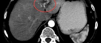
How long does a histological analysis of stomach tissue take?
If cancer is suspected, gastric histology is necessary. How long does this analysis take? Only the attending physician can answer this question. On average, histology takes about 2 weeks. This applies to planned studies, for example, when removing a polyp.
During surgery, urgent histological examination of the tissue may be necessary. In this case, the analysis takes no more than half an hour.