A malignant neoplasm, most often formed from squamous epithelial cells, is esophageal cancer. The tumor usually occurs in people after 45–55 years of age, predominantly in men. The explanation is the fact that they often abuse tobacco, alcohol, marinades and spices, which leads to the degeneration of esophageal cells into cancer. The prognosis will be favorable if a person seeks medical help in a timely manner - before the tumor reaches a significant size and secondary foci of cancer appear.
Causes of esophageal cancer
The causes of cancer of the esophageal mucosa, as well as other oncological pathologies, are not precisely known. The effect of irritating factors on the mucous membrane plays a huge role. Chemical, mechanical or thermal effects provoke the development of an inflammatory process - esophagitis, and subsequently cell dysplasia begins. Cellular changes due to the influence of negative factors increase and lead to malignant degeneration of organ tissue and the development of oncology.
The development of esophageal cancer can be triggered by numerous factors:
- Heredity.
- Human papillomavirus (HPV).
- Injury to an organ caused by foreign objects or swallowing solid food.
- Organ burn. This can be systematic eating of very hot dishes, and accidental consumption of hot liquids that cause a chemical burn. Usually these are alkalis, the consequences of consumption of which may appear years later.
- Unhealthy diet. Food oversaturated with spicy marinades, molds, and nitrates. A deficiency of fresh vegetables/fruits, as well as Se and other minerals, negatively affects the digestive system.
- Vitamin deficiencies. A deficiency of vitamins A, B, E causes the body's defenses to weaken. Cells are reborn.
- Frequent consumption of alcohol is one of the main risk factors. Alcoholics suffer from cancer of the digestive system 12 times more often. Strong alcohol burns the mucous membrane and makes it thinner.
- Tobacco smoking is another factor that causes cancer. Tobacco smoke contains carcinogens that cause negative processes in epithelial cells. Smokers get cancer 4 times more often.
Since a cancerous tumor usually occurs against the background of chronic esophagitis, modern medicine considers pathologies in which prolonged inflammation in the esophagus is observed as predisposing to oncology or precancerous conditions (such conditions include Barrett’s esophagus). Cancer of the digestive system may be associated with negative changes in the p53 gene, which, just like in pancreatic cancer, provoke an increase in the abnormal p53 protein, which does not cope with its functions and does not protect tissues from tumor formation. The cause of the development of oncology can also be HPV (human papillomavirus) - this microscopic organism, in particular, was identified in cancer patients living in China.
Causes of esophageal cancer
Diet is considered the main cause of esophageal cancer. Eating too hot, spicy food and alcohol can be a starting point for the development of cancer. Maintaining proper nutrition will reduce the risk of developing a dangerous disease.
Read here: Kidney cancer: symptoms and signs
The next factor influencing esophageal cancer is territorial affiliation. Studies have shown that residents of Japan, Iran, and Iraq are most susceptible to esophageal cancer due to eating mainly pickled, spicy foods. Low levels of vitamin consumption also contribute to the development of the disease in these areas. This is especially true for vitamins A and C.
According to medical research, esophageal cancer occurs three times more often in women who smoke than in non-smokers. And drinking alcohol increases the risk of developing a malignant tumor by 12 times.
Also affect the appearance of the disease and concomitant diseases. These include Barrett's esophagus.
Classification
Esophageal cancer is classified according to the international TNM nomenclature for malignant neoplasms:
- by stage (T0 - precancer, carcinoma, non-invasive epithelial tumor, T1 - cancer affects the mucous membrane, T2 - tumor grows into the submucosal layer, T3 - layers up to the muscular layer are affected, T4 - tumor penetration through all layers of the esophageal wall into the surrounding tissues);
- by the distribution of metastases in regional lymph nodes (N0 – no metastases, N1 – there are metastases);
- by the spread of metastases in distant organs (M1 – yes, M0 – no metastases).
Cancer can also be classified into stages from first to fourth, depending on the extent of the tumor in the wall and its metastasis.
Classification of esophageal tumors
Experts, trying to more accurately determine the structure, location and shape of a tumor in the tissues of the esophagus, resort to various existing classifications of cancer in this organ. However, the most popular and in demand are the following classifications of cancer in the esophagus:
- According to the characteristics of tumor growth:
- the growth of the primary focus of cancer directly into the lumen of the esophageal tube is an exophytic variant of cancer, the tumor seems to rise above the surrounding part of the mucosa;
- if the tumor forms in the thickness of the tissues of the esophagus or in the submucosal layer of the organ - an endophytic form of cancer;
- in cases where it is difficult to say unambiguously in which direction the tumor growth predominates, we will be talking about a mixed form of cancer.
- According to the structure and structure, esophageal cancer can be:
- squamous - cancer cells arise from elements of the squamous epithelium covering the esophagus from the inside, as a rule, this is either a superficial form of cancer or a deeply invasive variant of the tumor;
- adenocarcinoma - a cancer focus is formed from gland cells capable of secreting mucus in the esophagus; the tumor is prone to rapid progression and severe complications.
Squamous cell carcinoma of the esophagus is also usually divided into two more forms - keratinizing or non-keratinizing. According to the location of the tumor in the esophagus - in its upper part, middle, or lower segment.
The prognosis for five-year survival of patients directly depends on the stage at which the cancer was diagnosed. A favorable outcome is possible if the first signs of cancer were correctly assessed and treatment measures were carried out in full. Whereas an advanced case of cancer leads to rapid death.
The first symptoms of esophageal cancer
The danger of esophageal cancer is that 40% of cases of the disease are asymptomatic. The tumor is discovered accidentally during a chest x-ray. Very often, signs of the disease appear in the later stages, when treatment is difficult. Therefore, it is very important not to miss the first symptoms of cancer.
The first signs of esophageal cancer:
- Dysphagia is difficulty swallowing food. Appears when the tumor has blocked 70% of the esophagus. First, discomfort occurs when hard food passes through the esophagus, and then when swallowing liquid. Unlike stomach cramps, dysphagia is permanent.
- Pain behind the sternum. Often this is a burning sensation that appears while eating and radiates to the back. This indicates that an ulcer has appeared on the surface of the tumor.
- Esophageal vomiting. Regurgitation of small undigested portions of food.
- Unpleasant putrid odor from the mouth. Its appearance is due to the fact that food stagnates in the esophagus.
- Weight loss is caused by insufficient supply of nutrients to the body due to narrowing of the esophagus.
Retention of food above the narrowing of the esophagus provokes vomiting, belching of saliva and mucus. When pain appears behind the sternum with radiation to the area between the shoulder blades, when eating food and/or salivation, this means that esophagitis has developed - the formation has begun to grow into neighboring organs. If the tumor is localized in the area of the cardia (the transition between the esophagus and the stomach), the first symptom may be problems with swallowing and moving food, as well as regular belching of air and bad breath.
When a malignant neoplasm grows beyond the boundaries of the digestive system, it can put pressure on the respiratory tract, and breathing problems will appear. It also presses or grows into the nerve trunks, which are located next to the wall of the esophagus, the person wheezes, he begins to cough, and Horner's syndrome develops.
A sign of the last stage of cancer is unbearable pain and disruption of the functioning of neighboring organs. If negative symptoms develop, diagnosis must be carried out. Therefore, it is very important that diagnosis is carried out at an early stage, this increases the chances of recovery. Do not ignore strange signs and sensations; as soon as symptoms of the disease develop, you should urgently visit a doctor.
Forecast
Modern methods of therapy and innovative technologies in medicine make it possible to completely recover from the disease.
To do this, you should seek help from specialists in a timely manner so that doctors can provide quality medical care in the early stages of development.
The big problem is the asymptomatic course of the disease. This complicates the recovery process, since patients live with a dangerous disease inside without knowing it.
Quite often, patients turn to specialists with an already advanced form of cancer. This is due to the lack of a clinical picture in the early stages.
The main task is to eliminate the tumor surgically, preventing metastases from spreading.
If metastases form, there is no point in performing surgical treatment. In this case, radiation or chemotherapy is prescribed.
These methods allow you to prolong the patient’s life by a year. If these procedures were performed, followed by surgery, life expectancy increases by 5 years.
Therefore, when answering the question of how stage 3 esophageal cancer is eliminated, how long people live with it and what the prognosis is, it is worth paying attention to these indicators.
Stages of cancer
Modern medicine defines 4 stages of esophageal cancer:
- Stage 0. Malignant cells are located on the surface and do not spread into the submucosal layer.
- Stage 1. The tumor spreads deep into the mucosa, but does not affect the muscle layer. There is no metastasis. The sick person does not experience negative symptoms, but the tumor is noticeable during endoscopy.
- Stage 2. Sometimes problems with swallowing may develop, but usually the pathology occurs without symptoms. Stage 2 is divided into substages. Substage IIA. The neoplasm grows into the muscles and connective tissue layer of the organ, but does not affect surrounding organs, and there is no metastasis. Substage IIB is characterized by the fact that the tumor grows into the mucosa, and there are metastases in nearby lymph nodes.
- Stage 3. Doctors diagnose severe problems swallowing food, weight loss, and other symptoms of cancer. Metastasis to surrounding organs and nearby lymph nodes is observed. Treatment is very difficult and the prognosis is poor.
Stages and grades of esophageal cancer
Modern medicine defines 4 stages of esophageal cancer:
- At the first stage, the patient may not notice any changes in his body. When eating solid food, he has to drink liquid so that the food can reach the stomach.
- In the second stage of esophageal cancer, the patient may begin to have problems with nutrition. Many patients at this stage of cancer switch to liquid foods, purees and cereals.
- At the third stage of esophageal cancer, patients experience a narrowing of the food passage, which makes even the process of swallowing liquid difficult and painful.
- At the fourth stage of cancer, the patient experiences complete obstruction of the esophagus.
Esophageal cancer stage 1
The first stage of esophageal cancer is very often not accompanied by pronounced symptoms. The malignant neoplasm is very small in size and practically does not bother the patient. At this time, damage occurs to the mucous membranes of the walls of the esophagus, as well as the submucosa. A cancerous tumor at the first stage does not grow into the muscular layer of the esophagus and therefore responds very well to surgical treatment. Patients do not experience a narrowing of the lumen of the esophagus; they can eat well, as they do not experience discomfort either during meals or after meals.
Esophageal cancer stage 2
At the 2nd stage of development of esophageal cancer, the following organs are affected:
- mucous membranes of the walls of the esophagus;
- muscular membranes;
- submucosa.
At this time, the malignant neoplasm does not extend beyond the affected esophagus. In many patients, the lumen of the esophagus narrows, and therefore they have to switch to liquid food. When examining a patient, specialists can detect single metastases that affect regional lymph nodes.
Esophageal cancer stage 3
At the 3rd stage of development, the malignant neoplasm grows into all layers of the walls of the esophagus. In patients, the tumor affects the serosa, as well as the peri-esophageal tissue. As cancer develops, the lumen of the esophagus narrows and patients have problems with nutrition, as swallowing solid foods becomes problematic for them. In parallel, tumor metastasis occurs (metastases are found in regional lymph nodes). Organs adjacent to the esophagus are not damaged at this stage of cancer development.
Esophageal cancer stage 4
At stage 4 of esophageal cancer, patients experience metastasis of the tumor, which affects both regional and distant lymph nodes. The cancerous tumor spreads to the periesophageal tissue. A malignant neoplasm also affects the walls of the esophagus, the serosa and nearby organs. Most patients at this stage of cancer develop an esophageal-tracheal or esophageal-bronchial fistula.
Diagnostics
The patient is prescribed a number of diagnostic measures that will determine the exact type of tumor, its stage of development and location:
- X-ray (this is done using a contrast agent, which makes the esophagus visible on the X-ray). Using this study, specialists determine the location of the malignant neoplasm, its shape and size. Thanks to the x-ray, the oncologist can foresee possible complications that the type of cancer being examined will cause;
- Laparoscopy. This type of diagnosis makes it possible to identify metastases in the patient’s internal organs;
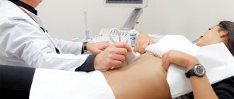
- Ultrasound examination. Through this study, specialists determine the exact size of the malignant neoplasm, as well as the presence of lymph nodes that are affected by metastases;
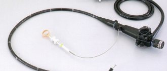
- Tomography (performed using an optical sensor). This technique was developed relatively recently by scientists and almost immediately began to be used in specialized medical institutions. Using an endoscope, a specialist examines the structure of the tumor. Thanks to the latest equipment, it is possible to determine the structure of tumor tissue to a depth of 1.5-2 mm. All information collected by the sensor is transferred to a computer, after which it is decrypted by a specialist. In the case where such equipment is installed in a medical institution, patients may not undergo a biopsy, since the data obtained about the tumor is sufficient to prescribe therapy. Patients are also prescribed positron emission tomography. Immediately before the examination, the patient is injected with glucose (radioactive). Its property is that it can selectively accumulate in cancer cells. The patient is placed in the center of a specially equipped room, and a scanner begins to rotate around him, which takes pictures of the cancerous tumor (it recognizes tumors whose size is 5-10 mm);
- Laparoscopy. With this diagnostic technique, the patient is punctured in the abdominal cavity (in the navel area) with a laparoscope needle, after which a tube with an optical device is inserted into the hole. Specialists have the opportunity to determine the location of a malignant neoplasm, its exact size, and also take biological material, which is immediately transferred for histological studies;
- Bronchoscopy. It is prescribed when the doctor suspects metastases in the larynx, trachea, bronchial tree, etc.;
- Esophagogastroduodenoscopy. When conducting this type of examination, specialists carefully examine not only the esophagus, but also other organs of the digestive tract. Thanks to the endoscope, it is possible to examine the inner surface of the esophagus, as well as take biological material for laboratory research (this is carried out under a microscope). Using esophagogastroduodenoscopy, it is possible to identify a malignant neoplasm at an early stage of development and promptly prescribe treatment to the patient, etc.
Patients are required to undergo a complete laboratory examination, which includes:
- blood chemistry;
- clinical blood test;
- general urine analysis;
- histological analysis of biopsy;
- tumor markers SCC, CYFRA 21-1, TRA.
Treatment
There are several ways to treat the disease. All of them are aimed at stopping the growth of the tumor and its complete elimination.
The techniques can be used individually or in combination. All measures depend on the degree of damage, the patient’s condition, and diagnostic results.
Surgery
The technique is applicable only in cases where the area of localization of the problem is in the lower or middle parts of the esophagus.
A significant advantage of this technique is the ability to expand the lumen of the organ in order to normalize adequate nutrition.
There are several types of intervention:
- Part of the affected area of the organ is removed, taking 5 cm from both parts on the sides. In some cases, the upper part of the gastric cavity must be removed. Both organs are stitched together.
- Removal is carried out when the middle part of the esophagus is affected. A hole is made in the anterior part of the peritoneum, into the stomach cavity, to provide nutrition by probing. The esophagus is removed. Lymph nodes are often treated. If the outcome of the operation is positive and there are no metastases, after 12 months an artificial organ is created based on the tissues of the small intestine.
Endoscopy
This technology is used in the early development of the disease. The advantage is low trauma. The procedure is carried out in several ways.
The endoscope is inserted through the mouth. A special video camera is attached to the tip of the device for detailed monitoring of the situation.
A laser or surgical sling may be attached. Using special cylindrical instruments, the lumen is expanded.
It is very important to choose the right treatment method; this will allow you to regain the ability to eat solid food fragments in most cases.
Radiation therapy
In some cases, the use of this technique has positive results.
The use of gamma therapy is used as an independent technique or in combination. It stops the growth of cancer cells and helps reduce the size of the tumor.
This technique is not so dangerous for neighboring tissues, since the rays act directly on the tumor. It is often combined with chemotherapy.
This helps to reduce the size of formations and stops the spread of metastases. Applicable to patients who cannot undergo surgery.
Chemotherapy
The treatment method is aimed at suppressing the tumor at the cellular level through exposure to poisonous and toxic substances.
This leads to cell death and stopping their growth. It is not enough to use only one drug.
The technique involves the introduction of a comprehensive set of medications, which should be prescribed exclusively by the attending physician. It is possible to enhance the effect by combining it with radiation therapy.
Combination
Combining radiation with chemotherapy gives the most powerful effect and positive results.
Both procedures are performed several weeks before surgery. This method allows you to enhance the therapeutic effect and gives a significant chance of recovery.
Particular attention is paid to patients to ensure preparatory measures for surgical intervention.
Since the patients’ bodies are severely depleted, they are prescribed courses of vitamin complexes, substances with protein and nutritional mixtures.
If it is possible to feed through the oral cavity, small portions of high-calorie protein foods are administered.
Purified still water, fruit drinks, and juice are offered as drinks. As an alternative, feeding tubes are used.
Under no circumstances should you delay visiting a doctor; this can be dangerous not only for your health, but also for your life.
It is possible to cure oncology, but this will require long and difficult treatment.
Early contact with specialists gives a better chance of recovery while saving life.
Complications
This oncological disease rarely occurs without severe impairment. Typically, complications appear already in the second stage of development of the pathological condition. The most common consequence of tumor formation is esophageal obstruction. In this case, the lumen is blocked by the existing tumor, which is why food from the upper section cannot enter the stomach. In the later stages of development of the oncological process, the patient is not able to eat even pureed foods, which leads to rapid depletion of the body.
Another common complication of this cancer is hemorrhage. The disintegration of the tumor and the formation of an ulcer inevitably predisposes to injury to the affected area of the esophagus. Any rough food can cause excessive bleeding. In some cases, this complication poses a serious threat to the patient's life. Due to the impaired ability to eat food and the gradually developing fear of attacks of suffocation, which characterize conditions when swallowed contents get stuck in the esophagus, rapid loss of body weight is observed. Developing cachexia significantly weakens the body.
In more rare cases, tumor disintegration leads to perforation of the trachea.
Thus, a fistula is formed. Through it, small pieces of food, as well as liquid from the esophagus, can penetrate the trachea. This complication is characterized by the appearance of a severe cough while eating.
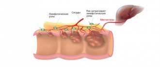
Metastases usually spread from a malignant tumor through the lymphatic system and blood vessels. In later stages, they can enter the brain, heart, lungs, liver and other vital organs, which inevitably leads to the appearance of severe symptoms on their part.
Symptoms
The manifestation of symptoms of benign and malignant disease differs from each other. The observed signs of pathology depend on the location, size and presence of complications. And also in terms of the sequential appearance of symptoms, at what stage of development the tumor is.
Early development of the disease
Detection of a tumor during esophagogastroscopy
Typically, the condition of patients with a benign tumor does not suffer. Rarely does weight loss occur due to anxiety. Growth occurs gradually and for a long time in the pathology of the esophagus, symptoms may not be indicated. Detection occurs during X-ray and endoscopic examinations. Rarely, a tumor can create esophageal obstruction.
In 50% of patients, dysphagia—impaired passage of food—shows up slowly over a number of years.
If the neoplasm reaches a significant size, pain, a feeling of pressure behind the sternum, and dyspeptic symptoms are noted. Sometimes the patient feels an improvement in the passage of food, against the background of a decrease in spasm. If the localization of benign neoplasms of the esophagus is indicated in the cervical region and has a long stalk, then regurgitation of the formation and asphyxia may be observed. Bleeding may occur with polyps. The significant size of the tumor leads to compression of the mediastinal organs, which is manifested by shortness of breath, cough, cyanosis and increased heart rate.
Malignancy of the disease
Among malignant tumors, the most common are cancer and sarcoma of the esophagus. The designated signs of damage to the esophagus are divided into local (the occurrence depends on damage to the walls), secondary (metastases to neighboring organs), and general symptoms. A late sign of the disease is the occurrence of dysphagia, against the background of lumen stenosis. This is due to a lesion blocking at least half the diameter of the esophagus. The following signs may precede the appearance of dysphagia: a feeling of a foreign object when making swallowing movements, a feeling of “scratching” behind the sternum, a feeling of food sticking to the esophageal mucosa.
Chest pain may be one of the signs of an esophageal tumor
After the onset of dysphagia, the foreign body sensation is initially indicated only when swallowing solid food. There is a feeling of delay in a certain section of the esophagus. A sip of water can reduce the severity of the symptom. With further development of the disease, the symptoms of obstruction increase, and even drinking water becomes difficult.
And also with a malignant course of the disease, there is aching pain behind the sternum. The symptom of pain may spread to the back and neck. Pain occurs due to compression of the esophageal mucosa by food against the background of a narrowed passage. If the pain is regular, and eating food does not affect the severity of symptoms or increases them, then this indicates that the tumor has penetrated into nearby organs and tissues. Periesophagitis and mediastinitis develop.
Significant obstruction of the lumen by a tumor and dysphagia lead to regurgitation of food. Sometimes patients artificially induce vomiting to alleviate the condition. Against the background of insufficient nutrition, nervous tension, and symptoms of intoxication, rapid loss of body weight occurs. The patient notes increased salivation.
Complicated forms of malignant progression are usually marked by severe symptoms of pain, dysphagia, and increased intoxication. When the recurrent nerve is irritated or compressed, hoarseness occurs. Affected sympathetic nerve nodes are referred to as Horner's syndrome. Compression of the vagus nerve by the tumor leads to a decrease in heart rate, coughing and vomiting. The growth of a tumor from the esophagus into the larynx leads to a change in voice timbre, shortness of breath and wheezing. If the tumor has penetrated the mediastinum, purulent mediastinitis occurs. By growing into a blood vessel, a malignant tumor can cause fatal bleeding. The connection of the esophagus with the trachea or bronchi through a fistula leads to coughing when drinking liquid. This leads to pneumonia, abscess or gangrene of the lungs.
How to treat esophageal cancer?
The doctor selects treatment methods for esophageal cancer for his patient, guided by the stage of the pathology, the size of the tumor, and the patient’s age. Surgical methods, chemotherapy, and radiation are used. Radiation sessions and chemotherapy may be prescribed before and after surgery.
Surgery involves removing part of the esophagus or the entire organ with tumor-altered tissue. If necessary, part of the stomach is also removed. The esophagus is replaced with part of the intestine or a gastrostomy is formed. There are several types of surgeries performed on patients with esophageal cancer.
Operation
Surgical removal of a tumor of the esophagus is traditionally performed in several ways:
- Extirpation - we are talking about cutting out the affected area with the surrounding fatty tissue and regional lymph nodes. This method is applicable only for 5% of patients due to high morbidity, the presence of serious concomitant pathologies and late diagnosis. After surgery, reconstructive plastic surgery of the larynx is necessary.
- Lewis method - indicated for tumors affecting the middle part of the esophagus. After subtotal resection, gastric tissue plastic surgery is performed as part of one intervention.
- Garlock method - used when cancer is localized in the lower third of the larynx, where there is a transition to the stomach. Resection of the affected areas and the lesser omentum is performed, after which an esophageal-gastric anastomosis is formed.
If possible, the operation is performed endoscopically, choosing one of the following options:
- Resection of the mucosa - a saline solution that lifts the tumor is injected into the submucosal layer located under the tumor, after which the tissue is removed with a polypectomy loop.
- Non-thermal photodynamic destruction - a laser is used that matches the radiation spectrum of the photosensitizer used. The drug is administered 2-3 days before the procedure in order to accumulate it in cancer-affected cells. After treating the tumor with a beam, the drug is activated and pathological structures are destroyed.
- Dilatation using a special medical instrument (bougies, balloon catheters) to expand the narrowed section of the esophagus. The effect of such therapy is short-lived, so it is only advisable as a preliminary stage of endoscopy.
- Recanalization is indicated for both partial and complete occlusion of the lumen of the larynx, if the tumor is localized in the upper third of the organ. The courses involve burning out the formation, the number of sessions is 4. Another option for destroying the malignant structure is the introduction of 96% ethyl alcohol into it every 7 days. The procedure is repeated three times.
- Endoprosthesis replacement helps to consolidate the effect of recanalization and return to normal nutrition. The indication for surgery is the presence of fistulas, which are eliminated by installing plastic tubular prostheses and self-expanding metal stents.
Treatment method
The treatment method for esophageal tumors is based on the use of combination therapy, which is prescribed individually by the treating oncologist. The following techniques are used, discussed in more detail below.
Surgical intervention
Abdominal surgery involves partial or complete removal of the esophagus. If there are affected lymph nodes, the surgeon also removes them. Tissue from the large or small intestine is used to restore the organ.
Surgical removal of the esophagus is possible if the tumor is located in the middle or lower part of the organ. In some cases, the upper part is removed, followed by suturing of the stomach and restoration of normal functioning after the necessary rehabilitation.
Statistics show that the mortality rate for esophageal cancer and surgery does not exceed more than 10%. Contraindications to surgical intervention:
- The presence of a large number of metastases in the lymph nodes and neighboring organs;
- Age category of patients over 70 years;
- Severe chronic diseases in the body;
- Disturbances in the functioning of the cardiovascular system and respiratory organs.
Currently reading: Main causes of pain in the esophagus: proper treatment and reviews from patients
If the malignant tumor is localized in the middle part of the esophagus, then during surgery a special hole is made through which a special probe is inserted and the patient is fed.
With such a diagnosis, it is best recommended to completely remove the esophagus with affected lymph nodes. After 12 months, a re-examination is carried out to identify metastases and, if they are absent, artificial organ replacement is performed (using tissue of the small intestine).
Carrying out endoscopic surgery
Endoscopic surgery is performed for uncomplicated disease. The essence of the technique is that an endoscope equipped with the necessary instruments is inserted into the patient and bougienage is performed.
This operation allows you to restore the lumen in the esophagus and eliminate severe narrowing. In this case, before carrying out the manipulation, a thorough examination of the body and passing the necessary tests is necessary.
Implementation of radiation therapy
Radiation therapy is one of the effective methods of treating esophageal cancer in the presence of contraindications to surgery (respiratory and cardiovascular diseases).
Such treatment is often prescribed in the postoperative period to eliminate possible relapses of the disease and prevent further spread of metastases to neighboring organs and lymph nodes.
If the tumor is inoperable, radiation therapy helps to significantly reduce the formation. The advantage of the technique is that it affects only the affected areas, since healthy cells are not negatively affected.
Chemotherapy
Chemotherapy is one of the main methods of eliminating malignant tumors and is based on the use of drugs of cytostatic origin containing toxic substances that can destroy cancer cells.
Moreover, such therapy also affects healthy cells in the body, which is accompanied by a large number of side effects (hair loss, development of stomatitis, significant decrease in immunity). These phenomena are reversible, so their recovery is possible immediately after treatment.
Chemotherapy is necessary if the following forms are present:
- Small cell cancer;
- A poorly differentiated form of the oncological process.
Treatment is carried out according to the following algorithm:
- In the second and third stages of cancer, the prescribed drugs destroy cancer cells and prevent further development of the tumor;
- In the fourth stage of cancer, treatment is palliative in nature and is aimed at prolonging the patient’s life while slowing the growth of the resulting tumor.
The most popular drugs prescribed for chemotherapy: Vindesine, Mitomycin, Farmorubicin, Bleomycin, 5-fluorouracil.
Diet and nutrition
Proper nutrition is important during the recovery period for esophageal cancer.
It is necessary to select dishes in such a way that they fully provide the body with all the components necessary for the normal functioning of internal organs. In this case, you should avoid eating rough food.
Basic nutritional recommendations for esophageal cancer include:
- Eating pureed food. This facilitates its passage through the esophagus and increases the absorption of nutrients.
- Dishes should not contain particles that could block the narrowed lumen.
- The total weight of food consumed per day should not exceed 3 kg.
- The amount of liquid is limited to 6 glasses, and the liquid included in soups is also taken into account.
- The number of meals should be at least 6. In this case, the portions should be small.
- The temperature of the food should be medium. Excessively hot and cold foods increase discomfort.
Almost all patients with esophageal cancer experience nutritional deficiencies, which negatively affects the functioning of internal organs and their mental state.
Therefore, it is necessary to adhere to the proposed nutritional principles constantly. The doctor may also recommend a course of use of vitamin-mineral complexes, which will have a positive effect on overall well-being and reduce the likelihood of developing anemia and hypovitaminosis.
Causes
It is impossible to single out one main reason leading to the formation of cancer cells in the walls of the esophagus.
The disease can occur under the influence of many provoking factors, and the risk of its development increases many times over if the human body is simultaneously affected by a whole group of negative conditions.
Most often, esophageal cancer is diagnosed:
- In people whose smoking experience amounts to tens of years. Tobacco contains carcinogenic substances that settle on the walls of the esophagus and lead to abnormal changes in epithelial cells. It has been established that in people who smoke, malignant neoplasms of the esophagus develop 4 times more often.
- With alcohol abuse. Drinks containing alcohol burn the esophagus, and this leads to an atypical proliferation of squamous epithelium. In chronic alcoholics, esophageal cancer is detected 12 times more often.
- With improper and irrational nutrition. The development of esophageal cancer is affected by constant consumption of pickled, too spicy and hot foods. Eating foods containing mold and lack of fresh plant foods in the diet has an adverse effect. The listed dietary habits are typical for residents of Central Asia, Japan, China, and some regions of Siberia, therefore in these areas the number of patients with esophageal cancer is tens and hundreds of times higher.
- After thermal and chemical burns of esophageal tissue. A burn can also be a consequence of constantly eating too hot food. If concentrated alkalis are accidentally ingested, a cancerous tumor in the esophagus can be discovered several years later.
- In people with vitamin deficiencies. The mucous layer of the esophagus needs vitamins such as A and E in sufficient quantities; they participate in creating the natural protective barrier of the organ. If vitamin deficiency is observed for a long time, then the cells of the organ cease to perform their function and degenerate.
There is also a hereditary predisposition to the development of cancer in the esophagus. Scientists were able to isolate a mutation in the p53 gene, which leads to the production of an abnormal protein. This protein disrupts the natural defenses of esophageal tissue against cancer cells.
Human papillomavirus is detected in the blood of many patients with malignant tumors of the esophagus, so it can be suggested that this microorganism can also give impetus to cell degeneration.
Esophagitis and a condition such as Barrett's esophagus can precede the development of cancer. Esophagitis is characterized by the constant reflux of hydrochloric acid into the esophagus, which irritates the walls of the organ.
Stomach diseases and obesity lead to esophagitis. Barrett's esophagus is a complication of esophagitis and is manifested by the replacement of stratified epithelium with columnar epithelium.
How long do people live with esophageal cancer: life prognosis
A complete cure for esophageal cancer is possible. The sooner the patient seeks help, the higher the chances that the tumor can be completely destroyed and prevent relapses.
Esophageal cancer has a relatively slow course and, in comparison with other oncological diseases, an average degree of malignancy. Often the disease manifests itself only in the later stages and when the patient seeks help, the disease is already advanced. If left untreated, the prognosis is always unfavorable and the life expectancy is about 6-8 months. From the moment the disease occurs, life expectancy without treatment is 5-6 years.
If the tumor has grown greatly and metastasized, then there is no point in operating on it. In this case, radiation therapy prolongs life by up to 12 months in 10% of cases. Modern techniques make it possible to improve these indicators.
In patients who underwent surgery and underwent a course of chemotherapy and radiation therapy, survival rate of more than 5 years is:
- at stage I more than 90%;
- at stage II - 50%;
- at stage III - about 10%.
The success of treatment depends on the characteristics of the disease: the rate of tumor growth, its prevalence, the presence of metastases and concomitant diseases, and the general condition of the person.
Life prognosis for esophageal cancer in women
The doctor, after analyzing the symptoms of esophageal cancer in women, studying the test results, and after prescribing the necessary treatment, can give an approximate prognosis of the patient’s future condition.
Depending on the stage of esophageal cancer and the development of the tumor, as well as a set of parameters such as the woman’s age, the presence of concomitant diseases, and other indicators, this may indicate possible life expectancy:
- after a full course of treatment at stage 1 of the oncological process, about 90% of patients can expect a full recovery;
- at stage 2 this figure is reduced to 50%;
- at stage 3, no more than 10% of patients survive the five-year mark;
- in the last stage, the average life expectancy rarely exceeds 8 months.
A preventive medical examination, as well as early diagnosis of the disease, will allow for simplified treatment with high results of effectiveness.
What is the prognosis for grade 3?
In stage 3 esophageal cancer, the tumor has grown through all layers of the esophagus and affected surrounding organs.
At this stage, metastases are detected in the nearest lymph nodes. If, for health reasons, the patient can undergo surgery, then it will be an extensive intervention. The surgeon will remove a significant portion of the esophagus and lymph nodes. In this case, about 10% of patients live more than 5 years.
If the tumor has affected vital organs, then supportive (palliative) treatment is prescribed. In this case, life expectancy is 8-12 months.
Therapeutic measures
The main treatment for esophageal cancer is surgery.
Given the fact that the risk of complications with a benign formation is high, treatment of an identified tumor of the esophagus is carried out by surgical intervention. If the formation is small and located on a thin stalk, then removal is performed using an endoscope. With an intramural location, treatment is carried out by enucleation (removal of only the affected area) of the formation, without violating the integrity of the mucous membrane of the esophagus. Treatment using alternative medicine can have a certain effect only after medical intervention.
Treatment of malignant tumors is performed with surgery, supported by radiation and chemotherapy. The dose of radiation therapy before surgery is 3000-5000 rad. The effect of the manipulations will be high if the tumor has not yet had time to grow into neighboring organs and tissues and there are no metastases.
The most common surgical treatment involves resection of the affected area of the esophagus and creation of a replacement graft from the greater curvature of the stomach.
If the body is inoperable, supportive operations are performed. To restore the passage of food, a gastrostomy tube is placed. There is no point in using radiation therapy when metastases are detected in distant lymph nodes. Chemotherapy is used: 5-fluorouracil, ftorafur with methotrexate and colhamine. The effect of drug therapy is minimal.
Carrying out chemotherapy for esophageal cancer
Prevention
To prevent cancer, it is enough to start watching your diet and give up bad habits. It is necessary to limit the consumption of spicy and hot foods, pickled foods. People with a predisposition to the disease should pay special attention to nutrition.
When detected in time, the disease is successfully cured. If you follow the advice and recommendations of doctors, oncology can be avoided.
Lung cancer Rectal cancer Esophageal hernia Esophageal candidiasis Stomach cancer Gastroesophageal reflux disease